
The family Echinostomatidae is the most numerous within the class Trematoda, Echinostoma paraensei Lie & Basch, 1967 is a widely distributed member of this family, with a typical life cycle with six larval stages, many of them studied by some authors (Kanev et al., 2000KANEV, I., STERNER, M., RADEV, V. and FRIED, B., 2000. An overview of the biology of echinostomes. In: B. FRIED and T.K. GRACZYC. Echinostomes as experimental models for biological research. Netherlands: Kluwer Academic Publishers, p. 1-29.; Kostadinova and Gibson, 2000KOSTADINOVA, A. and GIBSON, D.I., 2000. The systematics of the echinostomes. In: B. FRIED and T.K. GRACZYC. Echinostomes as experimental models for biological research. Netherlands: Kluwer Academic Publishers, p. 31-57.; Pinheiro et al., 2004PINHEIRO, J., MALDONADO JÚNIOR, A.J., ATTIAS, M. and LANFREDI, R.M., 2004. Morphology of the rediae of Echinostoma paraensei (Trematoda: Echinostomatidae) from its intermediate host Lymnaea columella (Mollusca, Gastropoda). Parasitology Research, vol. 93, no. 3, pp. 171-177. PMid:15127294. http://dx.doi.org/10.1007/s00436-004-1110-z.
http://dx.doi.org/10.1007/s00436-004-111...
, 2005PINHEIRO, J., MALDONADO JÚNIOR, A.J., ATTIAS, M. and LANFREDI, R.M., 2005. Ultrastructure of the miracidium of Echinostoma paraensei Lie and Basch, 1967 (Trematoda, Echinostomatidae). Parasitology Research, vol. 97, no. 5, pp. 367-372. PMid:16151745.; Toledo et al., 2007TOLEDO, R., MUÑOZ-ANTOLI, C. and FRIED, B., 2007. The use of Echinostomes to study host-parasite relationships between larval trematodes and invertebrates and cold-blooded vertebrate hosts. Parasitology Research, vol. 100, no. 6, pp. 1177-1185. PMid:17279393. http://dx.doi.org/10.1007/s00436-007-0470-6.
http://dx.doi.org/10.1007/s00436-007-047...
). The taxonomic features of the Echinostoma genus are complex, so are those of E. paraensei, described as a synonymy with E. caproni by Kanev (1985)KANEV, I., 1985. On the morphology, biology, ecology and taxonomy of the Echinostoma revulutum group (Trematoda: Echinostomatidae: Echinostoma). Bulgaria: University of Sophia. PhD Thesis. and Christensen et al. (1990)CHRISTENSEN, N.O., FRIED, B. and KANEV, I., 1990. Taxonomy of 37- collar spined Echinstoma (Trematoda: Ec2hinostomatidae) in studies on the population regulation in experimental rodent hosts. Angewandte Parasitologie, vol. 31, no. 3, pp. 127-130. PMid:2291497..
The E. paraensei cercariae were obtained from ‘Laboratório de Biologia e Parasitologia de Mamíferos e Reservatório’, IOC, FIOCRUZ, Rio de Janeiro state.
The specimens were observed using a Quanta 250 scanning electron microscope at 12.50 kV.
The cercariae present elongated body, with muscular striations all over it, which probably accompany the musculature. For E. paraensei, the fin-fold pattern is demonstrated in Figure 1, with two dorsal, two ventral, and two lateral folds. Fin folds present a different striation pattern, lengthwise, differing from the body muscular striation.

SEM of Echinostoma paraensei cercaria detailing the six fin folds. The main image shows the ventral fin folds (vff), the lateral fin folds (lff), and the dorsal fin folds (dff).
The body of E. paraensei cercariae presents a well developed, detached collar spine that partially surrounds the oral sucker, as indicated in Figure 2. This sucker is not prominent off the body as the ventral fin fold, which is protuberant. The tail is long with a thicker tip that narrows at the end.
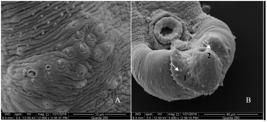
The collar spine of Echinostoma paraensei. (A) shows that the collar ends at the ventro-lateral region of the cercaria body; (B) a view of the spine collar, completely detached from the body and around the oral sucker.
The perioral region presents a sequence of papillae, forming a circle around this region (Figure 3). The papilla emerges from a pore situated in a bulbous protuberance (Figure 3B). These papillae are unicilliated and are also present in the collar, surrounding it and completing a full circle in the ventral region. There are more of them distributed in the tegument of the ventral and dorsal regions; they decrease in number along the cercarial body and completely disappear at the acetabulum region. The position and structure of these papillae suggest their sensorial role.
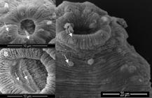
Details of the papillae of the oral and ventral suckers. The main image shows uniciliated papillae. (A) detail of the uniciliated papillae of the oral sucker, surrounding it; (B) the papillae of the ventral sucker. These papillae do not present any cillia, forming bulbous protrusions in the internal region of the sucker.
Internally, in the acetabulum, a range of papillae can be observed as bulbous projections surrounding the inner edge of the sucker, as shown in Figure 2. These papillae do not present any cilia, but a small round projection. The differences between the papillae of these regions may be related to the function they hold in each of them: in the anterior region, they have a sensory function because they are used to explore the environment in search for food; whereas in the acetabular portion, their function may be associated with recognition of the site of helminth infection and fixation, because the frontal part must be more involved in perception and orientation in order to find the second intermediate host.
The presence of a tegumentary fold on the tail of E. revolutum was also observed by Lie and Basch (1967)LIE, K.J. and BASCH, P.F., 1967. The life history of Echinostoma paraensei sp. n. (Trematoda: Echinostomatidae). The Journal of Parasitology, vol. 53, no. 6, pp. 1192-1199. PMid:6078607. http://dx.doi.org/10.2307/3276679.
http://dx.doi.org/10.2307/3276679...
, being used as a taxonomic criteria in the description of this species. According to Kanev et al. (1993)KANEV, I., EISENHUT, U., OSTROWSKI DE NUNEZ, M., MANGA GONZALEZ, M.Y., TZOLOV, D., DIMITROV, V. and RADEV, V., 1993. Description of the tail and fin folds of Echinostoma revolutum cercariae from its type locality (Trematoda: Echinostomatidae). Annales de Parasitologie Humaine et Comparée, vol. 68, no. 3, pp. 125-127. http://dx.doi.org/10.1051/parasite/1993683125.
http://dx.doi.org/10.1051/parasite/19936...
, Echinostoma revolutum (Froelich, 1802) cercariae have been described with different numbers and arragenment of fin folds. These data also speculate that the large dorso-ventral fin folds are associated with swimming, and the small ventro-lateral and single papila-like folds are probably sensorial.
The sheath that surrounds the basis of the spine is probably involved in the retraction of the spines. Maldonado Júnior et al. (2001)MALDONADO JÚNIOR, A., LOKER, E.S., MORGAN, J.A., REY, L. and LANFREDI, R.M., 2001. Description of the adult worm of a new Brazilian isolate of Echinostoma paraensei (Platyhelminthes: Digenea) from its natural vertebrabe host Nectomys squamipes by light and scanning electron microscopy and molecular analysis. Parasitology Research, vol. 87, no. 10, pp. 840-848. PMid:11688891. http://dx.doi.org/10.1007/s004360100451.
http://dx.doi.org/10.1007/s004360100451...
also described a protrusion around the spine in the adult worm. This characteristic may represent a way to avoid the loss of spines, considering that contraction can protect the spines from the attrition on rough surfaces. It can also protect them from the flow of food that occurs in the intestine of the host.
There was a lack of morphological studies on the cercaria of E. paraensei, as well as on descriptions of their external ensorial structures. This work shows and describes these features for the first time, complementing the studies on the Echinostomatidae family.
-
(With 3 figures)
References
- CHRISTENSEN, N.O., FRIED, B. and KANEV, I., 1990. Taxonomy of 37- collar spined Echinstoma (Trematoda: Ec2hinostomatidae) in studies on the population regulation in experimental rodent hosts. Angewandte Parasitologie, vol. 31, no. 3, pp. 127-130. PMid:2291497.
- KANEV, I., 1985. On the morphology, biology, ecology and taxonomy of the Echinostoma revulutum group (Trematoda: Echinostomatidae: Echinostoma) Bulgaria: University of Sophia. PhD Thesis.
- KANEV, I., EISENHUT, U., OSTROWSKI DE NUNEZ, M., MANGA GONZALEZ, M.Y., TZOLOV, D., DIMITROV, V. and RADEV, V., 1993. Description of the tail and fin folds of Echinostoma revolutum cercariae from its type locality (Trematoda: Echinostomatidae). Annales de Parasitologie Humaine et Comparée, vol. 68, no. 3, pp. 125-127. http://dx.doi.org/10.1051/parasite/1993683125
» http://dx.doi.org/10.1051/parasite/1993683125 - KANEV, I., STERNER, M., RADEV, V. and FRIED, B., 2000. An overview of the biology of echinostomes. In: B. FRIED and T.K. GRACZYC. Echinostomes as experimental models for biological research Netherlands: Kluwer Academic Publishers, p. 1-29.
- KOSTADINOVA, A. and GIBSON, D.I., 2000. The systematics of the echinostomes. In: B. FRIED and T.K. GRACZYC. Echinostomes as experimental models for biological research Netherlands: Kluwer Academic Publishers, p. 31-57.
- LIE, K.J. and BASCH, P.F., 1967. The life history of Echinostoma paraensei sp. n. (Trematoda: Echinostomatidae). The Journal of Parasitology, vol. 53, no. 6, pp. 1192-1199. PMid:6078607. http://dx.doi.org/10.2307/3276679
» http://dx.doi.org/10.2307/3276679 - MALDONADO JÚNIOR, A., LOKER, E.S., MORGAN, J.A., REY, L. and LANFREDI, R.M., 2001. Description of the adult worm of a new Brazilian isolate of Echinostoma paraensei (Platyhelminthes: Digenea) from its natural vertebrabe host Nectomys squamipes by light and scanning electron microscopy and molecular analysis. Parasitology Research, vol. 87, no. 10, pp. 840-848. PMid:11688891. http://dx.doi.org/10.1007/s004360100451
» http://dx.doi.org/10.1007/s004360100451 - PINHEIRO, J., MALDONADO JÚNIOR, A.J., ATTIAS, M. and LANFREDI, R.M., 2004. Morphology of the rediae of Echinostoma paraensei (Trematoda: Echinostomatidae) from its intermediate host Lymnaea columella (Mollusca, Gastropoda). Parasitology Research, vol. 93, no. 3, pp. 171-177. PMid:15127294. http://dx.doi.org/10.1007/s00436-004-1110-z
» http://dx.doi.org/10.1007/s00436-004-1110-z - PINHEIRO, J., MALDONADO JÚNIOR, A.J., ATTIAS, M. and LANFREDI, R.M., 2005. Ultrastructure of the miracidium of Echinostoma paraensei Lie and Basch, 1967 (Trematoda, Echinostomatidae). Parasitology Research, vol. 97, no. 5, pp. 367-372. PMid:16151745.
- TOLEDO, R., MUÑOZ-ANTOLI, C. and FRIED, B., 2007. The use of Echinostomes to study host-parasite relationships between larval trematodes and invertebrates and cold-blooded vertebrate hosts. Parasitology Research, vol. 100, no. 6, pp. 1177-1185. PMid:17279393. http://dx.doi.org/10.1007/s00436-007-0470-6
» http://dx.doi.org/10.1007/s00436-007-0470-6
Publication Dates
-
Publication in this collection
21 Sept 2017 -
Date of issue
May-Aug 2018
History
-
Received
29 July 2016 -
Accepted
29 Nov 2016


