Abstract
The objective of the present study was to analyse the bioactive compounds of the leaves of Conocarpus lancifolius (C. lancifolius). The GC-MS analysis of the hot methanolic extract of the leaves (HMEL) of C. lancifolius exhibited the bioactive compounds such as 1-(3-Methoxy-2-nitrobenzyl) iso quinoline, morphin-4-ol-6,7-dione, 1-bromo-N-methyl-, phytol, hexadecanoic acid, 2,3-dihydroxypropyl ester, 2,2':4',2”-terthiophene, ethyl iso-allocholate, caryophyllene oxide, campesterol, epiglobulol, cholestan-3-ol, 2-methylene-, (3á,5à)-, dasycarpidan-1-methanol, acetate (ester) and oleic acid, eicosyl ester. The FT-IR analysis of HMEL of C. lancifolius showed a unique peak at 3184, 2413, 1657 cm-1 representing coumaric acid, chlorogenic acid and ferulic acid. The HMEL of C. lancifolius was actively inhibiting the proliferation of breast cancer cells MCF-7 ATCC at the concentration of 72.66 ± 8.21 µg/ml as IC50 value. The HMEL of C. lancifolius also revealed a good spectrum of activity against Gram-positive and Gram-negative bacterial cultures screened in this work. The activity observed has shown more or less similar effects against screened bacteria. However, the magnitude of potentiality was significantly lesser compared to standard ciprofloxacin disc at p< 0.001 level (99% confidence intervals). Furthermore, the study demonstrating the bioactive compounds can be isolated from the leaves of C. lancifolius.
Keywords:
plants; leaves; bioactive molecules; multiple drug resistance; anticancer; antibacterial
Resumo
O objetivo do presente estudo foi analisar os compostos bioativos das folhas de Conocarpus lancifolius (C. lancifolius). A análise por GC-MS do extrato metanólico quente das folhas (HMEL) de C. lancifolius exibiu os compostos bioativos como 1- (3-Metoxi-2-nitrobenzil) isoquinolina, morfina-4-ol-6,7- diona, 1-bromo-N-metil-, fitol, ácido hexadecanoico, 2,3-di-hidroxipropil éster, 2,2 ': 4', 2 ” - tertiofeno, isoalocolato de etil, óxido de cariofileno, campesterol, epiglobulol, colestano -3-ol, 2-metileno-, (3á, 5à) -, dasycarpidan-1-metanol, acetato (éster) e ácido oleico, éster eicosílico. A análise FT-IR de HMEL de C. lancifolius mostrou um pico único em 3184, 2413, 1657 cm-1 representando ácido cumarico, ácido clorogênico e ácido ferúlico. O HMEL de C. lancifolius inibiu ativamente a proliferação de células de câncer de mama MCF-7 ATCC na concentração de 72,66 ± 8,21 µg / ml como valor de IC50. O HMEL de C. lancifolius também revelou bom espectro de atividade contra culturas de bactérias Gram-positivas e Gram-negativas rastreadas neste trabalho. A atividade observada mostrou efeitos mais ou menos semelhantes contra bactérias rastreadas. No entanto, a magnitude da potencialidade foi significativamente menor em comparação com o disco de ciprofloxacina padrão em nível de p < 0,001 (intervalos de confiança de 99%). Além disso, o estudo demonstrando os compostos bioativos pode ser isolado das folhas de C. lancifolius.
Palavras-chave:
plantas; folhas; moléculas bioativas; resistência a múltiplos medicamentos; anticâncer; antibacteriano
1. Introduction
Multiple drug resistance (MDR) is one of the foremost therapeutic problems associated with anticancer drug therapy and antibiotics / antibacterial therapy. Therefore, several researchers across the world aimed to develop novel bioactive molecules against cancer and infectious bacterial organisms. Nature is blessed with abundant medicinal plants and trees with unique medicinal values. Traditionally, herbs and medicinal plants were the main resources for curing many diseases across the world in ancient times. Therefore, natural products play a significant role in maintaining a healthy life (Moni et al., 2019MONI, S.S., HADI SULTAN, M., MAKEEN, H.A., JABEEN, A., SANOBAR, S., SIDDIQUI, R., UR REHMAN, Z., ALAM, M.S., AHMAD, S., ELMOBARK, M.E. and MOOCHIKKAL, R., 2019. Phytochemical and spectral analysis of the methanolic extracts of leaves of Murraya koenigii of Jazan, Saudi Arabia. Natural Product Research, pp. 1-5. http://dx.doi.org/10.1080/14786419.2019.1679137. PMid:31631708.
http://dx.doi.org/10.1080/14786419.2019....
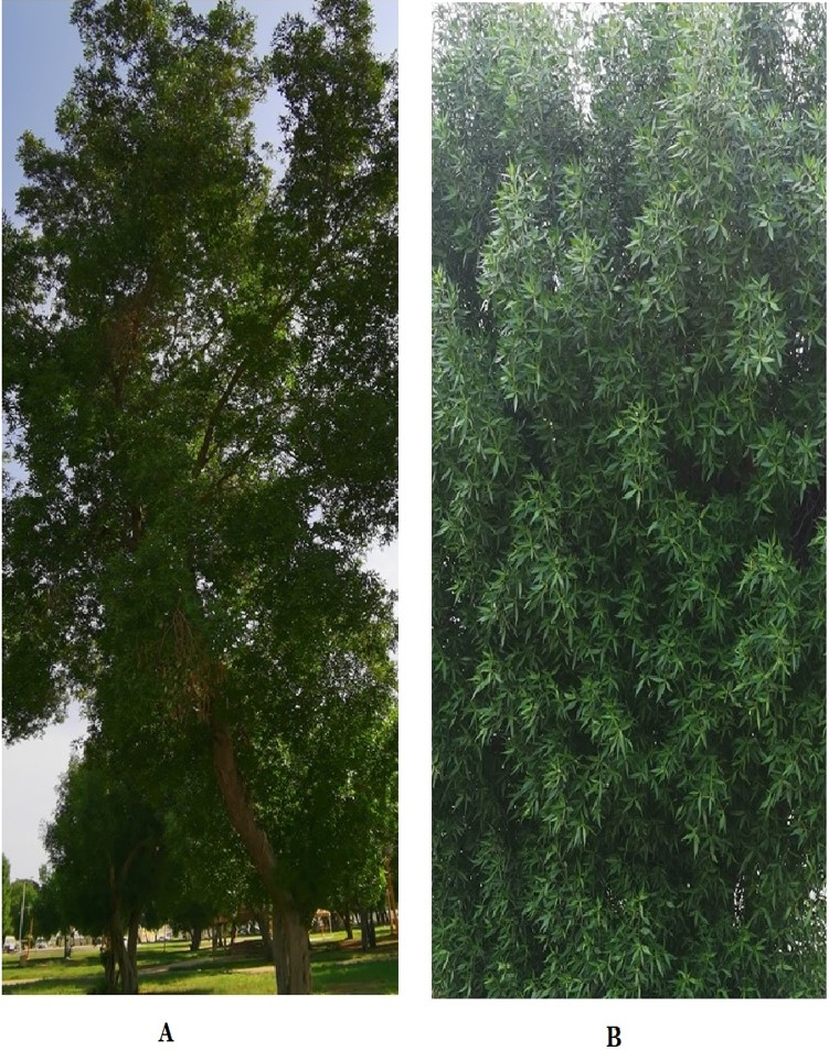
). C. lancifolius is a greenish tree that grows around 10 to 30 meters and is widely distributed in Jazan, Saudi Arabia (Figure 1). C. lancifolius, one of the two species in the genus Conocarpus, is a tree that belongs to the family Combretaceae. The tree is fast-growing, heat tolerant, and therefore has been grown in most of the parks and roadsides in Jazan region. It has been reported for medicinal value, especially anti-diabetic and cytotoxicity property (Al-Taweel et al., 2016AL-TAWEEL, A.M., PERVEEN, S., FAWZY, G.A., MEHMOOD, R., KHAN, A. and KHAN, S.I., 2016. New ellagic acid derivative from the fruits of heat-tolerant plant Conocarpus lancifolius Engl. and their anti-inflammatory, cytotoxic, PPAR agonistic activities. Pakistan Journal of Pharmaceutical Sciences, vol. 29, no. 5, (suppl.), pp. 1833-1837. PMid:28476711.). Studies suggested that C. lancifolius is used as a food for cattle goats, sheep, and camels in some countries; its wood is used to produce burning fuel, coal and build ships (Wensvoort, 2008WENSVOORT, J., 2008. Browse silage in the United Arab Emirates. Wildlife Middle East News, vol. 3, no. 1, pp. 1-2.). It has been reported as a medicinal plant for the therapy of diabetes mellitus, anaemia, flu, fever, skin ulcers, and syphilis (El-Sayed et al., 2012EL-SAYED, S.A.H., SALIH, A.B., MOHAMED, M.S., MORTADA, M.E.S. and EMAN, A.E.W., 2012. Phytochemical studies and evaluation of antioxidant, anticancer and antimicrobial properties of Conocarpus erectus L. Growing in Taif, Saudi Arabia. European Journal of Medicinal Plants, vol. 2, no. 2, pp. 93-112. http://dx.doi.org/10.9734/EJMP/2012/1040.
http://dx.doi.org/10.9734/EJMP/2012/1040...
; Malik et al., 2014MALIK, S., BASHIR, A.C., MUHAMMAD, U. and KHURRAM, A., 2014. Antidiabetic potential of Conocarpus lancifolius. Bangladesh Journal of Pharmacology, vol. 9, pp. 244-249. http://dx.doi.org/10.3329/bjp.v9i2.18556.
http://dx.doi.org/10.3329/bjp.v9i2.18556...
). However, there are very limited reports showing the medicinal value of C. lancifolius grown in Jazan, Saudi Arabia. This made us to analyse the bio constituents and their medicinal properties to document its therapeutic value.

The tree Conocarpus lancifolius located in Jazan city, Kingdom of Saudi Arabia (A) Aerial view of the tree shows many bunches of stems with leaves (B) Landscape view of the tree which shows the bunches of the leaves arises from the stem of tree.
2. Materials and methods
2.1. Collection, identification, and extraction
Jazan city of Jizan province is located in the southwest part of Saudi Arabia, situated in the Red sea's coastal region. It has a natural habitat of C. lancifolius and is planted around the city. The tree branches of C. lancifolius was collected by breaking from its main branch and pooled. The collected samples were packed in polythene biohazard yellow bags, tied, and transported to the working laboratory. The leaves of C. lancifolius were plucked out from the collected branches and washed thoroughly in normal tap water to remove the impurities. Later the leaves were further washed in Millipore water and air-dried by spreading on the clean floor at room temperature under the shade for 3 weeks. A herbarium curator identified the washed specimens in the Herbarium of Jazan University (JAZUH) and the reference number was 1213 JAZUH. The voucher specimen was also deposited at the herbarium of Jazan University for future reference.
The air-dried leaves were cut into small pieces and powdered using a grinder. The coarsely powdered samples obtained were pooled and kept in an airtight container. Extraction was carried out using a continuous percolation technique with methanol as a solvent. 200g of dried leaves powder was subjected to Soxhlet extraction with methanol at 60° C for 4 hours. After extraction, the extracts were transferred in a separate glass beaker and kept open for the evaporation of the solvent via an air-drying process. After complete air drying of the leaves extract, dried samples were collected by scraping from the glass beakers and pooled and weighed. The dried powder of the methanolic extract of the leaves (HMEL) of C. lancifolius was subjected to various phytochemical analysis, FT-IR and GC-MS studies to determine the various bio compounds present in C. lancifolius.
2.2. Spectral analysis of the HMEL
2.2.1. Gas chromatography-Mass spectrometry (GC-MS) analysis
A Thermo Scientific GC-MS, AS 3000 autosampler, trace ultra-GC, and ISQ detector were used to identify the structural aspects of the methanolic extract of bioactive compounds. The instrument equipped with a TR 5MS capillary column with dimensions of 30 m length x 0.25 mm diameter (internal diameter) x 0.25 µm (film thickness) was used for the separation of the components. Helium, at a flow rate of 1.2 mL/min (constant flow mode), was used as the carrier gas. A volume of 2 µL of sample extract was injected in spitless mode. The injection port was set at 320 ºC, and the temperature of the oven was initially set at 70 ºC for 5 minutes. The oven temperature was subsequently ramped to 205º C at the rate of 5º C/min for 5 minutes; 280º C at the rate of 5 o C/min for 5 minutes, 290º C at the rate of 5 ºC/min for 5 minutes, and finally to 300º C at the rate of 5ºC/min for 5 minutes. The maximum oven temperature was set at 320 º C. The mass spectrometer was operated in an electron ionization (EI) mode within the mass range of 60-900 amu with 0.6 scan times (min). The MS transfer line temperature and ion source temperature were kept at 320o C and 350oC, respectively, with an electron multiplier voltage of 1 Kv. The mass spectrophotometer was operated and spectral analysis was performed using Xcalibur software. Interpretation of the mass spectrum was determined by using inbuilt software libraries such as NIST, REPLIB, and MAINLIB. The constituent percentages were measured based on the peak area.
2.2.2. Fourier transform infrared spectroscopy studies (FT-IR)
The leaf powder was analyzed through FT-IR spectroscopy (Nicolet iS10 FT-IR spectrophotometer) using the pressed pellet technique. The pellet sample's spectra were obtained through the FT-IR spectrophotometer against reference KBr pellet in the range of 400 - 4000 cm-1 with a resolution of 4 cm-1.
2.3. Cytotoxic effect of the HMEL
In this work, the human breast cancer cell line MCF-7 cell line was cultured and maintained in RPMI-1640 medium at pH 7.4. Further, 10% fetal bovine serum (FBS) was added with RPMI-1640 medium as a supplement. Penicillin (100 U/ml) and streptomycin (100 g/ml) were added to the medium. The cells were incubated in a CO2 incubator (Heraeus, GmbH, Germany) at 37ºC with 90% humidity and 5% CO2. The Cells were treated with HMEL of C. lancifolius by dissolving in DMSO while the untreated control cultures received only the vehicle (DMSO < 1%).
The cytotoxic profile of the HMEL of C. lancifolius, was assessed by MTT assay (Syam et al., 2012SYAM, S., ABDELWAHAB, S., AL-MAMARY, M.A. and MOHAN, S., 2012. Synthesis of chalcones with anticancer activities. Molecules (Basel, Switzerland), vol. 17, no. 6, pp. 6179-6195. http://dx.doi.org/10.3390/molecules17066179. PMid:22634834.
http://dx.doi.org/10.3390/molecules17066...
; Gloria et al., 2019GLORIA, I., BERTOCCHI, M., ANDREANI, G., FARRUGGIA, G., CAPPADONE, C., SALAROLI, R., FORNI, M. and BERNARDINI, C., 2019. Cytotoxic Effects of Artemisia annua L. and Pure Artemisinin on the D-17 Canine Osteosarcoma Cell Line. Oxidative Medicine and Cellular Longevity, vol. 2019, pp. 1615758. http://dx.doi.org/10.1155/2019/1615758. PMid:31354901.
http://dx.doi.org/10.1155/2019/1615758...
). Briefly, 1 × 10⁶ cells/ml was seeded in 96 well microtiter plates for both, and various gradient concentrations of samples were plated out in triplicates and incubated in a CO2 inhibitor for 48 h. Each plate included untreated cell controls and blank cell-free control. After incubation, MTT (5 mg/ml) was added to each well, and the plates were incubated for a further 4 h after which the media was removed. DMSO (100 µl) was added into each well to solubilize the formazan crystals. The absorbance was read at a wavelength of 570 nm using a microtiter plate reader (BioTek Instruments, USA). The percentage of cellular viability was calculated with the appropriate controls taken into account. The experiment was done in triplicate, and the following formula was used to calculate the inhibitory rate of cell proliferation: Growth inhibition = (OD control – OD treated) / OD control × 100. The cytotoxicity of the sample on cancer cells was expressed as IC50 values, the sample concentration reducing the absorbance of treated cells by 50% concerning untreated cells.
2.4. Morphological changes in treated cells
Human breast cancer cells MCF-7 was cultured in RPMI-1640 medium at pH 7.4 by placing in 24 well culture plates and incubated overnight in a CO2 incubator. The cells were then treated in two batches with methanolic extract of C. lancifolius at IC50 and kept for 48 hr. After the treatment, the first batch treated cells were directly photographed and observed for morphological changes under 20 & 40X magnifications using Nikon Eclipse TS 100 inverted phase-contrast microscope, Nikon cooperation, Japan. Eclipse TS 100 microscope uses eco illumination that provides sufficient brightness for imaging through phase contrast. The morphological characters were confirmed in the second batch by performing a neutral red dye test. The test was performed by removing the media, and cells were washed with cold, sterile PBS. The washed cells were treated with 0.5% neutral red at a volume of 100 µl to each well and stained for 2 minutes. The stains were washed out with PBS and immediately observed under phase contrast inverted microscope under 20 & 40X magnification using Nikon Eclipse TS 100 inverted phase-contrast microscope, Nikon cooperation, Japan.
2.5. Antibacterial studies
2.5.1. Bacterial strains used and standardization
An equal set of Gram-positive and Gram-negative bacteria was employed in this study, in line with the earlier report (Okhundedaev et al., 2019OKHUNDEDAEV, B.S., BACHER, M., MUKHAMATKHANOVA, R.F., SHAMYANOV, I.J., ZENGIN, G., BÖHMDORFER, S., MAMADALIEVA, N.Z. and ROSENAU, T., 2019. Flavone glucosides from Artemisia juncea. Natural Product Research, vol. 33, no. 15, pp. 2169-2175. http://dx.doi.org/10.1080/14786419.2018.1490901. PMid:30422004.
http://dx.doi.org/10.1080/14786419.2018....
). Briefly, 24 h cultures of Staphylococcus aureus, Streptococcus pyogenes, Bacillus subtilis, Klebsiella pneumoniae, Escherichia coli, and Pseudomonas aeruginosa were prepared in nutrient broth and incubated at 37 ºC. The cultures were standardized using serial dilution-agar plate analysis. The cultures were briefly diluted serially using sterilized nutrient broth, resulting in working bacterial culture with a concentration gradient of 10-1 to 10-9. Spread plate technique was followed, 100 µl of the working bacterial cultures were spread on agar plates with individual organisms in their respective plates. The working bacterial cultures' potential viability was determined in terms of colony-forming unit per ml (CFU/ml) and tabulated.
2.5.2. Determination of Minimum inhibitory concentration
The minimum inhibitory concentration was performed by the broth tube dilution method. The gradient concentrations of samples were prepared by diluting the Millipore water samples to get a predetermined concentration of 1250, 1000, 750, 500, 250, 125 µg/100 ml. The minimum concentration of inhibiting the bacterial growth was determined based on the visibility of bacterial growth in the broth, and the concentration was noted and used for antibacterial sensitivity studies.
2.5.3. Antibacterial sensitivity studies
A specified quantity of Muller Hinton (MH) agar was dissolved in Millipore water and sterilized in an autoclave. The agar plates were prepared aseptically and the spread plate technique was followed to seed the organisms on individual MH agar plates. The plates were covered with their lids, and the culture was allowed to diffuse through the media for 30 min. Wells of diameter 10 mm were made using a sterile standard borer for the individual organism, the extracts were placed in the respective wells and plates were incubated at 37ºC for 24 h. The antibacterial effects of bioactive extracts were quantified by measuring the zone of inhibition around each well in millimetres (mm).
The Kirby-Bauer technique was employed with standard ciprofloxacin disc (5µg/disc). The technique was performed as described earlier in agar well diffusion procedure, but the ciprofloxacin disc was placed on the agar surface rather than in punched well and the plates were incubated at 37 ºC for 24 h. The spectrum of the antibacterial effect of ciprofloxacin disc was determined by measuring the zone of inhibition around the disc and the values were expressed in millimetres (mm).
3. Results
The phytochemical analysis of the HMEL of C. lancifolius showed various compounds such as carbohydrates, amino acids, alkaloids, tannins, steroids, and saponins. The GC-MS spectral chromatogram (Figure 2) showed possible bioactive compounds in the HMEL of C. lancifolius. Figure 3 depicting the structure of bioactive constituents in the HMEL of C. lancifolius. The potential bioactive compounds are 1-(3-Methoxy-2-nitrobenzyl) isoquinoline, Morphin-4-ol-6,7-dione, 1-bromo-N-methyl-, Phytol, Hexadecanoic acid, 2,3-dihydroxypropyl ester, Ethyl iso-allocholate, Caryophyllene oxide, Epiglobulol, Cholestan-3-ol, 2-methylene-, (3á,5à)-, Dasycarpidan-1-methanol, acetate (ester), Campesterol and Oleic acid, eicosyl ester. The FT-IR spectroscopy of the HMEL of C. lancifolius was displayed with unique fingerprint regions of various bioactive compounds (Figure 4). The respective compounds of FT-IR analysis were tabulated (Table 1) and represents the presence of possible compounds amino acids, coumaric acid, ferulic acid, Fatty acids, aromatic hydrocarbons, quercetin, tannins, cutins and polysaccharides.

GC-MS spectral chromatogram of the methanolic extract of the leaves (HMEL) of Conocarpus lancifolius.

Bioactive components of the hot methanolic extract of the leaves (HMEL) of Conocarpus lancifolius. (1) 1-(3-Methoxy-2-nitrobenzyl) isoquinoline, MF: C17H14N2O3; MW: 294; RT: 54.41 (2) Morphin-4-ol-6,7-dione, 1-bromo-N-methyl-, MF: C17H18BrNO3; MW: 363; RT: 61.07 (3) Phytol, MF: C20H40O; MW: 296; RT: 35.85 (4) Hexadecanoic acid, 2,3-dihydroxypropyl ester, MF: C19H38O4; MW: 320; RT: 46.15 (5) 2,2':4',2”-Terthiophene, MF: C12H8S3; MW: 248; RT: 50.45 (6) Ethyl iso-allocholate, MF: C26H44O5; MW: 436; RT: 56.67 (7) Caryophyllene oxide, MF: C15H24O; MW: 220; RT: 24.32 (8) Epiglobulol, MF: C15H26O; MW: 222; RT: 25.19 (9) Cholestan-3-ol, 2-methylene-, (3á,5à)-, MF: C28H48O; MW: 400; RT: 27.22 (10) Dasycarpidan-1-methanol, acetate (ester), MF: C20H26N2O2; MW: 326; RT: 32.05 (11) Campesterol, MF: C28H48O; MW: 400; RT: 58.60 (12) Oleic acid, eicosyl ester, MF: C38H74O2; MW: 562; RT: 58.08. MF – Molecular Formula; MW – Molecular Weight; RT – Retention Time (Minutes).

FT-IR spectroscopy of the hot methanolic extract of the leaves (HMEL) of Conocarpus lancifolius.
Phytochemical composition of the hot methanolic extract of the leaves (HMEL) of Conocarpus lancifolius through FT-IR analysis.
The results of Table 2 suggest that the HMEL of C. lancifolius was able to inhibit the proliferation of MCF-7 breast cancer cells. The IC50 value of the HMEL of C. lancifolius showed growth inhibition at the concentration of 72.66 ± 8.21 µg/ml (Figure 5). The morphological changes further confirmed the anticancer activity of HMEL of C. lancifolius in MCF-7. The non-treated cells had shown typical morphology of healthy MCF-7 cells (Figure 6A & C). The morphology of control cells was intact, showing clear nucleation and cell differentiation. In contrast, the cells that were treated with extracts had shown detached cells, dead cells, and shrunken cells with condensed or fragmented apoptotic nuclei (Figure 6B & D). The spectrum of antibacterial effect of HMEL of C. lancifolius against selected human pathogenic bacteria including Gram-positive Staphylococcus aureus, Streptococcus pyogenes, and Bacillus subtilis; Gram-negative bacteria Escherichia coli, Klebsiella pneumonia, and Pseudomonas aeruginosa was measured. The study revealed that HMEL of C. lancifolius exhibited an equal spectrum of activity against Gram-positive and Gram-negative bacteria. The zone of inhibition was expressed in millimetre (mm), which was recorded in Table 3.
Cytotoxic effect of methanolic extract of the leaves (HMEL) of Conocarpus lancifolius against treated MCF-7 cells.

The dose response curve of the hot methanolic extract of the leaves (HMEL) of Conocarpus lancifolius against MCF-7 cells.

Morphology of MCF-7 cells unstained and stained with neutral red after 48 h treatment with of the hot methanolic extract of the leaves (HMEL) of Conocarpus lancifolius. (A) Microscopical view of unstained healthy cells at 1 × 10⁶ cells / ml concentration mounted at 20 × magnification under phase contrast microscope (B) Microscopical view of stained healthy cells at 1 × 10⁶ cells / ml concentration mounted at 20 × magnification under phase contrast microscope (C) Microscopical view of unstained cells after the treatment at 1 × 10⁶ cells / ml concentration mounted at 40 × magnification under phase contrast microscope (D) Microscopical view of stained cells after the treatment at 1 × 10⁶ cells / ml concentration mounted at 40 × magnification under phase contrast microscope The arrow arrows of the figure B & D indicate the nuclei with apoptotic nuclear morphology in both un treated and drug treated cells.
The antibacterial activity of the hot methanolic extract of the leaves (HMEL) of Conocarpus lancifolius against selected human pathogenic bacteria.
4. Discussion
In the present study, HMEL of C. lancifolius exhibited 1-(3-Methoxy-2-nitrobenzyl) isoquinoline, a derivative of the alkaloid isoquinoline. In 2013 researchers reported the antibacterial efficacy of novel isoquinoline derivatives (Galán et al., 2013GALÁN, A., MORENO, L., PÁRRAGA, J., SERRANO, A., SANZ, M.J., CORTES, D. and CABEDO, N., 2013. Novel isoquinoline derivatives as antimicrobial agents. Bioorganic & Medicinal Chemistry, vol. 21, no. 11, pp. 3221-3230. http://dx.doi.org/10.1016/j.bmc.2013.03.042. PMid:23601815.
http://dx.doi.org/10.1016/j.bmc.2013.03....
). Phytol is acyclic diterpene alcohol that has been detected in this study. Phytol is produced by all photosynthetic plants closely associated with chlorophyll (Islam et al., 2018ISLAM, M.T., ALI, E.S., UDDIN, S.J., SHAW, S., ISLAM, M.A., AHMED, M.I., CHANDRA SHILL, M., KARMAKAR, U.K., YARLA, N.S., KHAN, I.N., BILLAH, M.M., PIECZYNSKA, M.D., ZENGIN, G., MALAINER, C., NICOLETTI, F., GULEI, D., BERINDAN-NEAGOE, I., APOSTOLOV, A., BANACH, M., YEUNG, A.W.K., EL-DEMERDASH, A., XIAO, J., DEY, P., YELE, S., JÓŹWIK, A., STRZAŁKOWSKA, N., MARCHEWKA, J., RENGASAMY, K.R.R., HORBAŃCZUK, J., KAMAL, M.A., MUBARAK, M.S., MISHRA, S.K., SHILPI, J.A. and ATANASOV, A.G., 2018. Phytol: a review of biomedical activities. Food and Chemical Toxicology, vol. 121, pp. 82-94. http://dx.doi.org/10.1016/j.fct.2018.08.032. PMid:30130593.
http://dx.doi.org/10.1016/j.fct.2018.08....
). Phytol is also reported as a major constituent of plant-derived essential oils, and it has potent antimicrobial properties (Murbach Teles Andrade et al., 2014MURBACH TELES ANDRADE, B.F., NUNES BARBOSA, L., DA SILVA PROBST, I. and FERNANDES JÚNIOR, A., 2014. Antimicrobial activity of essential oils. The Journal of Essential Oil Research, vol. 26, no. 1, pp. 34-40. http://dx.doi.org/10.1080/10412905.2013.860409.
http://dx.doi.org/10.1080/10412905.2013....
). Hexadecanoic acid, 2,3-dihydroxypropyl ester is a fatty acid derivative that has been detected in HMEL of C. lancifolius. Hexadecanoic acid, commonly termed palmitate, has been reported for its antibacterial and antifungal properties (Chandrasekaran et al., 2011CHANDRASEKARAN, M., SENTHIL KUMAR, A. and VENKATESALU, V., 2011. Antibacterial and antifungal efficacy of fatty acid methyl esters from leaves of Sesuvium portulacastrum L. European Review for Medical and Pharmacological Sciences, vol. 15, no. 7, pp. 775-780. PMid:21780546.; Rahuman et al., 2000RAHUMAN, A.A., GOPALAKRISHNAN, G., GHOUSE, B.S., ARUMUGAM, S. and HIMALAYAN, B., 2000. Effect of Feronia limonia on mosquito larvae. Fitoterapia, vol. 71, no. 5, pp. 553-555. http://dx.doi.org/10.1016/S0367-326X(00)00164-7. PMid:11449505.
http://dx.doi.org/10.1016/S0367-326X(00)...
). The presence of palmitic acid has been reported in the leaves of C. lancifolius in 2011 (Redha et al., 2011REDHA, A., AL-MANSOUR, N., SULEMAN, P., AFZAL, M. and AL-HASAN, R., 2011. Leaf Traits and histochemistry of trichomes of Conocarpus lancifolius a Combretaceae in semi-arid conditions. American Journal of Plant Sciences, vol. 2, no. 2, pp. 165-174. http://dx.doi.org/10.4236/ajps.2011.22018.
http://dx.doi.org/10.4236/ajps.2011.2201...
). 2,2':4',2”-terthiophene is an oligomer of the heterocycle thiophene reported for optical properties and anticancer effect (Chiara et al., 2010CHIARA, B.D., FERRO, R., GIUSEPPE DI, S. and RICCARDO, T., 2010. Structural and optical properties of conjugated molecules in Perhydrotriphenylene (PHTP) and in other channel-forming inclusion compounds. ChemInform, vol. 32, no. 43. http://dx.doi.org/10.1002/chin.200143264.
http://dx.doi.org/10.1002/chin.200143264...
; Zhang et al., 2009ZHANG, P., LIANG, D., JIN, W., QU, H., CHENG, Y., LI, X. and MA, Z., 2009. Cytotoxic thiophenes from the root of Echinops grijisii Hance. Zeitschrift fur Naturforschung. C, Journal of biosciences, vol. 64, no. 3-4, pp. 193-196. http://dx.doi.org/10.1515/znc-2009-3-407. PMid:19526711.
http://dx.doi.org/10.1515/znc-2009-3-407...
). Ethyl iso-allocholate, otherwise called ethyl cholate, was observed in the HMEL of C. lancifolius, reported for the antibacterial property (Malathi et al., 2016MALATHI, K., ANBARASU, A. and RAMAIAH, S., 2016. Ethyl Iso-allocholate from a medicinal rice Karungkavuni inhibits dihydropteroate synthase in Escherichia coli: a molecular docking and dynamics study. Indian Journal of Pharmaceutical Sciences, vol. 78, no. 6, pp. 780-788. http://dx.doi.org/10.4172/pharmaceutical-sciences.1000184.
http://dx.doi.org/10.4172/pharmaceutical...
). However, a recent study has demonstrated reduced tumor growth (Thakur and Ahirwar, 2019THAKUR, R.S. and AHIRWAR, B., 2019. A steroidal derivative from Trigonella foenum graecum L. that induces apoptosis in vitro and in vivo. Journal of Food and Drug Analysis, vol. 27, no. 1, pp. 231-239. http://dx.doi.org/10.1016/j.jfda.2018.05.001. PMid:30648576.
http://dx.doi.org/10.1016/j.jfda.2018.05...
). Caryophyllene oxide is a bicyclic sesquiterpene observed in the HMEL of C. lancifolius and has been shown as an anticancer molecule (Pan et al., 2016PAN, Z., WANG, S.K., CHENG, X.L., TIAN, X.W. and WANG, J., 2016. Caryophyllene oxide exhibits anticancer effects in MG-63 human osteosarcoma cells via the inhibition of cell migration, generation of reactive oxygen species and induction of apoptosis. Bangladesh Journal of Pharmacology, vol. 11, no. 4, pp. 817-823. http://dx.doi.org/10.3329/bjp.v11i4.27517.
http://dx.doi.org/10.3329/bjp.v11i4.2751...
). Epiglobulol is a member of the class of compounds known as 5,10-cycloaromadendrane sesquiterpenoids that have been observed in the HMEL of C. lancifolius, which has been reported for antimicrobial activity (Tan et al., 2008TAN, M., ZHOU, L., HUANG, Y., WANG, Y., HAO, X. and WANG, J., 2008. Antimicrobial activity of globulol isolated from the fruits of Eucalyptus globulus Labill. Natural Product Research, vol. 22, no. 7, pp. 569-575. http://dx.doi.org/10.1080/14786410701592745. PMid:18569693.
http://dx.doi.org/10.1080/14786410701592...
). Cholestan-3-ol, 2-methylene-, (3á,5à)-, a saturated tetracyclictriterpene have been determined in the HMEL of C. lancifolius. Cholestan-3-ol has already been reported in Canscora perfoliata Lam. (Gentianaceae) (Thanga Krishna et al., 2012THANGA KRISHNA, K.S., MUTHUKUMARASAMY, S. and MOHAN, V.R., 2012. GC-MS determination of bioactive components of Canscora perfoliata Lam. (Gentianaceae). Journal of Applied Pharmaceutical Science, vol. 2, no. 8, pp. 210-214.). Dasycarpidan-1-methanol, acetate (ester) an alkaloidal compound has been identified in the HMEL of C. lancifolius in this work which is not yet reported. This compound has been reported for exhibiting anti-inflammatory, antibacterial, antifungal, and anticancer properties (Abeer Fauzi Al et al., 2017ABEER FAUZI AL, R., ASHWAK, F.K. and IMAD, H.H., 2017. Phytochemical screening of methanolic leaves extract of Malva Sylvestris. International Journal of Pharmacognosy and Phytochemical Research, vol. 9, no. 4, pp. 537-552. http://dx.doi.org/10.25258/phyto.v9i2.8127.
http://dx.doi.org/10.25258/phyto.v9i2.81...
). Campesterol and Oleic acid, eicosyl ester were identified in the HMEL of C. lancifolius with high retention time. Campesterol is a phytosterol that has not been reported in C. lancifolius. However, the compound has been reported for antioxidant properties (Kmiecik et al., 2011KMIECIK, D., KORCZAK, J., RUDZIŃSKA, M., KOBUS-CISOWSKA, J., GRAMZA-MICHAŁOWSKA, A. and HĘŚ, M., 2011. β-Sitosterol and campesterol stabilisation by natural and synthetic antioxidants during heating. Food Chemistry, vol. 128, no. 4, pp. 937-942. http://dx.doi.org/10.1016/j.foodchem.2011.03.122.
http://dx.doi.org/10.1016/j.foodchem.201...
). Oleic acid, eicosyl ester is a monounsaturated omega-9 fatty acid, that has been reported for having mosquito repellent activity (Gurunathan et al., 2016GURUNATHAN, A., SENGUTTUVAN, J. and PAULSAMY, S., 2016. Evaluation of mosquito repellent activity of isolated oleic acid, eicosyl ester from Thalictrum javanicum. Indian Journal of Pharmaceutical Sciences, vol. 78, no. 1, pp. 103-110. http://dx.doi.org/10.4103/0250-474X.180259. PMid:27168688.
http://dx.doi.org/10.4103/0250-474X.1802...
). The FT-IR spectrum depicted a large parabola-shaped peak observed at 3184 cm-1 with aromatic C-H stretching, vibrations, with weak intensity representing the presence of Coumaric acid, aliphatic compounds (Fachriyah et al., 2019FACHRIYAH, E., AYU, T. and KUSRINI, D., 2019. Identification of Phenolic acid from ethanol extract leaves binahong (Anredera cordifolia (ten) stennis) and antioxidant activity test. Journal of Physics: Conference Series, vol. 1217, pp. 012051. http://dx.doi.org/10.1088/1742-6596/1217/1/012051.
http://dx.doi.org/10.1088/1742-6596/1217...
). However, alkane stretching indicated the presence of steroids, tannins, saponins. The peak at 2413 cm-1 with medium intensity showed O-H (Carboxylic acid), which indicates the presence of Coumaric acid, Chlorogenic acid, and ferulic acid (Gurunathan et al., 2016GURUNATHAN, A., SENGUTTUVAN, J. and PAULSAMY, S., 2016. Evaluation of mosquito repellent activity of isolated oleic acid, eicosyl ester from Thalictrum javanicum. Indian Journal of Pharmaceutical Sciences, vol. 78, no. 1, pp. 103-110. http://dx.doi.org/10.4103/0250-474X.180259. PMid:27168688.
http://dx.doi.org/10.4103/0250-474X.1802...
). The peak at 1657 cm-1 with strong intensity showed C=O (Carboxylic group) with stretching vibration, which demonstrated Quercetin, Coumaric acid, and ferulic acid (Gurunathan et al., 2016GURUNATHAN, A., SENGUTTUVAN, J. and PAULSAMY, S., 2016. Evaluation of mosquito repellent activity of isolated oleic acid, eicosyl ester from Thalictrum javanicum. Indian Journal of Pharmaceutical Sciences, vol. 78, no. 1, pp. 103-110. http://dx.doi.org/10.4103/0250-474X.180259. PMid:27168688.
http://dx.doi.org/10.4103/0250-474X.1802...
).
The cytotoxic effect of HMEL of C. lancifolius may be due to the presence of hexadecanoic acid, 2,3-dihydroxypropyl. Earlier research findings suggested that the hexadecenoic acid was exhibiting cytotoxicity to human leukemia cells at the concentration of 12.5 to 50 µg/ml, but did not show any cytotoxicity to normal human dermal fibroblast (Al-Wahaibi et al., 2020AL-WAHAIBI, L.H.N., MAHMOOD, A., KHAN, M. and ALKHATHLAN, H.Z., 2020. Comparative study on the essential oils of Artemisia judaica and A. herba-alba from Saudi Arabia. Arabian Journal of Chemistry, vol. 13, no. 1, pp. 2053-2065. http://dx.doi.org/10.1016/j.arabjc.2018.03.004.
http://dx.doi.org/10.1016/j.arabjc.2018....
). The anticancer property may also be due to the presence of ethyl iso-allocholate, 2,2':4',2”-terthiophene, caryophyllene oxide, and dasycarpidan-1-methanol, acetate (ester). Ethyl iso-allocholate has been reported for its potent anticancer property and showed the highest cytotoxicity against A549 lung cancer cells in vitro and in vivo (Pan et al., 2016PAN, Z., WANG, S.K., CHENG, X.L., TIAN, X.W. and WANG, J., 2016. Caryophyllene oxide exhibits anticancer effects in MG-63 human osteosarcoma cells via the inhibition of cell migration, generation of reactive oxygen species and induction of apoptosis. Bangladesh Journal of Pharmacology, vol. 11, no. 4, pp. 817-823. http://dx.doi.org/10.3329/bjp.v11i4.27517.
http://dx.doi.org/10.3329/bjp.v11i4.2751...
; Fachriyah et al., 2019FACHRIYAH, E., AYU, T. and KUSRINI, D., 2019. Identification of Phenolic acid from ethanol extract leaves binahong (Anredera cordifolia (ten) stennis) and antioxidant activity test. Journal of Physics: Conference Series, vol. 1217, pp. 012051. http://dx.doi.org/10.1088/1742-6596/1217/1/012051.
http://dx.doi.org/10.1088/1742-6596/1217...
). Studies have demonstrated the cytotoxic effect of thiophenes. Studies also suggested that caryophyllene oxide has proved to have an anticancer effect against MG-63 human osteosarcoma cells (Pan et al., 2016PAN, Z., WANG, S.K., CHENG, X.L., TIAN, X.W. and WANG, J., 2016. Caryophyllene oxide exhibits anticancer effects in MG-63 human osteosarcoma cells via the inhibition of cell migration, generation of reactive oxygen species and induction of apoptosis. Bangladesh Journal of Pharmacology, vol. 11, no. 4, pp. 817-823. http://dx.doi.org/10.3329/bjp.v11i4.27517.
http://dx.doi.org/10.3329/bjp.v11i4.2751...
). The spectrum of antibacterial activity was due to the presence of phytol, hexadecanoic acid, 2,3-dihydroxypropyl ester, and dasycarpidan-1-methanol, acetate (ester). An earlier study suggested that alkaloidal extract from Leaves of C. lancifolius exhibited an antibacterial effect (Hayssam et al., 2013HAYSSAM, M.A., MOHAMED, Z.M.S. and AHMED, A.M., 2013. In-vitro antibacterial activities of alkaloids extract from leaves of Conocarpus lancifolius Engl. Journal of Pure & Applied Microbiology, vol. 7, no. 3, pp. 1903-1907.). The HMEL of C. lancifolius has exhibited several potentially bioactive compounds that were reflected in the cytotoxicity and antibacterial studies.
Conclusion
The need to develop novel drug molecules is increasing in demand gradually and steadily because of the development of MDR against cancer and bacterial infections. Plant resources are remarkable sources for the development of medicinal agents from time immortal. The pharmaceutical importance of C. lancifolius is not yet explored properly. The present study suggested the presence of certain significant compounds in HMEL of C. lancifolius. The bioactive compounds are hexadecanoic acid, sesquiterpenoids, phytol and dasycarpidan-1-methanol, ethyl iso-allocholate, 2,2':4',2”-terthiophene and caryophyllene oxide. The respective compounds were responsible for antimicrobial and cytotoxicity activities. The results obtained in this study are highly promising to develop novel anticancer and antibacterial drug molecules, which will be a significant advancement towards welfare of human society.
Abbreviations
HMEL, hot methanolic extract of the leaves; GC-MS, Gas chromatography–mass spectrometry; FT-IR, Fourier transform - infrared spectroscopy; MDR, Multiple drug resistance; MTT, 3-[4,5-dimethylthiazole-2-yl]-2,5-diphenyltetrazolium bromide; IC, Inhibitory concentration
Acknowledgements
The authors are thankful to Dr. Remesh Moochikkal, Department of Biology, Faculty of Science, Jazan University, Saudi Arabia.
References
- ABEER FAUZI AL, R., ASHWAK, F.K. and IMAD, H.H., 2017. Phytochemical screening of methanolic leaves extract of Malva Sylvestris. International Journal of Pharmacognosy and Phytochemical Research, vol. 9, no. 4, pp. 537-552. http://dx.doi.org/10.25258/phyto.v9i2.8127
» http://dx.doi.org/10.25258/phyto.v9i2.8127 - AL-TAWEEL, A.M., PERVEEN, S., FAWZY, G.A., MEHMOOD, R., KHAN, A. and KHAN, S.I., 2016. New ellagic acid derivative from the fruits of heat-tolerant plant Conocarpus lancifolius Engl. and their anti-inflammatory, cytotoxic, PPAR agonistic activities. Pakistan Journal of Pharmaceutical Sciences, vol. 29, no. 5, (suppl.), pp. 1833-1837. PMid:28476711.
- AL-WAHAIBI, L.H.N., MAHMOOD, A., KHAN, M. and ALKHATHLAN, H.Z., 2020. Comparative study on the essential oils of Artemisia judaica and A. herba-alba from Saudi Arabia. Arabian Journal of Chemistry, vol. 13, no. 1, pp. 2053-2065. http://dx.doi.org/10.1016/j.arabjc.2018.03.004
» http://dx.doi.org/10.1016/j.arabjc.2018.03.004 - CHANDRASEKARAN, M., SENTHIL KUMAR, A. and VENKATESALU, V., 2011. Antibacterial and antifungal efficacy of fatty acid methyl esters from leaves of Sesuvium portulacastrum L. European Review for Medical and Pharmacological Sciences, vol. 15, no. 7, pp. 775-780. PMid:21780546.
- CHIARA, B.D., FERRO, R., GIUSEPPE DI, S. and RICCARDO, T., 2010. Structural and optical properties of conjugated molecules in Perhydrotriphenylene (PHTP) and in other channel-forming inclusion compounds. ChemInform, vol. 32, no. 43. http://dx.doi.org/10.1002/chin.200143264
» http://dx.doi.org/10.1002/chin.200143264 - EL-SAYED, S.A.H., SALIH, A.B., MOHAMED, M.S., MORTADA, M.E.S. and EMAN, A.E.W., 2012. Phytochemical studies and evaluation of antioxidant, anticancer and antimicrobial properties of Conocarpus erectus L. Growing in Taif, Saudi Arabia. European Journal of Medicinal Plants, vol. 2, no. 2, pp. 93-112. http://dx.doi.org/10.9734/EJMP/2012/1040
» http://dx.doi.org/10.9734/EJMP/2012/1040 - FACHRIYAH, E., AYU, T. and KUSRINI, D., 2019. Identification of Phenolic acid from ethanol extract leaves binahong (Anredera cordifolia (ten) stennis) and antioxidant activity test. Journal of Physics: Conference Series, vol. 1217, pp. 012051. http://dx.doi.org/10.1088/1742-6596/1217/1/012051
» http://dx.doi.org/10.1088/1742-6596/1217/1/012051 - GALÁN, A., MORENO, L., PÁRRAGA, J., SERRANO, A., SANZ, M.J., CORTES, D. and CABEDO, N., 2013. Novel isoquinoline derivatives as antimicrobial agents. Bioorganic & Medicinal Chemistry, vol. 21, no. 11, pp. 3221-3230. http://dx.doi.org/10.1016/j.bmc.2013.03.042 PMid:23601815.
» http://dx.doi.org/10.1016/j.bmc.2013.03.042 - GLORIA, I., BERTOCCHI, M., ANDREANI, G., FARRUGGIA, G., CAPPADONE, C., SALAROLI, R., FORNI, M. and BERNARDINI, C., 2019. Cytotoxic Effects of Artemisia annua L. and Pure Artemisinin on the D-17 Canine Osteosarcoma Cell Line. Oxidative Medicine and Cellular Longevity, vol. 2019, pp. 1615758. http://dx.doi.org/10.1155/2019/1615758 PMid:31354901.
» http://dx.doi.org/10.1155/2019/1615758 - GURUNATHAN, A., SENGUTTUVAN, J. and PAULSAMY, S., 2016. Evaluation of mosquito repellent activity of isolated oleic acid, eicosyl ester from Thalictrum javanicum. Indian Journal of Pharmaceutical Sciences, vol. 78, no. 1, pp. 103-110. http://dx.doi.org/10.4103/0250-474X.180259 PMid:27168688.
» http://dx.doi.org/10.4103/0250-474X.180259 - HAYSSAM, M.A., MOHAMED, Z.M.S. and AHMED, A.M., 2013. In-vitro antibacterial activities of alkaloids extract from leaves of Conocarpus lancifolius Engl. Journal of Pure & Applied Microbiology, vol. 7, no. 3, pp. 1903-1907.
- ISLAM, M.T., ALI, E.S., UDDIN, S.J., SHAW, S., ISLAM, M.A., AHMED, M.I., CHANDRA SHILL, M., KARMAKAR, U.K., YARLA, N.S., KHAN, I.N., BILLAH, M.M., PIECZYNSKA, M.D., ZENGIN, G., MALAINER, C., NICOLETTI, F., GULEI, D., BERINDAN-NEAGOE, I., APOSTOLOV, A., BANACH, M., YEUNG, A.W.K., EL-DEMERDASH, A., XIAO, J., DEY, P., YELE, S., JÓŹWIK, A., STRZAŁKOWSKA, N., MARCHEWKA, J., RENGASAMY, K.R.R., HORBAŃCZUK, J., KAMAL, M.A., MUBARAK, M.S., MISHRA, S.K., SHILPI, J.A. and ATANASOV, A.G., 2018. Phytol: a review of biomedical activities. Food and Chemical Toxicology, vol. 121, pp. 82-94. http://dx.doi.org/10.1016/j.fct.2018.08.032 PMid:30130593.
» http://dx.doi.org/10.1016/j.fct.2018.08.032 - KMIECIK, D., KORCZAK, J., RUDZIŃSKA, M., KOBUS-CISOWSKA, J., GRAMZA-MICHAŁOWSKA, A. and HĘŚ, M., 2011. β-Sitosterol and campesterol stabilisation by natural and synthetic antioxidants during heating. Food Chemistry, vol. 128, no. 4, pp. 937-942. http://dx.doi.org/10.1016/j.foodchem.2011.03.122
» http://dx.doi.org/10.1016/j.foodchem.2011.03.122 - MALATHI, K., ANBARASU, A. and RAMAIAH, S., 2016. Ethyl Iso-allocholate from a medicinal rice Karungkavuni inhibits dihydropteroate synthase in Escherichia coli: a molecular docking and dynamics study. Indian Journal of Pharmaceutical Sciences, vol. 78, no. 6, pp. 780-788. http://dx.doi.org/10.4172/pharmaceutical-sciences.1000184
» http://dx.doi.org/10.4172/pharmaceutical-sciences.1000184 - MALIK, S., BASHIR, A.C., MUHAMMAD, U. and KHURRAM, A., 2014. Antidiabetic potential of Conocarpus lancifolius. Bangladesh Journal of Pharmacology, vol. 9, pp. 244-249. http://dx.doi.org/10.3329/bjp.v9i2.18556
» http://dx.doi.org/10.3329/bjp.v9i2.18556 - MONI, S.S., HADI SULTAN, M., MAKEEN, H.A., JABEEN, A., SANOBAR, S., SIDDIQUI, R., UR REHMAN, Z., ALAM, M.S., AHMAD, S., ELMOBARK, M.E. and MOOCHIKKAL, R., 2019. Phytochemical and spectral analysis of the methanolic extracts of leaves of Murraya koenigii of Jazan, Saudi Arabia. Natural Product Research, pp. 1-5. http://dx.doi.org/10.1080/14786419.2019.1679137 PMid:31631708.
» http://dx.doi.org/10.1080/14786419.2019.1679137 - MURBACH TELES ANDRADE, B.F., NUNES BARBOSA, L., DA SILVA PROBST, I. and FERNANDES JÚNIOR, A., 2014. Antimicrobial activity of essential oils. The Journal of Essential Oil Research, vol. 26, no. 1, pp. 34-40. http://dx.doi.org/10.1080/10412905.2013.860409
» http://dx.doi.org/10.1080/10412905.2013.860409 - OKHUNDEDAEV, B.S., BACHER, M., MUKHAMATKHANOVA, R.F., SHAMYANOV, I.J., ZENGIN, G., BÖHMDORFER, S., MAMADALIEVA, N.Z. and ROSENAU, T., 2019. Flavone glucosides from Artemisia juncea. Natural Product Research, vol. 33, no. 15, pp. 2169-2175. http://dx.doi.org/10.1080/14786419.2018.1490901 PMid:30422004.
» http://dx.doi.org/10.1080/14786419.2018.1490901 - PAN, Z., WANG, S.K., CHENG, X.L., TIAN, X.W. and WANG, J., 2016. Caryophyllene oxide exhibits anticancer effects in MG-63 human osteosarcoma cells via the inhibition of cell migration, generation of reactive oxygen species and induction of apoptosis. Bangladesh Journal of Pharmacology, vol. 11, no. 4, pp. 817-823. http://dx.doi.org/10.3329/bjp.v11i4.27517
» http://dx.doi.org/10.3329/bjp.v11i4.27517 - RAHUMAN, A.A., GOPALAKRISHNAN, G., GHOUSE, B.S., ARUMUGAM, S. and HIMALAYAN, B., 2000. Effect of Feronia limonia on mosquito larvae. Fitoterapia, vol. 71, no. 5, pp. 553-555. http://dx.doi.org/10.1016/S0367-326X(00)00164-7 PMid:11449505.
» http://dx.doi.org/10.1016/S0367-326X(00)00164-7 - REDHA, A., AL-MANSOUR, N., SULEMAN, P., AFZAL, M. and AL-HASAN, R., 2011. Leaf Traits and histochemistry of trichomes of Conocarpus lancifolius a Combretaceae in semi-arid conditions. American Journal of Plant Sciences, vol. 2, no. 2, pp. 165-174. http://dx.doi.org/10.4236/ajps.2011.22018
» http://dx.doi.org/10.4236/ajps.2011.22018 - SYAM, S., ABDELWAHAB, S., AL-MAMARY, M.A. and MOHAN, S., 2012. Synthesis of chalcones with anticancer activities. Molecules (Basel, Switzerland), vol. 17, no. 6, pp. 6179-6195. http://dx.doi.org/10.3390/molecules17066179 PMid:22634834.
» http://dx.doi.org/10.3390/molecules17066179 - TAN, M., ZHOU, L., HUANG, Y., WANG, Y., HAO, X. and WANG, J., 2008. Antimicrobial activity of globulol isolated from the fruits of Eucalyptus globulus Labill. Natural Product Research, vol. 22, no. 7, pp. 569-575. http://dx.doi.org/10.1080/14786410701592745 PMid:18569693.
» http://dx.doi.org/10.1080/14786410701592745 - THAKUR, R.S. and AHIRWAR, B., 2019. A steroidal derivative from Trigonella foenum graecum L. that induces apoptosis in vitro and in vivo. Journal of Food and Drug Analysis, vol. 27, no. 1, pp. 231-239. http://dx.doi.org/10.1016/j.jfda.2018.05.001 PMid:30648576.
» http://dx.doi.org/10.1016/j.jfda.2018.05.001 - THANGA KRISHNA, K.S., MUTHUKUMARASAMY, S. and MOHAN, V.R., 2012. GC-MS determination of bioactive components of Canscora perfoliata Lam. (Gentianaceae). Journal of Applied Pharmaceutical Science, vol. 2, no. 8, pp. 210-214.
- WENSVOORT, J., 2008. Browse silage in the United Arab Emirates. Wildlife Middle East News, vol. 3, no. 1, pp. 1-2.
- ZHANG, P., LIANG, D., JIN, W., QU, H., CHENG, Y., LI, X. and MA, Z., 2009. Cytotoxic thiophenes from the root of Echinops grijisii Hance. Zeitschrift fur Naturforschung. C, Journal of biosciences, vol. 64, no. 3-4, pp. 193-196. http://dx.doi.org/10.1515/znc-2009-3-407 PMid:19526711.
» http://dx.doi.org/10.1515/znc-2009-3-407
Publication Dates
-
Publication in this collection
26 July 2021 -
Date of issue
2023
History
-
Received
10 Oct 2020 -
Accepted
26 Jan 2021