Abstract
Vascular Doppler ultrasound is a noninvasive method that can help in diagnostic and therapeutic planning in case of pedal arterial obstructive disease. The dorsalis pedis artery is the direct continuation of the anterior tibial artery and follows a straight course along the dorsum of the foot, leading medially to the first intermetatarsal space, where it gives off its terminal branches. The posterior tibial artery forks distal to the medial malleolus and gives rise to the lateral plantar and medial plantar arteries. The medial plantar artery has a smaller caliber and runs medially in the sole of the foot, while the lateral plantar artery is of larger caliber, following a lateral course in the plantar region and forming the deep plantar arch, which anastomoses with the dorsalis pedis artery via the deep plantar artery. The arteries of the foot can be assessed noninvasively with Doppler, providing an adequate level of anatomical detail.
Keywords:
Doppler ultrasonography, duplex; tibial arteries; vascular surgery procedures
Resumo
A ultrassonografia vascular com Doppler é um método não invasivo útil no diagnóstico e planejamento terapêutico da doença oclusiva das artérias podais. A artéria pediosa dorsal é a continuação direta da artéria tibial anterior e tem trajeto retilíneo no dorso do pé, dirigindo-se medialmente ao primeiro espaço intermetatarsiano, onde dá origem a seus ramos terminais. A artéria tibial posterior distalmente ao maléolo medial se bifurca e dá origem às artérias plantar lateral e plantar medial. A plantar medial apresenta menor calibre e segue medialmente na planta do pé, enquanto a plantar lateral é mais calibrosa, seguindo um curso lateral na região plantar e formando o arco plantar profundo, o qual se anastomosa com a artéria pediosa dorsal através da artéria plantar profunda. A avaliação das artérias podais pode ser realizada de maneira não invasiva com exame de eco-Doppler, com adequado nível de detalhamento anatômico.
Palavras-chave:
ultrassonografia Doppler; artérias da tíbia; procedimentos cirúrgicos vasculares
INTRODUCTION
Critical lower limb ischemia is the end stage of peripheral arterial disease (PAD) when arterial perfusion is insufficient to meet basic metabolic demand, characterized clinically by pain at rest or presence of trophic ulcers.11 Conte MS, Bradbury AW, Kolh P, et al. Global vascular guidelines on the management of chronic limb-threatening ischemia. J Vasc Endovasc Surg. 2019;69(6S):3S-125S.e40.,22 Manzi M, Cester G, Palena LM, Alek J, Candeo A, Ferraresi R. Vascular imaging of the foot: the first step toward endovascular recanalization. Radiographics. 2011;31(6):1623-36. http://dx.doi.org/10.1148/rg.316115511. PMid:21997985.
http://dx.doi.org/10.1148/rg.316115511...
Critical lower limb ischemia can be treated by surgical revascularization or with endovascular treatment; of which endovascular treatment is notable for its minimally invasivity and the improved options in terms of materials.33 Higashimori A, Iida O, Yamauchi Y, et al. Outcomes of one straight-line flow with and without pedal arch in patients with critical limb ischemia. Catheter Cardiovasc Interv. 2016;87(1):129-33. http://dx.doi.org/10.1002/ccd.26164. PMid:26489531.
http://dx.doi.org/10.1002/ccd.26164...
,44 Rashid H, Slim H, Zayed H, et al. The impact of arterial pedal arch quality and angiosome revascularization on foot tissue loss healing and infrapopliteal bypass outcome. J Vasc Surg. 2013;57(5):1219-26. http://dx.doi.org/10.1016/j.jvs.2012.10.129. PMid:23523278.
http://dx.doi.org/10.1016/j.jvs.2012.10....
Although use of Doppler ultrasound (vascular ultrasonography with Doppler - duplex scanning) to assess the arterial system is routine in the context of critical ischemia and PAD, adequate characterization of the pedal arteries (dorsalis pedis artery [DPA], plantar arteries, and deep plantar arch) with Doppler ultrasonography is not yet conducted routinely and assessment of these arteries is very often delegated to arteriography.11 Conte MS, Bradbury AW, Kolh P, et al. Global vascular guidelines on the management of chronic limb-threatening ischemia. J Vasc Endovasc Surg. 2019;69(6S):3S-125S.e40.,55 Meyer A, Schinz K, Lang W, Schmid A, Regus S, Rother U. Outcomes and influence of the pedal arch in below-the-knee angioplasty in patients with end-stage renal disease and critical limb ischemia. Ann Vasc Surg. 2016;35:121-9. http://dx.doi.org/10.1016/j.avsg.2016.01.039. PMid:27238998.
http://dx.doi.org/10.1016/j.avsg.2016.01...
6 Sommerset J, Teso D, Feliciano B, et al. Innovative arterial duplex examination: a guide to evaluate flow in the foot using pedal acceleration time. J Vasc Ultrasound. 2019;43(1):11-7. http://dx.doi.org/10.1177/1544316719827328.
http://dx.doi.org/10.1177/15443167198273...
-77 Hofmann WJ, Magometschnigg H. Pedal artery bypass. Acta Chir Belg. 2004;104(6):654-8. http://dx.doi.org/10.1080/00015458.2004.11679638. PMid:15663270.
http://dx.doi.org/10.1080/00015458.2004....
However, now that technological advances in equipment and training of vascular ultrasonographers have yielded improvements, assessment of the pedal arteries with Doppler ultrasonography can be useful both for surgical and endovascular planning44 Rashid H, Slim H, Zayed H, et al. The impact of arterial pedal arch quality and angiosome revascularization on foot tissue loss healing and infrapopliteal bypass outcome. J Vasc Surg. 2013;57(5):1219-26. http://dx.doi.org/10.1016/j.jvs.2012.10.129. PMid:23523278.
http://dx.doi.org/10.1016/j.jvs.2012.10....
,88 Hingorani AP, Ascher E, Marks N, et al. Limitations of and lessons learned from clinical experience of 1,020 duplex arteriography. Vascular. 2008;16(3):147-53. http://dx.doi.org/10.2310/6670.2008.00014. PMid:18674463.
http://dx.doi.org/10.2310/6670.2008.0001...
and for patient follow-up after treatment.66 Sommerset J, Teso D, Feliciano B, et al. Innovative arterial duplex examination: a guide to evaluate flow in the foot using pedal acceleration time. J Vasc Ultrasound. 2019;43(1):11-7. http://dx.doi.org/10.1177/1544316719827328.
http://dx.doi.org/10.1177/15443167198273...
The pedal arteries can also be used for retrograde arterial access via distal puncture for tibial or femoropopliteal revascularization.99 Manzi M, Palena LM. Treating calf and pedal vessel disease: the extremes of intervention. Semin Intervent Radiol. 2014;31(4):313-9. http://dx.doi.org/10.1055/s-0034-1393967. PMid:25435656.
http://dx.doi.org/10.1055/s-0034-1393967...
,1010 Mustapha JA, Diaz-Sandoval LJ, Saab F. Tibial-pedal arterial access & retrograde interventions for advanced peripheral arterial disease & critical limb ischemia. Interv Cardiol (Lond). 2015;7(5):451-67. http://dx.doi.org/10.2217/ica.15.33.
http://dx.doi.org/10.2217/ica.15.33...
Knowledge of the ultrasonographic anatomy of the pedal arteries is indispensable for all of these applications. This review article describes the ultrasonographic anatomy and technique for assessing the pedal arteries with Doppler ultrasonography.
ANATOMY1111 Gray H. The arteries of the lower extremity. In: Bartleby.com. Anatomy of the human body. Bartleby.com; 1918. [atualizado 2000; citado 2019 nov 11]. https://www.bartleby.com/107/
https://www.bartleby.com/107/...
A. Dorsal region
The DPA is the direct continuation of the anterior tibial artery (ATA) after it passes the tibiotalar (ankle) joint. Its primary branches are the lateral tarsal and arcuate arteries. At the level of the first intermetatarsal space, the DPA gives off its terminal branches, the deep plantar artery and the first dorsal metatarsal artery.
B. Plantar region
The posterior tibial artery (PTA) forks distal of the medial malleolus, giving rise to the lateral plantar (LPA) and medial plantar (MPA) arteries (Figure 1). The MPA is of smaller caliber and runs medially along the plantar foot, whereas the LPA is of larger caliber, following a lateral path along the plantar foot, forming the deep plantar arch (DPA), which anastomoses with the DPA via the deep plantar artery, at the level of the base of the first intermetatarsal space.

The posterior tibial artery forks distal of the medial malleolus, giving rise to the lateral plantar and medial plantar arteries.
Ultrasound examination technique
The paths of the pedal arteries were marked on the skin to facilitate location. Color and spectral Doppler images of the pedal arteries were acquired in B mode using multifrequency linear transducers and a Philips Affiniti 70 ultrasound scanner (Philips Healthcare, Eindhoven, Holland).
A. Dorsalis pedis artery
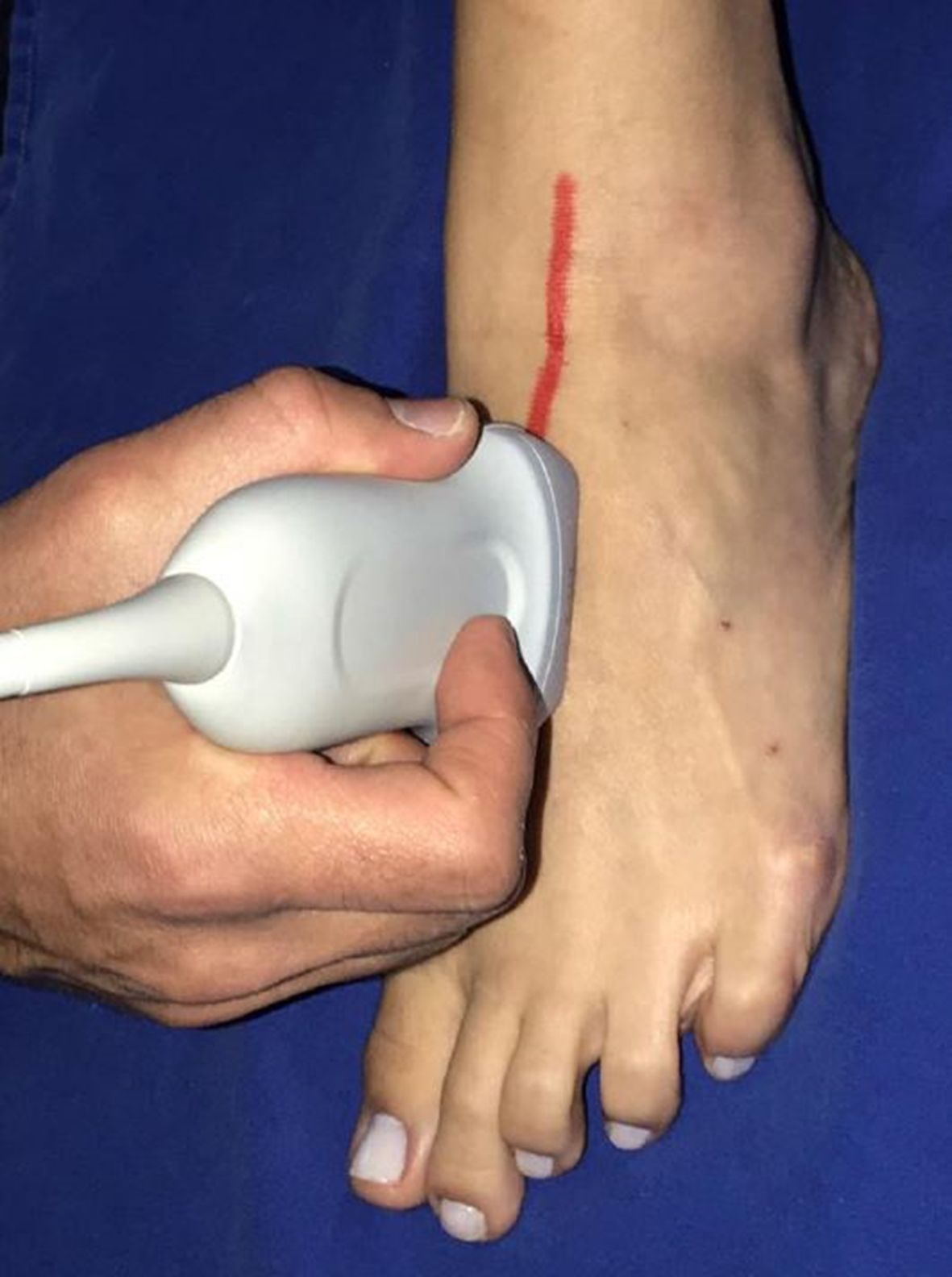
The assessment initiates with the anterior tibial artery distal of the ankle, located anteriorly of the tibia, documenting patency and spectral curve pattern. Proceeding distally along the dorsum of the foot, the DPA is scanned, adjusting transducer frequency and depth, because the DPA is of small caliber and very superficial. The DPA follows a straight path along the dorsum of the foot, running medially to the first intermetatarsal space, where it gives rise to its terminal branches. Its topographical anatomy should be imagined as running in the direction of the first interdigital space (Figure 2).

B. Plantar arteries and deep plantar arch
Next, the PTA is identified at the level of the medial malleolus, documenting patency and spectral curve pattern. Using cross-sectional images of the PTA and proceeding distally to the medial margin of the foot, the bifurcation of the PTA at the MPA and LPA is observed (Figure 3).

The MPA is the smaller caliber branch, following a path more directly in the direction of the great toe. Longitudinal images of the MPA are obtained with the transducer pointing in the direction of the great toe from the more proximal portion of the foot (Figure 4).

Longitudinal images of the medial plantar artery are acquired with the transducer pointing in the direction of the great toe from the more proximal part of the foot.
The LPA is the larger caliber, lateral branch and follows a path in the direction of the fifth metatarsal (Figure 5). The base of the fifth metatarsal is an important landmark for assessment of the LPA because the LPA gives rise to the DPA at this point. The LPA is located about 2.5 cm medial of the base of the fifth metatarsal (Figure 6).

The lateral plantar artery is the lateral branch with a larger caliber than the medial plantar artery, following a path in the direction of the base of the fifth metatarsal.

Location of lateral plantar artery, around 2.5 cm medial of the base of the fifth metatarsal.
The landmark used for assessment of the DPA is once more the base of the fifth metatarsal. The LPA is identified and then the transducer is turned so that its largest axis is aligned in the direction of the first intermetatarsal space, providing longitudinal images of the DPA (Figure 7). It is important to note that, when analyzed from the plantar surface, the DPA is located deeper from the plantar fascia than the plantar lateral artery, since it runs to an anastomosis with the dorsalis pedis artery. In order to position the transducer in the direction of the first intermetatarsal space, a parallel line should be imagined joining the heads of the second to fifth metatarsals, placing the transducer against the base of the fifth metatarsal (Figure 7).

Longitudinal images of the deep plantar arch, obtained by following the lateral plantar artery and then turning the transducer so that its longer axis is in the direction of the first intermetatarsal space.
In view of all of the above, the following facts are worthy of consideration. Doppler ultrasonography is the noninvasive method of choice for assessment of the lower limb arterial system, achieving excellent correlation with arteriography (gold standard), if conducted by experienced professionals.66 Sommerset J, Teso D, Feliciano B, et al. Innovative arterial duplex examination: a guide to evaluate flow in the foot using pedal acceleration time. J Vasc Ultrasound. 2019;43(1):11-7. http://dx.doi.org/10.1177/1544316719827328.
http://dx.doi.org/10.1177/15443167198273...
,88 Hingorani AP, Ascher E, Marks N, et al. Limitations of and lessons learned from clinical experience of 1,020 duplex arteriography. Vascular. 2008;16(3):147-53. http://dx.doi.org/10.2310/6670.2008.00014. PMid:18674463.
http://dx.doi.org/10.2310/6670.2008.0001...
However, in daily practice, Doppler ultrasonography is used much less than arteriography for assessment of the pedal arteries, possibly because of vascular ultrasonographists’ unfamiliarity with the ultrasonographic arterial anatomy of the foot. Now that the resolution of ultrasound scanners has improved, ultrasonographic characterization of the pedal arteries is possible and reproducible and, with adequate training, it does not take very long to perform.
Assessment of the plantar arch can be of prognostic value and it has been documented that a patent plantar arch is predictive of the patency of reconstructions and of healing of trophic ulcers.1212 Roedersheimer LR, Feins R, Green RM. Doppler evaluation of the pedal arch. Am J Surg. 1981;142(5):601-4. http://dx.doi.org/10.1016/0002-9610(81)90435-9. PMid:7304817.
http://dx.doi.org/10.1016/0002-9610(81)9...
,1313 Haine A, Haynes AG, Limacher A, et al. Patency of the arterial pedal-plantar arch in patients with chronic kidney disease or diabetes mellitus. Ther Adv Cardiovasc Dis. 2018;12(5):145-53. http://dx.doi.org/10.1177/1753944718756605. PMid:29431578.
http://dx.doi.org/10.1177/17539447187566...
There is also a correlation between the severity of chronic renal failure (reduction in glomerular filtration rate) and plantar arch patency.1313 Haine A, Haynes AG, Limacher A, et al. Patency of the arterial pedal-plantar arch in patients with chronic kidney disease or diabetes mellitus. Ther Adv Cardiovasc Dis. 2018;12(5):145-53. http://dx.doi.org/10.1177/1753944718756605. PMid:29431578.
http://dx.doi.org/10.1177/17539447187566...
It can be difficult to detect flow in the distal tibial arteries with digital arteriography in severe ischemia cases,1414 Alson MD, Lang EV, Kaufman JA. Pedal arterial imaging. J Vasc Interv Radiol. 1997;8(1 Pt 1):9-18. http://dx.doi.org/10.1016/S1051-0443(97)70507-4. PMid:9025033.
http://dx.doi.org/10.1016/S1051-0443(97)...
and in such cases ultrasonography with Doppler may detect patent vessels that are suitable for revascularization, increase limb salvage rates.77 Hofmann WJ, Magometschnigg H. Pedal artery bypass. Acta Chir Belg. 2004;104(6):654-8. http://dx.doi.org/10.1080/00015458.2004.11679638. PMid:15663270.
http://dx.doi.org/10.1080/00015458.2004....
,88 Hingorani AP, Ascher E, Marks N, et al. Limitations of and lessons learned from clinical experience of 1,020 duplex arteriography. Vascular. 2008;16(3):147-53. http://dx.doi.org/10.2310/6670.2008.00014. PMid:18674463.
http://dx.doi.org/10.2310/6670.2008.0001...
,1515 Eiberg JP, Hansen MA, Jørgensen LG, Rasmussen JB, Jensen F, Schroeder TV. In-situ bypass surgery on arteriographically invisible vessels detected by Doppler-ultrasound for limb salvage. J Cardiovasc Surg (Torino). 2004;45(4):375-9. PMid:15365518.,1616 Hofmann WJ, Walter J, Ugurluoglu A, Czerny M, Forstner R, Magometschnigg HH. Preoperative high-frequency duplex scanning of potential pedal target vessels. J Vasc Surg. 2004;39(1):169-75. http://dx.doi.org/10.1016/S0741-5214(03)01044-9. PMid:14718835.
http://dx.doi.org/10.1016/S0741-5214(03)...
During arteriography, the pedal arteries are assessed routinely, with a lateral projection which can be used to characterize the DPA, the LPA, and the plantar arch.
This assessment can also be performed using Doppler ultrasonography, rapidly and noninvasively, both for diagnosis and for follow-up after treatment. It is concluded that the pedal arteries can be noninvasively assessed with Doppler ultrasonography, affording an excellent level of anatomic detail.
-
How to cite: Takahashi LA, França GJ, Del Valle CE, Ferreira LRC. Assessment of the pedal arteries with Duplex Scanning. J Vasc Bras. 2020;19:e20200068. https://doi.org/10.1590/1677-5449.200068
-
Financial support:
None. -
The study was carried out at Hospital de Clínicas (HC), Universidade Federal do Paraná (UFPR), Curitiba, PR, Brazil.
REFERÊNCIAS
-
1Conte MS, Bradbury AW, Kolh P, et al. Global vascular guidelines on the management of chronic limb-threatening ischemia. J Vasc Endovasc Surg. 2019;69(6S):3S-125S.e40.
-
2Manzi M, Cester G, Palena LM, Alek J, Candeo A, Ferraresi R. Vascular imaging of the foot: the first step toward endovascular recanalization. Radiographics. 2011;31(6):1623-36. http://dx.doi.org/10.1148/rg.316115511 PMid:21997985.
» http://dx.doi.org/10.1148/rg.316115511 -
3Higashimori A, Iida O, Yamauchi Y, et al. Outcomes of one straight-line flow with and without pedal arch in patients with critical limb ischemia. Catheter Cardiovasc Interv. 2016;87(1):129-33. http://dx.doi.org/10.1002/ccd.26164 PMid:26489531.
» http://dx.doi.org/10.1002/ccd.26164 -
4Rashid H, Slim H, Zayed H, et al. The impact of arterial pedal arch quality and angiosome revascularization on foot tissue loss healing and infrapopliteal bypass outcome. J Vasc Surg. 2013;57(5):1219-26. http://dx.doi.org/10.1016/j.jvs.2012.10.129 PMid:23523278.
» http://dx.doi.org/10.1016/j.jvs.2012.10.129 -
5Meyer A, Schinz K, Lang W, Schmid A, Regus S, Rother U. Outcomes and influence of the pedal arch in below-the-knee angioplasty in patients with end-stage renal disease and critical limb ischemia. Ann Vasc Surg. 2016;35:121-9. http://dx.doi.org/10.1016/j.avsg.2016.01.039 PMid:27238998.
» http://dx.doi.org/10.1016/j.avsg.2016.01.039 -
6Sommerset J, Teso D, Feliciano B, et al. Innovative arterial duplex examination: a guide to evaluate flow in the foot using pedal acceleration time. J Vasc Ultrasound. 2019;43(1):11-7. http://dx.doi.org/10.1177/1544316719827328
» http://dx.doi.org/10.1177/1544316719827328 -
7Hofmann WJ, Magometschnigg H. Pedal artery bypass. Acta Chir Belg. 2004;104(6):654-8. http://dx.doi.org/10.1080/00015458.2004.11679638 PMid:15663270.
» http://dx.doi.org/10.1080/00015458.2004.11679638 -
8Hingorani AP, Ascher E, Marks N, et al. Limitations of and lessons learned from clinical experience of 1,020 duplex arteriography. Vascular. 2008;16(3):147-53. http://dx.doi.org/10.2310/6670.2008.00014 PMid:18674463.
» http://dx.doi.org/10.2310/6670.2008.00014 -
9Manzi M, Palena LM. Treating calf and pedal vessel disease: the extremes of intervention. Semin Intervent Radiol. 2014;31(4):313-9. http://dx.doi.org/10.1055/s-0034-1393967 PMid:25435656.
» http://dx.doi.org/10.1055/s-0034-1393967 -
10Mustapha JA, Diaz-Sandoval LJ, Saab F. Tibial-pedal arterial access & retrograde interventions for advanced peripheral arterial disease & critical limb ischemia. Interv Cardiol (Lond). 2015;7(5):451-67. http://dx.doi.org/10.2217/ica.15.33
» http://dx.doi.org/10.2217/ica.15.33 -
11Gray H. The arteries of the lower extremity. In: Bartleby.com. Anatomy of the human body. Bartleby.com; 1918. [atualizado 2000; citado 2019 nov 11]. https://www.bartleby.com/107/
» https://www.bartleby.com/107/ -
12Roedersheimer LR, Feins R, Green RM. Doppler evaluation of the pedal arch. Am J Surg. 1981;142(5):601-4. http://dx.doi.org/10.1016/0002-9610(81)90435-9 PMid:7304817.
» http://dx.doi.org/10.1016/0002-9610(81)90435-9 -
13Haine A, Haynes AG, Limacher A, et al. Patency of the arterial pedal-plantar arch in patients with chronic kidney disease or diabetes mellitus. Ther Adv Cardiovasc Dis. 2018;12(5):145-53. http://dx.doi.org/10.1177/1753944718756605 PMid:29431578.
» http://dx.doi.org/10.1177/1753944718756605 -
14Alson MD, Lang EV, Kaufman JA. Pedal arterial imaging. J Vasc Interv Radiol. 1997;8(1 Pt 1):9-18. http://dx.doi.org/10.1016/S1051-0443(97)70507-4 PMid:9025033.
» http://dx.doi.org/10.1016/S1051-0443(97)70507-4 -
15Eiberg JP, Hansen MA, Jørgensen LG, Rasmussen JB, Jensen F, Schroeder TV. In-situ bypass surgery on arteriographically invisible vessels detected by Doppler-ultrasound for limb salvage. J Cardiovasc Surg (Torino). 2004;45(4):375-9. PMid:15365518.
-
16Hofmann WJ, Walter J, Ugurluoglu A, Czerny M, Forstner R, Magometschnigg HH. Preoperative high-frequency duplex scanning of potential pedal target vessels. J Vasc Surg. 2004;39(1):169-75. http://dx.doi.org/10.1016/S0741-5214(03)01044-9 PMid:14718835.
» http://dx.doi.org/10.1016/S0741-5214(03)01044-9
Publication Dates
-
Publication in this collection
11 Dec 2020 -
Date of issue
2020
History
-
Received
21 Nov 2019 -
Accepted
12 Aug 2020