Abstract
The present study reports a snakebite in a horse in the state of Pará, Brazil. At initial evaluation the animal was reluctant to walk and had tachycardia, tachypnea, severe lameness, bleeding on the pastern and swelling around the left hind leg. Blood samples from the bleeding sites, took on the first day, showed leukocytosis and neutrophilia, whereas biochemical values of urea and creatinine were significantly increased. The chosen treatment was snake antivenom, fluid therapy, antibiotics, anti-inflammatory agents and diuretic drugs. On the fourth day of therapy, the hematological values were within normal parameters. There was improvement related to the clinical lameness and swelling of the limb. However, a decrease in water intake and oliguria were observed. On the seventh day the animal died. Necropsy revealed areas of hemorrhagic edema in the left hind limb and ventral abdomen; the kidneys presented equimosis in the capsule, and when cut they were wet. Moreover, the cortex was pale, slightly yellow and the medullary striae had the same aspect. Based on these data, we concluded that the snakebite in the present study was caused by Bothrops spp. and that renal failure contributed to death.
snakebites; horses; Bothrops
CASE REPORT
Fatal bothropic snakebite in a horse: a case report
Silva NS; Silveira JAS; Albernaz TT; Campos KF; Oliveira CMC; Freitas NFQR; Bomjardim HA; Barbosa JD
Veterinary Diagnostic Center (CEDIVET), Federal University of Pará (UFPA), Castanhal, Pará State, Brazil
Correspondence to Correspondence to: Natália da Silva e Silva Central de Diagnóstico Veterinário (CEDIVET) Universidade Federal do Pará Maximino Porpino da Silva 1000, Castanhal, 68740-080, PA, Brasil Phone/fax: +55 91 3721 1686 Email: nataliasilva@ufpa.br.
ABSTRACT
The present study reports a snakebite in a horse in the state of Pará, Brazil. At initial evaluation the animal was reluctant to walk and had tachycardia, tachypnea, severe lameness, bleeding on the pastern and swelling around the left hind leg. Blood samples from the bleeding sites, took on the first day, showed leukocytosis and neutrophilia, whereas biochemical values of urea and creatinine were significantly increased. The chosen treatment was snake antivenom, fluid therapy, antibiotics, anti-inflammatory agents and diuretic drugs. On the fourth day of therapy, the hematological values were within normal parameters. There was improvement related to the clinical lameness and swelling of the limb. However, a decrease in water intake and oliguria were observed. On the seventh day the animal died. Necropsy revealed areas of hemorrhagic edema in the left hind limb and ventral abdomen; the kidneys presented equimosis in the capsule, and when cut they were wet. Moreover, the cortex was pale, slightly yellow and the medullary striae had the same aspect. Based on these data, we concluded that the snakebite in the present study was caused by Bothrops spp. and that renal failure contributed to death.
Key words: snakebites, horses, Bothrops.
INTRODUCTION
Farmers, ranchers, and also field veterinarians believe that snakebites are a common cause of death among farm animals, which provoke great economic losses. Fatal snakebites in cattle are less frequent than it is believed, that is, their importance has been overrated (1). Several factors indicate that concerning horses, a similar situation occurs. In Brazil, a study on snakebites in horses revealed only one fatal case caused by Bothrops spp. (2).
Sporadic accidents involving Bothrops snakes occur among cattle and horses. Infrequently, samples from dead animals are sent to the laboratory to establish the cause of death and, thus, there is little information on the clinical effects provoked by the venom of these snakes on such animals (3). In the state of Pará, farmers, ranchers and veterinarians have reported the occurrence of suspected cases of snakebite in horses, cattle and dogs.
The diagnosis of envenomation is difficult to define because owners do not usually record the exact time of the bite, nor can identify the snake. However, during anamnesis, epidemiological data, such as the presence of snakes in the region, and clinical signs of the animal may give a clue about the species of the snake, enabling the veterinarians to select the most appropriate treatment (4).
With respect to humans, in São Paulo, a more accurate assessment showed that 85.6% of the snakebites were caused by Bothrops (5); however, a study conducted in the south of Minas Gerais state found that most envenomations were caused by Crotalus durissus (6).
Given the scarcity of literature data on snakebites in farm animals, this paper aims to describe the clinical, pathological and laboratory changes in a case of snakebite in a horse in the city of Castanhal, Pará state, Brazil.
CASE REPORT
A 15-year old male mixed breed horse, weighing about 400 kg, belonged to a farm located in the city of Castanhal, state of Pará. According to the handler, during the day the animal was in a stall and at night it grazed on a paddock of star grass (Cynodon nlemfuensis). In the morning of August 18, 2009, when collecting the animal, the handler found that it had marked lameness; its left hind leg was swollen and bleeding in the pastern region. Soon, he suspected of snakebite, since another farm employee had been bitten by a snake and eventually poisonous snakes were found, known in the region as pit vipers.
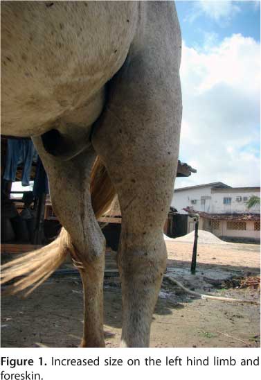
On clinical examination, it was observed that the animal had tachycardia, tachypnea, reluctance to walk, severe lameness, and bleeding in the pastern. A marked increase in volume around the left hind limb, extending to the region of the foreskin, was also noticed (Figure 1). During palpation the animal showed clinical signs of pain. Bleeding was also easily observed through the perforation left by the needles used for blood collection and drug administration (Figure 2).
Blood samples were collected using anticoagulants for complete blood count and without anticoagulants for assessment of renal function. These samples were collected from the jugular vein on the day of the bite (first day) and three days after (fourth day). The hematologic values were determined by automatic cell counter (DC 550®, CELM, Brazil) and serum levels of urea and creatinine were analyzed using colorimetric kits (Bioclín®, Doles, Brazil), through a spectrophotometer (Bioplus, Bio 2000, Brazil). These tests were performed at the Laboratory of Clinical Pathology of the Veterinary Diagnostic Center (CEDIVET) of the Federal University of Pará, in Castanhal.
The complete blood count on the first day showed leukocytosis and neutrophilia. On the fourth day, the values were within normal parameters, except for the decreased hematocrit. Serum levels of urea and creatinine increased progressively: on the first day the values were 84 mg/dL of urea and 28 mg/dL of creatinine, and on the fourth day respectively, 219 mg/dL and 5.3 mg/dL (reference range: 10 to 24 mg/dL of urea and 1.2 to 1.9 mg/dL of creatinine). From the fourth day on, it was observed that the animal was occasionally in sternal recumbency or in lateral extent, showing difficulty to get up.
The therapy comprised, since the first aid, antivenom, fluid therapy, antibiotics, anti-inflammatory agents and diuretic drugs was chosen. On the fourth day of treatment, the animal had an improvement in clinical aspects related to lameness and swelling of the limb. However, it showed a decrease in water intake and oliguria, symptoms that worsened over the course of the day. On the seventh day, the animal died.
At necropsy, there were areas of hemorrhagic edema around the left hind limb and ventral abdomen that reached the subcutaneous tissue and muscles (Figure 3). The kidneys had equimosis on the capsule and were wet at cutting, the cortex was pale, slightly yellowish and the medullary region showed the same striation appearance. The other organs did not show significant alterations.
Samples of different organs and tissues were collected and placed in 10% formalin. The material was processed routinely and stained with hematoxylin-eosin (HE) in the Department of Pathology of the Animal Health Project of Embrapa in association with the Federal Rural University of Rio de Janeiro. The histopathology revealed in the renal cortex areas where the urine tubules suffered coagulative necrosis and in the interstitial cortical tissue, edema with no purulent infiltrates, focal congestion and hemorrhage were observed. The liver presented severe congestion, mainly centrilobular. In the cuttings of the striated muscle there was no purulent infiltrates between the fibers. In the lung, there were areas of congestion.
DISCUSSION
The clinical and histopathological findings observed in the horse of this case are consistent with those previously described for Bothrops spp. envenomation. It is important to notice that although the bite was on the limb, the animal developed systemic problems and died. This was observed in an experiment, in which Bothrops spp. venom was inoculated, through the skin, on the left front leg of an animal that also developed systemic disturbances caused by hemorrhagic problems, resulting in death (7).
The increased size of the affected limb, in the present study, is probably due to local inflammation. Bothrops venom has proteolytic activity, which is believed to be responsible for the inflammation. However, it is not clear whether these local effects could be reproduced by purified components, such as the principles of bleeding or proteases, or even if the proteolytic activity of these components act to damage the tissue (8).
The decrease of hematocrit level, on the fourth day, may be attributed to the hemorrhagic edema and the bleeding, since some animals might develop anemia due to severe bleedings (9). In previous studies on animals with venom of Bothrops alternatus, the animals bled easily and had decreased hematocrit and hemoglobin (10).
The leukocytosis provoked by neutrophilia observed in the tests of the first day disappeared on the fourth day. This alteration may be due to the stress experienced by the animal. Nevertheless, this result differs from that described by Chiacchio et al. (9), who reported Bothrops bites in three horses that revealed no changes in white blood cell count.
The increase in urea and creatinine levels may be explained by the acute renal failure that may occur due to several factors, such as disseminated intravascular coagulation, hypotension, direct nephrotoxic action of the venom, and hemolysis (11, 12). Tachycardia and tachypnea may be due to the stress and pain (3). These findings did not agree with those by Chiacchio et al. (9), who described that the envenomed horses had no alterations in heart and breathing rates. In the current study, the presence of hemorrhagic edema observed at autopsy corroborates the experimental Bothrops envenomation in cattle (10, 13).
In tests performed in cattle, Bothrops venom - regardless of inoculation route, dose and application site - produced edema (14). Moreover, another work found that in cases of Bothrops envenomation in cattle, sheep and horses, the swelling was due to severe bleeding and not to edema (10). In the current report, necrosis was not observed in the assumed bite site, which corroborates studies on experimental inoculation of Bothrops venom in horses, sheep, goats, pigs, cats, rats and mice (15).
Despite the treatment that was initiated on the first day, it was only partially effective, since the horse had recovered from some clinical signs, but died on the seventh day. We believe that the renal failure contributed to the death. Based on these data, we may suggest that the snakebite was caused by Bothrops spp.
Submission status
Received: May 27, 2011.
Accepted: August 10, 2011.
Abstract: published online: August 17, 2011.
Full paper published online: November 30, 2011.
Conflicts of interest
There is no conflict.
- 1. Tokarnia CH, Peixoto PV. A importância dos acidentes ofídicos como causa de mortes em bovinos no Brasil. Pesq Vet Bras. 2006;26(2):55-68.
- 2. Raposo JB, Méndez MC, Baialardi CEG, Raffi MB. Acidente ofídico em equino no sul do Brasil - relato de caso. Rev Fac Zoo Vet Agro. 2000/2001;7/8(1):51-7.
- 3. Grunert E, Grunert D. Beobachtungen von Bothrops- Schlangenbissverletzungen bei Rind und Pferd in Rio Grande do Sul, Brasilien. Vet Med Nachrichten. 1969;3(1):217-32.
- 4. Ferreira Jr RS, Barraviera B. Management of venomous snakebites in dogs and cats in Brazil. J Venom Anim Toxins incl Trop Dis. 2004;10(2):112-32.
- 5. Takaoka NY, Albuquerque MJ, Pires de Campos VAS, Gualtieri VB, Kats G, Jorge MT, Ribeiro LA. Distribuição dos acidentes por Bothrops, Crotalus e Micrurus segundo os Escritórios Regionais de Saúde (ERSAS) do Estado de São Paulo. Rev Soc Bras Med Trop. 1994;27(10):118.
- 6. Franco RL, Rocha CC, Jorge MT, Ribeiro LA. Snakebites in southern Minas Gerais state, Brazil. J Venom Anim Toxins. 2001;7(1):56-68.
- 7. Freitas NFQR, Sousa MGS, Bomjardim HA, Reis ASB, Lopes CTA, Silveira JAS, et al. Quadro clínico-patológico do envenenamento botrópico experimental em equinos. Rev Bras Med Vet Equina. 2011;35:202.
- 8. Ferreira Jr RS, Barraviera B. Tissue necrosis after canine bothropic envenoming: a case report. J Venom Anim Toxins. 2001;7(2):302-12.
- 9. Chiacchio SB, Martins GTB, Amorim RM, Gonçalves RC, Barraviera B, Ferreira Jr RS. Triple bothropic envenomation in horses caused by a single snake. J Venom Anim Toxins incl Trop Dis. 2011;17(1):111-7.
- 10. Caldas SA, Tokarnia CH, França TN, Brito MF, Graça FAS, Coelho CD, et al. Aspectos clínico-patológicos e laboratoriais do envenenamento experimental por Bothrops alternatus em bovinos. Pesq Vet Bras. 2008;28(6):303-12.
- 11. Rezende NA, Amaral CFS, Bambirra EA, Lachatt JJ, Coimbra TM. Functional and histopathological renal changes induced in rats by Bothrops jararaca venom. Braz J Med Biol Res. 1989;22(3):407-16.
- 12. Boer-Lima PA, Gontijo JA, da Cruz-Hofling MA. Histological and functional renal alterations caused by Bothrops moojeni snake venom in rats. Am J Trop Med Hyg. 1999;61(5):698-706.
- 13. Novaes AP, Lucas S, Abe AS, Fernandes W, Puorto G, Almeida IL. Envenenamento botrópico em bovinos: tratamento opcional. Vet News. 1997;30(1):9-12.
- 14. Araujo P, Rosenfeld G, Belluomini HE. Toxicidade de venenos ofídicos II. Doses mortais para bovinos. Arq Inst Biol. 1963;30(2):43-8.
- 15. Araujo P, Belluomini HE. Toxicidade de venenos ofídicos. Sensibilidade específica de animais domésticos e de laboratório. Mem Inst Butantan. 1960-2;30(1):143-56.
Publication Dates
-
Publication in this collection
05 Dec 2011 -
Date of issue
2011
History
-
Accepted
10 Aug 2011 -
Received
27 May 2011