Abstract
Background
Cathepsin D (CatD) is a lysosomal proteolytic enzyme expressed in almost all tissues and organs. This protease is a multifunctional enzyme responsible for essential biological processes such as cell cycle regulation, differentiation, migration, tissue remodeling, neuronal growth, ovulation, and apoptosis. The overexpression and hypersecretion of CatD have been correlated with cancer aggressiveness and tumor progression, stimulating cancer cell proliferation, fibroblast growth, and angiogenesis. In addition, some studies report its participation in neurodegenerative diseases and inflammatory processes. In this regard, the search for new inhibitors from natural products could be an alternative against the harmful effects of this enzyme.
Methods
An investigation was carried out to analyze CatD interaction with snake venom toxins in an attempt to find inhibitory molecules. Interestingly, human CatD shows the ability to bind strongly to snake venom phospholipases A2 (svPLA2), forming a stable muti-enzymatic complex that maintains the catalytic activity of both CatD and PLA2. In addition, this complex remains active even under exposure to the specific inhibitor pepstatin A. Furthermore, the complex formation between CatD and svPLA2 was evidenced by surface plasmon resonance (SPR), two-dimensional electrophoresis, enzymatic assays, and extensive molecular docking and dynamics techniques.
Conclusion
The present study suggests the versatility of human CatD and svPLA2, showing that these enzymes can form a fully functional new enzymatic complex.
Keywords:
Cathepsin D; Phospholipases A2; Snake venom; Enzyme complex
Keywords:
Cathepsin D; Phospholipases A2; Snake venom; Enzyme complex
Background
Cathepsins compose a family of lysosomal proteases mainly found in acidic endo/lysosomal compartments and are implicated in a broad spectrum of physiologic processes, such as intracellular protein degradation, energy metabolism, hormonal regulation, bone resorption, and immune responses [11. Yadati T, Houben T, Bitorina A, Shiri-Sverdlov R. The ins and outs of cathepsins: Physiological function and role in disease management. Cells. 2020 Jul 13;9(7):1679. doi: 10.3390/cells9071679.
https://doi.org/10.3390/cells9071679...
]. These proteins belong to three protease families, characterized based on differences in the following amino acids at their active site: aspartic proteases (D and E), serine proteases (A and G), or cysteine proteases (B, C, H, F, K, L, O, S, V, X, and W) [11. Yadati T, Houben T, Bitorina A, Shiri-Sverdlov R. The ins and outs of cathepsins: Physiological function and role in disease management. Cells. 2020 Jul 13;9(7):1679. doi: 10.3390/cells9071679.
https://doi.org/10.3390/cells9071679...
-44. Brix K, Dunkhorst A, Mayer K, Jordans S. Cysteine cathepsins: cellular roadmap to different functions. Biochimie. 2008 Feb;90(2):194-207. doi: 10.1016/j.biochi.2007.07.024.
https://doi.org/10.1016/j.biochi.2007.07...
].
Furthermore, cathepsins are essential to maintaining cell homeostasis [55. Ward C, Martinez-Lopez N, Otten EG, Carroll B, Maetzel D, Singh R, Sarkar S, Korolchuk VI. Autophagy, lipophagy and lysosomal lipid storage disorders. Biochim Biophys Acta. 2016 Apr;1861(4):269-84. doi: 10.1016/j.bbalip.2016.01.006.
https://doi.org/10.1016/j.bbalip.2016.01...
]. The inactivation, loss of function, and overexpression of these proteases can result in inappropriate degradation and abnormal accumulation of lysosomal waste [11. Yadati T, Houben T, Bitorina A, Shiri-Sverdlov R. The ins and outs of cathepsins: Physiological function and role in disease management. Cells. 2020 Jul 13;9(7):1679. doi: 10.3390/cells9071679.
https://doi.org/10.3390/cells9071679...
, 66. Stoka V, Turk V, Turk B. Lysosomal cathepsins and their regulation in aging and neurodegeneration. Ageing Res Rev. 2016 Dec;32:22-37. doi: 10.1016/j.arr.2016.04.010.
https://doi.org/10.1016/j.arr.2016.04.01...
]. In addition, extracellular oversecretion of cathepsins is associated with uncontrolled cell proliferation, invasion, and differentiation, which in turn may bring about the onset of fatal pathologies, including atherosclerosis, cancer, and tissue fibrosis [66. Stoka V, Turk V, Turk B. Lysosomal cathepsins and their regulation in aging and neurodegeneration. Ageing Res Rev. 2016 Dec;32:22-37. doi: 10.1016/j.arr.2016.04.010.
https://doi.org/10.1016/j.arr.2016.04.01...
-1212. Conus S, Perozzo R, Reinheckel T, Peters C, Scapozza L, Yousefi S, Simon HU. Caspase-8 is activated by cathepsin D initiating neutrophil apoptosis during the resolution of inflammation. J Exp Med. 2008 Mar 17;205(3):685-98. doi: 10.1074/jbc.M111.306399.
https://doi.org/10.1074/jbc.M111.306399...
].
Due to its physio-pathological functions, cathepsin D (CatD) is one of the most studied lysosomal proteases [1313. Mattson MP. Neuronal Life-and-Death Signaling, Apoptosis, and Neurodegenerative Disorders. Antioxid Redox Signal. 2006 Nov-Dec;8(11):1997-2006. doi: 10.1038/35040009.
https://doi.org/10.1038/35040009...
-1515. Garcia M, Platet N, Liaudet E, Laurent V, Derocq D, Brouillet J, Rochefort H. Biological and clinical significance of cathepsin D in breast cancer metastasis. Stem Cells. 1996 Nov;14(6):642-50. doi: 10.1002/stem.140642.
https://doi.org/10.1002/stem.140642...
]. CatD is an aspartic endopeptidase with two conserved Asp residues in its active site; these residues tend to deprotonate, indicating that the pH-optimum of activity resides at pH values below 5 [1616. Metcalf P, Fusek M. Two crystal structures for cathepsin D: the lysosomal targeting signal and active site. EMBO J. 1993 Apr;12(4):1293-302. doi: 10.1002/j.1460-2075.1993.tb05774.x.
https://doi.org/10.1002/j.1460-2075.1993...
]. In addition, CatD has three distinct regions that are characteristic of aspartic proteases, an N-terminal domain (residues 1-188), a C-terminal domain (residues 189-346), and an interdomain, antiparallel/3-sheet formed by the N-terminus (residues 1-7), the C-terminus (residues 330-346), as well as the linker residues between domains (160-200) [1717. Baldwin ET, Bhat TN, Gulnik S, Hosur M V., Sowder RC, Cachau RE, Collins J, Silva AM, Erickson JW. Crystal structures of native and inhibited forms of human cathepsin D: implications for lysosomal targeting and drug design. Proc Natl Acad Sci U S A. 1993 Jul 15;90(14):6796-800. doi: 10.1073/pnas.90.14.6796.
https://doi.org/10.1073/pnas.90.14.6796...
].
Considered a multifunctional enzyme due to its involvement in various biological processes, CatD operates in both cytosolic and extracellular environments [1313. Mattson MP. Neuronal Life-and-Death Signaling, Apoptosis, and Neurodegenerative Disorders. Antioxid Redox Signal. 2006 Nov-Dec;8(11):1997-2006. doi: 10.1038/35040009.
https://doi.org/10.1038/35040009...
, 1818. Zaidi N, Maurer A, Nieke S, Kalbacher H. Cathepsin D: A cellular roadmap. Biochem Biophys Res Commun. 2008 Nov 7;376(1):5-9. doi: 10.1016/j.bbrc.2008.08.099.
https://doi.org/10.1016/j.bbrc.2008.08.0...
-2222. Khalkhali-Ellis Z, Hendrix MJC. Two faces of cathepsin D: Physiological guardian angel and pathological demon. Biol Med (Aligarh). 2014 Jul;6(2):1000206. doi: 10.4172/0974-8369.1000206.
https://doi.org/10.4172/0974-8369.100020...
]. Studies have shown that CatD is involved in the activation of precursors of biologically active proteins in pre-lysosomal compartments of specialized cells [1111. Liaudet-Coopman E, Beaujouin M, Derocq D, Garcia M, Glondu-Lassis M, Laurent-Matha V, Prébois C, Rochefort H, Vignon F. Cathepsin D: newly discovered functions of a long-standing aspartic protease in cancer and apoptosis. Cancer Lett. 2006 Jun 18;237(2):167-79. doi: 10.1016/j.canlet.2005.06.007.
https://doi.org/10.1016/j.canlet.2005.06...
, 2020. Masson O, Bach AS, Derocq D, Prébois C, Laurent-Matha V, Pattingre S, Liaudet-Coopman E. Pathophysiological functions of cathepsin D: Targeting its catalytic activity versus its protein binding activity? Biochimie. 2010;92:1635-43. doi: 10.1016/j.biochi.2010.05.009.
https://doi.org/10.1016/j.biochi.2010.05...
, 2323. Gocheva V, Joyce JA. Cysteine cathepsins and the cutting edge of cancer invasion. Cell Cycle. 2007 Jan 1;6(1):60-4. doi: 10.4161/cc.6.1.3669.
https://doi.org/10.4161/cc.6.1.3669...
]. This enzyme is indispensable for cellular functions such as cell migration, differentiation, growth, cycle progression, tissue remodeling, and neovascularization activation [66. Stoka V, Turk V, Turk B. Lysosomal cathepsins and their regulation in aging and neurodegeneration. Ageing Res Rev. 2016 Dec;32:22-37. doi: 10.1016/j.arr.2016.04.010.
https://doi.org/10.1016/j.arr.2016.04.01...
, 1111. Liaudet-Coopman E, Beaujouin M, Derocq D, Garcia M, Glondu-Lassis M, Laurent-Matha V, Prébois C, Rochefort H, Vignon F. Cathepsin D: newly discovered functions of a long-standing aspartic protease in cancer and apoptosis. Cancer Lett. 2006 Jun 18;237(2):167-79. doi: 10.1016/j.canlet.2005.06.007.
https://doi.org/10.1016/j.canlet.2005.06...
, 1212. Conus S, Perozzo R, Reinheckel T, Peters C, Scapozza L, Yousefi S, Simon HU. Caspase-8 is activated by cathepsin D initiating neutrophil apoptosis during the resolution of inflammation. J Exp Med. 2008 Mar 17;205(3):685-98. doi: 10.1074/jbc.M111.306399.
https://doi.org/10.1074/jbc.M111.306399...
, 1919. Benes P, Vetvicka V, Fusek M. Cathepsin D - Many functions of one aspartic protease. Crit Rev Oncol Hematol. 2008 Oct;68(1):12-28. doi: 10.1016/j.critrevonc.2008.02.008.
https://doi.org/10.1016/j.critrevonc.200...
, 2222. Khalkhali-Ellis Z, Hendrix MJC. Two faces of cathepsin D: Physiological guardian angel and pathological demon. Biol Med (Aligarh). 2014 Jul;6(2):1000206. doi: 10.4172/0974-8369.1000206.
https://doi.org/10.4172/0974-8369.100020...
, 2424. Fusek M, Vetvicka V. Dual role of cathepsin D: ligand and protease. Biomed Pap Med Fac Univ Palacky Olomouc Czech Repub. 2005 Jun;149(1):43-50. doi: 10.5507/pb.2005.003.
https://doi.org/10.5507/pb.2005.003...
-2727. Pimenta DC, Oliveira A, Juliano MA, Juliano L. Substrate specificity of human cathepsin D using internally quenched fluorescent peptides derived from reactive site loop of kallistatin. Biochim Biophys Acta. 2001 Jan 12;1544(1-2):113-22. doi: 10.1016/s0167-4838(00)00209-0.
https://doi.org/10.1016/s0167-4838(00)00...
]. Additionally, CatD is involved in initiating the apoptotic cascade [2828. Appelqvist H, Johansson A, Linderoth E, Johansson U, Antonsson B, Steinfeld R, Kagedal K, Ollinger K. Lysosome-mediated apoptosis is associated with cathepsin D-specific processing of bid at Phe24 , Trp48 , and Phe183. Ann Clin Lab Sci. 2012 Summer;42(3):231-42. , 2929. Charlotte Johansson A, Appelqvist AJH, Nilsson C, Kagedal K, Roberg K, Ollinger K. Regulation of apoptosis-associated lysosomal membrane permeabilization. Apoptosis. 2010 May;15(5):527-40. doi: 10.1007/s10495-009-0452-5
https://doi.org/10.1007/s10495-009-0452-...
] in lysosomal cell death pathways [2222. Khalkhali-Ellis Z, Hendrix MJC. Two faces of cathepsin D: Physiological guardian angel and pathological demon. Biol Med (Aligarh). 2014 Jul;6(2):1000206. doi: 10.4172/0974-8369.1000206.
https://doi.org/10.4172/0974-8369.100020...
, 2525. Johansson AC, Steen H, Öllinger K, Roberg K. Cathepsin D mediates cytochrome c release and caspase activation in human fibroblast apoptosis induced by staurosporine. Cell Death Differ. 2003 Nov;10(11):1253-9. doi: 10.1038/sj.cdd.4401290
https://doi.org/10.1038/sj.cdd.4401290...
].
CatD is directly related to the pathogenesis and progression of degenerative diseases [66. Stoka V, Turk V, Turk B. Lysosomal cathepsins and their regulation in aging and neurodegeneration. Ageing Res Rev. 2016 Dec;32:22-37. doi: 10.1016/j.arr.2016.04.010.
https://doi.org/10.1016/j.arr.2016.04.01...
, 3030. Castino R, Isidoro C, Murphy D. Autophagy-dependent cell survival and cell death in an autosomal dominant familial neurohypophyseal diabetes insipidus in vitro model. FASEB J. 2005 Jun;19(8):1024-6. doi: 10.1096/fj.04-3163fje.
https://doi.org/10.1096/fj.04-3163fje...
], such as lymphoid cell degeneration [3131. Saftig P, Hetman M, Schmahl W, Weber K, Heine L, Mossmann H, Koster A, Hess B, Evers M, von Figura K. Mice deficient for the lysosomal proteinase cathepsin D exhibit progressive atrophy of the intestinal mucosa and profound destruction of lymphoid cells. EMBO J. 1995 Aug 1;14(15):3599-608. doi: 10.1002/j.1460-2075.1995.tb00029.x.
https://doi.org/10.1002/j.1460-2075.1995...
], Parkinson’s [3232. Crabtree D, Dodson M, Ouyang X, Boyer-Guittaut M, Liang Q, Ballestas ME, Fineberg N, Zhang J. Over-expression of an inactive mutant cathepsin D increases endogenous alpha-synuclein and cathepsin B activity in SH-SY5Y cells. J Neurochem. 2014 Mar;128(6):950-61. doi: 10.1111/jnc.12497.
https://doi.org/10.1111/jnc.12497...
] and Alzheimer’s disease [3333. Di Domenico F, Tramutola A, Perluigi M. Cathepsin D as a therapeutic target in Alzheimer’s disease. Expert Opin Ther Targets. 2016 Dec;20(12):1393-5. doi: 10.1080/14728222.2016.1252334.
https://doi.org/10.1080/14728222.2016.12...
], atherosclerosis [3434. Zhao CF, Herrington DM. The function of cathepsins B, D, and X in atherosclerosis. Am J Cardiovasc Dis. 2016;6(4):163-70.], and different types of cancer [3535. Olson OC, Joyce JA. Cysteine cathepsin proteases: regulators of cancer progression and therapeutic response. Nat Rev Cancer. 2015 Dec;15(12):712-29. doi: 10.1038/nrc4027.
https://doi.org/10.1038/nrc4027...
, 3636. Palermo C, Joyce JA. Cysteine cathepsin proteases as pharmacological targets in cancer. Trends Pharmacol Sci. 2008 Jan;29(1):22-8. doi: 10.1016/j.tips.2007.10.011.
https://doi.org/10.1016/j.tips.2007.10.0...
]. For instance, some cell types under pathological conditions overexpress and secrete CatD to the extracellular environment via lysosomal release [2020. Masson O, Bach AS, Derocq D, Prébois C, Laurent-Matha V, Pattingre S, Liaudet-Coopman E. Pathophysiological functions of cathepsin D: Targeting its catalytic activity versus its protein binding activity? Biochimie. 2010;92:1635-43. doi: 10.1016/j.biochi.2010.05.009.
https://doi.org/10.1016/j.biochi.2010.05...
]; this makes CatD an important tumor marker in breast, bladder, and mouth cancers, among others [3535. Olson OC, Joyce JA. Cysteine cathepsin proteases: regulators of cancer progression and therapeutic response. Nat Rev Cancer. 2015 Dec;15(12):712-29. doi: 10.1038/nrc4027.
https://doi.org/10.1038/nrc4027...
, 3636. Palermo C, Joyce JA. Cysteine cathepsin proteases as pharmacological targets in cancer. Trends Pharmacol Sci. 2008 Jan;29(1):22-8. doi: 10.1016/j.tips.2007.10.011.
https://doi.org/10.1016/j.tips.2007.10.0...
]. Furthermore, due to the participation of cathepsins in a broad spectrum of diseases, these proteases are promising therapeutic targets for small molecules and peptide drugs [3333. Di Domenico F, Tramutola A, Perluigi M. Cathepsin D as a therapeutic target in Alzheimer’s disease. Expert Opin Ther Targets. 2016 Dec;20(12):1393-5. doi: 10.1080/14728222.2016.1252334.
https://doi.org/10.1080/14728222.2016.12...
, 3636. Palermo C, Joyce JA. Cysteine cathepsin proteases as pharmacological targets in cancer. Trends Pharmacol Sci. 2008 Jan;29(1):22-8. doi: 10.1016/j.tips.2007.10.011.
https://doi.org/10.1016/j.tips.2007.10.0...
].
In order to investigate human CatD inhibitors for the design and development of tools and agents of scientific and therapeutic interest, snake venoms belonging to the genera Bothrops, Crotalus, and Lachesis have been used as natural sources of biologically active molecules able to act selectively and specifically on different cellular targets [3737. Almeida JR, Resende LM, Watanabe RK, Carregari VC, Huancahuire-Vega S, da S. Caldeira CA, Coutinho-Neto A, Soares AM, Vale N, Gomes PAC, Marangoni S, Calderon LA, Silva SL. Snake venom peptides and low mass proteins: Molecular tools and therapeutic agents. Curr Med Chem. 2017;24(30):3254-82. doi: 10.2174/0929867323666161028155611.
https://doi.org/10.2174/0929867323666161...
, 3838. da Silva GM , de Souza DHB , Waitman KB, Ebram MC, Fessel MR, Zainescu IC, Portaro FC, Heras M, Andrade SA. Design, synthesis, and evaluation of Bothrops venom serine protease peptidic inhibitors. J Venom Anim Toxins incl Trop Dis. 2021;27. doi: 10.1590/1678-9199-JVATITD-2020-0066.
https://doi.org/10.1590/1678-9199-JVATIT...
]. Of all the bioactive molecules present in snake venoms, phospholipases A2 (svPLA2) are among the most frequently encountered and studied [3939. Murakami M, Taketomi Y, Girard C, Yamamoto K, Lambeau G. Emerging roles of secreted phospholipase A2 enzymes: Lessons from transgenic and knockout mice. Biochimie. 2010 Jun;92(6):561-82. doi: 10.1016/j.biochi.2010.03.015.
https://doi.org/10.1016/j.biochi.2010.03...
, 4040. Fortes-Dias CL, Macedo DHF, Barbosa RP, Souza-Silva G, Ortolani PL. Identification and characterization of the first endogenous phospholipase A2 inhibitor from a non-venomous tropical snake, Boa constrictor (Serpentes: Boidae). J Venom Anim Toxins incl Trop Dis. 2020;26. doi: 10.1590/1678-9199-JVATITD-2019-0044.
https://doi.org/10.1590/1678-9199-JVATIT...
]; these proteins have established physical-chemical properties and a variety of pharmacologic and toxic effects in snakebite envenomation, such as myonecrosis, anticoagulation, platelet aggregation inhibition, neurotoxicity, cardiotoxicity, hypotension and edema formation [4141. Burke JE, Dennis EA. Phospholipase A2 structure/function, mechanism, and signaling. J Lipid Res. 2009 Apr;50(Suppl):S237-42. doi: 10.1194/jlr.R800033-JLR200.
https://doi.org/10.1194/jlr.R800033-JLR2...
-4545. Harris J, Scott-Davey T. Secreted phospholipases A2 of snake venoms: Effects on the peripheral neuromuscular system with comments on the role of phospholipases A2 in disorders of the CNS and their uses in industry. Toxins (Basel). 2013 Dec 17;5(12):2533-71. doi: 10.3390/toxins5122533.
https://doi.org/10.3390/toxins5122533...
].
Interestingly, human CatD shows the ability to bind strongly to svPLA2s, forming a stable and functional complex that is able to remain active even at pH values higher than 5 and is also unaffected by the inhibitor pepstatin A. These results, presented and discussed below, demonstrate the multifunctionality and versatility of CatD, warranting many new possibilities for the understanding of cathepsin functions in cytosolic and extracellular environments during physiologic and pathologic processes. Therefore, the present study aims to demonstrate and characterize an enzymatic complex formed by human CatD and a snake venom phospholipase A2.
Methods
Cathepsin D
Cathepsin D (cod. C8696) was obtained from Sigma-Aldrich Ltda and prepared according to the manufacturer’s recommendations.
Snake venoms
All snake venoms used in this study were acquired from the Venom Bank at CEBio/Fiocruz Rondônia/UNIR (Centro de Estudos de Biomoléculas Aplicadas a Saúde), Porto Velho, RO, under local government authorization license number: IBAMA nº 27131-3 and CGEN/CNPq 010627/2011-1.
Phospholipases A2 (PLA2s)
The Bothropstoxin-I (BthTX-I) and Bothropstoxin-II (BthTX-II) from Bothrops jararacussu were obtained from the Venom Bank at CEBio (Centro de Estudos de Biomoléculas Aplicadas a Saúde/Fiocruz Rondônia/UNIR), located in Porto Velho, RO. PLA2 LmtTX from Lachesis muta provided by Diniz-Sousa et al. [4646. Diniz-Sousa R, Caldeira CAS, Kayano AM, Paloschi M V, Pimenta DC, Simões-Silva R, Ferreira AS, Zanchi FB, Matos NB, Grabner FP, Calderon LA, Zuliani JP, Soares AM. Identification of the molecular determinants of the antibacterial activity of LmutTX, a Lys49 phospholipase A2 homologue isolated from Lachesis muta muta snake venom (Linnaeus, 1766). Basic Clin Pharmacol Toxicol. 2018 Apr;122(4):413-23. doi: 10.1111/bcpt.12921.
https://doi.org/10.1111/bcpt.12921...
], PLA2 BnuTX-I from Bothrops urutu provided by Corrêa et al. [4747. Correa EA, Kayano AM, Diniz-Sousa R, Setubal SS, Zanchi FB, Zuliani JP, Matos NB, Almeida JR, Resende LM, Marangoni S, Silva SL, Soares AM, Calderon LA. Isolation, structural and functional characterization of a new Lys49 phospholipase A2 homologue from Bothrops neuwiedi urutu with bactericidal potential. Toxicon. 2016 Jun 1;115:13-21. doi: 10.1016/j.toxicon.2016.02.021.
https://doi.org/10.1016/j.toxicon.2016.0...
], PLA2 Braziliase-I and Braziliase-II from Bothrops brazili provided by Kayano et al. [4848. Kayano AM, Simões-Silva R, Medeiros PSM, Maltarollo VG, Honorio KM, Oliveira E, Albericio F, da Silva SL, Aguiar ACC, Krettli AU, Fernandes CFC, Juliani JP, Calderon LA, Stábeli RG, Soares AM. BbMP-1, a new metalloproteinase isolated from Bothrops brazili snake venom with in vitro antiplasmodial properties. Toxicon. 2015 Nov;106:30-41. doi: 10.1016/j.toxicon.2015.09.005.
https://doi.org/10.1016/j.toxicon.2015.0...
] and Sobrinho et al. [4949. Sobrinho JC, Kayano AM, Simões-Silva R, Alfonso JJ, Gomez AF, Gomez MCV, Zanchi FB, Moura LA, Souza VR, Fuly AL, Oliveira E, da Silva SL, Almeida JR, Zuliani JP, Soares AM. Anti-platelet aggregation activity of two novel acidic Asp49-phospholipases A2 from Bothrops brazili snake venom. Int J Biol Macromol. 2018 Feb;107(Pt A):1014-22. doi: 10.1016/j.ijbiomac.2017.09.069.
https://doi.org/10.1016/j.ijbiomac.2017....
].
Bothrops jararaca snake venom fractionation
B. jararaca venom was solubilized in 50mM ammonium bicarbonate buffer (AMBIC), pH 8.0 and applied to an anion exchange column (CM-Sepharose 10 x 30 cm). The fractions were eluted in a linear gradient of 500 mM AMBIC, pH 8.0 under a flow of 1 mL/min. Absorbances were measured at 215 and 280 nm. The fractions were subjected to salt removal in a 15mL filter (AMICON ULTRA-15) with a 50 kDa cutoff.
Binding assays
Surface plasmon resonance (SPR) molecular interaction assays were performed in a Biacore T200 system (GE Healthcare). Cathepsin D immobilization was done using a CM5 S-type sensor chip via amine coupling. The contact time of each cycle was set at 60 seconds, with a flow rate of 30 µL/min, followed by 60 seconds of dissociation time. For the regeneration stage at the end of each cycle, a 0.5% TFA solution was used with 30 seconds of contact time at a flow of 30 µL/min. All experiments were performed at 25 ºC, and binding assays were conducted in phosphate-saline buffer (PBS), pH 7.4 and analytes at a concentration of 100 µg/mL.
Protein quantification
The protein concentrations present in venom samples were determined using Bradford’s method [5050. Bradford MM. A rapid and sensitive method for the quantitation of microgram quantities of protein utilizing the principle of protein-dye binding. Anal Biochem. 1976 May 7;72:248-54. doi: 10.1006/abio.1976.9999.
https://doi.org/10.1006/abio.1976.9999...
]. For spectrophotometric measurements, the sample was aliquoted in a 1 mL disposable plastic cuvette along with 1:10 (v/v) Bradford reagent, which was incubated for 15 minutes. Absorbance was monitored at 595 nm using a Biomate 3 spectrophotometer. The calibration curve was performed using bovine albumin (Sigma).
SDS-PAGE
The relative mass of proteins was determined by SDS-PAGE using discontinuous gels, with a stacking gel (4% acrylamide in 0.5 M Tris-HCl buffer, pH 6.8) (Sigma Aldrich, USA) and a resolving gel (12.5% acrylamide in 1.5 M Tris-HCl buffer, pH 8.8). The experimental buffer solution used to fill the wells was 0.06 M Tris-Base, 0.5 M Glycine, and 10% SDS (Sigma Aldrich, USA). The samples with 1M DTT were preheated to 95 °C for 5 min and applied to the stacking gel wells along with the Molecular Weight standard (7 to 175 kDa - BioLabs P7709S, USA). In the electrophoretic run, a current of 15 mA per gel and free voltage was fixed for 1 hour and 40 minutes. After this, the gel was washed for 15 minutes with a fixing solution (ethyl alcohol 50% and acetic acid 12%) and then stained with Coomassie G-250 blue solution (Sigma Aldrich, USA) for 10-30 minutes. After this period, the gel was bleached in a bleaching solution (20% ethyl alcohol and 3% acetic acid). The gels’ images were scanned using Image Scanner III (GE Lifescience Health Care).
The 2D electrophoresis consisted of two steps: isoelectric focusing and 1D SDS-PAGE. For the first dimension, the sample was prepared in a rehydration solution (8 M urea, 2% CHAPS, 0.5/2% IPG buffer, 0.002% bromophenol blue, and 1 M DTT); this same solution was then incubated with a 7-cm strip (pH 3-10, linear) for 12-20 h. After rehydration, the strip was applied to an Ettan IPGphor 3 (GE Healthcare) isoelectric focusing system and later stored at − 80 °C. For the second dimension, the strip was washed with DTT and iodoacetamide diluted in 5 mL of equilibration buffer solution (6 M urea, 2% SDS, 30% glycerol, 50 mM Tris- HCl, pH 7.4, 0.002% bromophenol blue). Then, the strip was applied to a 15% polyacrylamide gel. The gel was stained with Coomassie Blue G-250 and scanned in a GE Image Scanner III apparatus.
Metalloprotease contamination analysis of BthTX-II
Proteolytic activity was evaluated according to the method described by Rodrigues and coworkers [5151. Rodrigues VM, Soares AM, Guerra-Sá R, Rodrigues V, Fontes MRM, Giglio JR. Structural and functional characterization of neuwiedase, a nonhemorrhagic fibrin(ogen)olytic metalloprotease from Bothrops neuwiedi snake venom. Arch Biochem Biophys. 2000 Sep 15;381:213-24. doi: 10.1006/abbi.2000.1958.
https://doi.org/10.1006/abbi.2000.1958...
], with adaptations, using casein as a substrate. Samples (12 µg/mL) were incubated with 250 µL of 2% casein in 0.1 M sodium citrate (pH 3, 4, 5, 6, 7) for 30 minutes at 37 °C, interrupted by the addition of 250 μL of 20% trichloroacetic acid (TCA). Similarly, sample contamination by metalloprotease at different pHs was analyzed by adding 10uL of ethylenediaminetetraacetic acid (EDTA). The solution was left to rest for 30 minutes at room temperature and then centrifuged at 10,000 x g for 15 minutes at 25 °C. The proteolytic activity was estimated based on the absorbance of the supernatant at 280 nm, with trypsin as a positive control.
Proteolytic activity on casein
The proteolytic activity was evaluated according to the method described by Rodrigues et al. [5151. Rodrigues VM, Soares AM, Guerra-Sá R, Rodrigues V, Fontes MRM, Giglio JR. Structural and functional characterization of neuwiedase, a nonhemorrhagic fibrin(ogen)olytic metalloprotease from Bothrops neuwiedi snake venom. Arch Biochem Biophys. 2000 Sep 15;381:213-24. doi: 10.1006/abbi.2000.1958.
https://doi.org/10.1006/abbi.2000.1958...
], with adaptations, using casein as a substrate. Samples (6.3 µg/mL) were incubated with 250 µL of 2% casein in 0.1 M sodium citrate (pH 3, 4, 5, 6, 7) for 30 minutes at 37 °C and then interrupted by the addition of 250 µL of 20% trichloroacetic acid (TCA). The solution was left to stand for 30 minutes at room temperature and then centrifuged at 10,000 x g for 15 minutes at 25 °C. Proteolytic activity was estimated based on the absorbance of the supernatant at 280 nm. The proteolytic activity monitored in SDS-PAGE electrophoresis was performed according to the protocol described above. Inhibition was carried out by means of exposure to high temperatures (90 ºC).
Phospholipasic activity on 4N3OBA
This procedure was carried out as described by Petrovic and coworkers [5252. Petrovic N, Grove C, Langton PE, Misso NLA, Thompson PJ. A simple assay for a human serum phospholipase A2 that is associated with high-density lipoproteins. J Lipid Res. 2001 Oct;42(10):1706-13. 10.1016/S0022-2275(20)32226-4
https://doi.org/10.1016/S0022-2275(20)32...
]. 5 mg of the substrate 4-nitro-3-octanoyloxy-benzoic acid (4N3OBA) (Enzo Lifescience, USA) was diluted in 5.4 mL of acetonitrile. 0.2 mL aliquots were dried and stored at -20 °C. Each tube containing 4N3OBA was diluted in 2 mL of sample buffer (0.01 M Tris-HCl at pH 8.0, 0.01 M CaCl2, and 0.1 M NaCl) (Sigma Aldrich, USA) and maintained on ice. In order to determine the phospholipasic activity, a total of 190 μL of 4N3OBA reagent combined with 10 μL of sample (cathepsin + BthTX-II, and inhibitor) was applied in a 1:1 ratio, pre-diluted in water and incubated at 37 °C; subsequently, the substrate was added to the samples and immediately incubated at 37 ºC. The absorbance was measured at 425 nm for 30 minutes (interval of 1 min). Phospholipase activity was considered directly proportional to the increase in absorbance values and expressed as the mean ± standard deviation; the results were submitted to analysis of variance (ANOVA) followed by Tukey’s post-test for p < 0.05.
In silico molecular interactions
All PLA2s used in the in vitro assays were assessed through molecular docking against cathepsin D (CatD). The available structures of CatD (4OD9), BthTX-I (3CXI), BthTX-II (2OQD), and Crotoxin B (3R0l) were extracted from the RCSB Protein Data Bank. The structures of Braziliase II (UniProtKB: P0DUN4) and LmutTX (UniProtKB: P0DUN7) were generated by means of comparative modeling using the Rosetta web server [5353. Song Y, DiMaio F, Wang RYR, Kim D, Miles C, Brunette T, Thompson J, Baker D. High resolution comparative modeling with RosettaCM. Structure. 2013 Oct 8;21(10):1735-42. doi: 10.1016/j.str.2013.08.005.
https://doi.org/10.1016/j.str.2013.08.00...
]. The structural conformation guiding the interaction and complexation of CatD and PLA2 were predicted through a consensus of 5 protein/protein docking tools (pyDock, ZDOCK, HDOCK, ClusPro, and GRAMX). The CatD + BthTX-II complex was subjected to molecular dynamics, with five replicas of 100 ns using GROMACS 2020.2 employing the CHARMM36-mar2019 force field [5454. Huang J, Rauscher S, Nawrocki G, Ran T, Feig M, De Groot BL, et al. CHARMM36m: An improved force field for folded and intrinsically disordered proteins. Nat Methods. 2016 Nov 7;14:71-3. doi:10.1038/nmeth.4067.
https://doi.org/10.1038/nmeth.4067...
]. All simulations were carried out with a neutral net charge box of 4 Å radius from the farthest atom, solvated with TIP3P water, and equilibrated with 100 mM NaCl. The system was minimized with the steeper descent minimization until it reaches the power levels below 100 kJ/mol/nm.
Then, the box was equilibrated under an isochoric-isothermal (NVT) ensemble for 1 ns, generating speeds according to the distribution of Maxwell-Boltzmann at 310.15 K using the V-Rescale thermostat [5555. Bussi G, Donadio D, Parrinello M. Canonical sampling through velocity rescaling. J Chem Phys. 2007 Jan 7;126(1):014101. doi: 10.1063/1.2408420.
https://doi.org/10.1063/1.2408420...
] followed by an isothermal-isobaric (NPT) ensemble using the Berendsen barostat at 1 bar [5656. Berendsen HJC, Postma JPM, Van Gunsteren WF, Dinola A, Haak JR. Molecular dynamics with coupling to an external bath. J Chem Phys. 1984 Jun 27;81(1984):3684-90. doi: 10.1063/1.448118
https://doi.org/10.1063/1.448118...
]. Subsequently, five replicas of unrestrained 100 ns simulations were executed using the Nose-Hoover Thermostat [5757. Hoover WG. Canonical dynamics: Equilibrium phase-space distributions. Phys Rev A. 1985 Mar;31(3):1695-7. doi: 10.1103/PhysRevA.31.1695.
https://doi.org/10.1103/PhysRevA.31.1695...
] and Parrinello-Rahman barostat [5858. Parrinello M, Rahman A. Polymorphic transitions in single crystals: A new molecular dynamics method. J Appl Phys. 1981;52:7182-90. doi: 10.1063/1.328693.
https://doi.org/10.1063/1.328693...
].
Nonbonded interactions were calculated within a radius of 12 Å using a switching function between 10 and 12 Å. Afterwards, the trajectories were analyzed, and radius of gyration and backbone RMSD measurements were extracted from the main interacting parties for stability assessment. Further, the trajectories were subjected to clusterization using the gromos method [5959. Daura X, Gademann K, Jaun B, Seebach D, van Gunsteren WF, Mark AE. Peptide folding: When simulation meets experiment. Angew Chemie Int Ed. 1999;38(1-2):236-40. doi: 10.1002/(SICI)1521-3773(19990115)38:1/2%3C236::AID-ANIE236%3E3.0.CO;2-M.
https://doi.org/10.1002/(SICI)1521-3773(...
] with an RMSD distribution of 2 Å. All images and interaction maps were created using UCSF Chimera 1.13.1 [6060. Pettersen EF, Goddard TD, Huang CC, Couch GS, Greenblatt DM, Meng EC, Ferrin TE. UCSF Chimera - A visualization system for exploratory research and analysis. J Comput Chem. 2004 Oct;25(13):1605-12. doi: 10.1002/jcc.20084.
https://doi.org/10.1002/jcc.20084...
].
Results
Snake venom binding assays
Snake venoms were screened as to their potential interactions with human CatD, aiming to generate an extensive analysis of binding responses featuring the unique molecular content found in each venom. In this fashion, the bioactive compounds with the most affinity towards CatD could be inferred based on venom composition. For this purpose, thirteen venoms from different species were used (Fig. 1 and Table 1). Among these species, Bothrops brazili, B. jararaca, B. jararacussu, and B. leucurus stood out as promising due to their association and dissociation profiles and the maximum number of responses reached.

SPR assays between snake venoms and human cathepsin D (CatD). Sensorgrams were generated in a screening round of snake venoms against CatD. All interactions are plotted according to the response upon binding in RU (resonance units).
Bothrops jararaca venom is one of the most well-characterized and studied venoms and showed a significant binding response (1,625 RU mg/mL) with CatD; for those reasons, it was selected for further analysis. In order to identify the venom components responsible for the majority of interaction signals, B. jararaca crude venom was fractionated through cation exchange chromatography (Fig. 2A). The chromatography resulted in 12 fractions that were later submitted to SPR assays against CatD.

Chromatographic profile of B. jararaca snake venom, SDS-PAGE, and binding assays of the isolated fractions. (A) The chromatographic profile demonstrates fractionation on a CM-Sepharose column previously equilibrated with 50 mM AMBIC, pH 8.0, and fractions eluted with a 0-100% gradient of 500 mM AMBIC, pH 8.0, at a constant flow rate of 1 mL/min, monitored at 215 (red) and 280 nm (blue). The twelve fractions collected were numbered from 1 to 12, the fractions of interest 10, 11, and 12 being indicated with asterisks (*). SDS-PAGE of the 12 fractions from B. jararaca venom. (B) MM: molecular mass, V: crude venom, and eight fractions named F1 to F8. (C) MM: molecular mass, and fractions from F9 to F12. (D) Fraction interaction responses: 10 (blue), 11 (green) and 12 (red) with responses of 125, 10 and 12 RUs, respectively.
The subsequent assays revealed that only fractions 10, 11, and 12 presented significant interactions with CatD, showing responses from 25, 12, and 10 RUs at a concentration of 50 mM (Fig. 2D). Next, the protein profile of each fraction was determined by SDS-PAGE, resulting in clear monophoretic bands around 13 kDa for all three fractions (Fig. 2C), compatible with svPLA2 mass and bands between 30 to 40 kDa, suggesting snake venom metalloproteases (SVMPs) in the fractions 10 and 11 (Fig. 2C). When these fractions were tested for their phospholipase activity, fractions 3, 8, 10, 11, and 12 showed relevant activity against the substrate 4N3OBA (results not shown), confirming the presence of phospholipases in the fractions of interest.
These data strongly suggested that human CatD has the ability to interact with svPLA2s. In order to investigate this tendency and evaluate the specific affinity between both proteins, six svPLA2s from the genera Bothrops and Lachesis were submitted to SPR assays at concentrations of 15 and 50 mM (Table 2).
The binding analysis via SPR spectroscopy revealed that the toxins tested (except BthTX-I and Braziliase I) displayed tight binding to immobilized CatD (Fig. 3). For instance, BthTX-II (an enzymatic Asp-49-PLA2) [6161. Andrião-Escarso SH, Soares AM, Rodrigues VM, Angulo Y, Díaz C, Lomonte B, Gutiérrez JM, Giglio JR. Myotoxic phospholipases A2 in Bothrops snake venoms: Effect of chemical modifications on the enzymatic and pharmacological properties of bothropstoxins from Bothrops jararacussu. Biochimie. 2000 Aug;82(8):755-63. doi: 10.1016/s0300-9084(00)01150-0.
https://doi.org/10.1016/s0300-9084(00)01...
] presented interaction showing dose-dependent SPR responses ranging from 420 to 1,420 at concentrations of 15 and 50 mM, respectively (Fig. 3C).

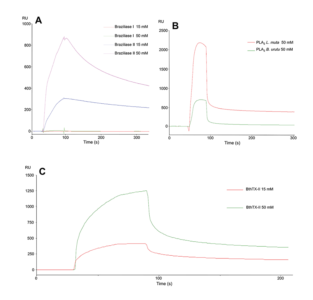
Binding assays between CatD and snake venom PLA2s. (A) Interactions of CatD with Braziliase-I and Braziliase-II (concentrations of 15 and 50 µM). (B) Responses were obtained from the interaction between CatD and svPLA2s from Bothrops neuwiedi urutu (BnuTX-I) and Lachesis muta (LmutTX). (C) Interaction test between CatD and BthTX-II (concentrations of 15 and 50 mM). The analyzed samples were submitted to salt removal in a 5 mL Hitrap desalting (GE) column.
Different from Braziliase-I, Braziliase-II showed a dose-dependent sensorgram of 245 RUs (15 mM) and 837 RUs (50 mM) with a prolonged dissociation phase suggesting a possible low dissociation rate constant (Kd) (Fig. 3A), which could be investigated through further analysis. Both BnuTX-I from B. urutu and LmutTX from L. muta also interacted with immobilized CatD (Fig. 3B), showing sensorgrams with different intensities of 552 and 2,180 RU at 50 mM [4646. Diniz-Sousa R, Caldeira CAS, Kayano AM, Paloschi M V, Pimenta DC, Simões-Silva R, Ferreira AS, Zanchi FB, Matos NB, Grabner FP, Calderon LA, Zuliani JP, Soares AM. Identification of the molecular determinants of the antibacterial activity of LmutTX, a Lys49 phospholipase A2 homologue isolated from Lachesis muta muta snake venom (Linnaeus, 1766). Basic Clin Pharmacol Toxicol. 2018 Apr;122(4):413-23. doi: 10.1111/bcpt.12921.
https://doi.org/10.1111/bcpt.12921...
, 4747. Correa EA, Kayano AM, Diniz-Sousa R, Setubal SS, Zanchi FB, Zuliani JP, Matos NB, Almeida JR, Resende LM, Marangoni S, Silva SL, Soares AM, Calderon LA. Isolation, structural and functional characterization of a new Lys49 phospholipase A2 homologue from Bothrops neuwiedi urutu with bactericidal potential. Toxicon. 2016 Jun 1;115:13-21. doi: 10.1016/j.toxicon.2016.02.021.
https://doi.org/10.1016/j.toxicon.2016.0...
]. In any case, both showed a similar shape in their association and dissociation curves.
Despite the high level of homology among svPLA2s, the binding analysis between CatD and these toxins exhibited interactions with different intensity profiles. Nevertheless, the binding profile of CatD towards svPLA2 displayed high similarity, suggesting a common recognition site. It is worth pointing out that overall, svPLA2s present a characteristic and consistent tridimensional structure, which could be the driving factor behind the ability of CatD to interact with the svPLA2s tested in this study [6161. Andrião-Escarso SH, Soares AM, Rodrigues VM, Angulo Y, Díaz C, Lomonte B, Gutiérrez JM, Giglio JR. Myotoxic phospholipases A2 in Bothrops snake venoms: Effect of chemical modifications on the enzymatic and pharmacological properties of bothropstoxins from Bothrops jararacussu. Biochimie. 2000 Aug;82(8):755-63. doi: 10.1016/s0300-9084(00)01150-0.
https://doi.org/10.1016/s0300-9084(00)01...
, 6262. Dennis EA, Cao J, Hsu YH, Magrioti V, Kokotos G. Phospholipase A2 enzymes: Physical structure, biological function, disease implication, chemical inhibition, and therapeutic intervention. Chem Rev. 2011 Oct 12;111(10):6130-85. doi: 10.1021/cr200085w.
https://doi.org/10.1021/cr200085w...
].
Enzymatic activity of the cathepsin D + BthTX-II complex
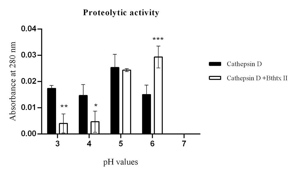
Initially, the apparent molecular mass and isoelectric point (pI) of the CatD + BthTX-II complex, as well as that of both enzymes separately, BthTX-II and CatD, were verified through two-dimensional electrophoresis (Fig. 4), determining a molecular mass of approximately 60 kDa and pI of 5.79 for the CatD + BthTX-II complex. Next, the proteolytic activity of CatD and of its complex with svPLA2 (BthTX-II) were evaluated using casein as a substrate at pH values of 3, 4, 5, 6, and 7, and Pepstatin A as a specific inhibitor. The optimal enzymatic activity of CatD was observed at pH 5, which is in agreement with previous studies [6363. Conner GE, Cathepsin D. Handb proteolytic enzym. Barrett A, Rawlings N, Woessner J editors. Elsevier; 2004. p. 43-52. doi: 10.1016/B978-0-12-382219-2.00281-7.
https://doi.org/10.1016/B978-0-12-382219...
]. On the other hand, the CatD + BthTX-II complex proved to be functional at different pH values reaching maximum activity at pH 6 (Fig. 5), revealing that the binding between these two proteins changes CatD’s functionalities, increasing its pH-dependent activity to higher values. Additionally, the CatD + BthTX-II complex is resistant to the inhibitor Pepstatin A at pH 6, suggesting the possibility of changes in enzyme specificity (Fig. 6A).

Two-dimensional SDS-PAGE: CatD, BthTX-II, and enzymatic complex. (A) Two-dimensional SDS-PAGE of CatD showing a pI of 4.74 and approximate molecular mass of 35 kDa. (B) Two-dimensional SDS-PAGE of BthTX-ll with a pI of 8.74, with an approximate molecular mass of 14 kDa. (C) Two-dimensional SDS-PAGE of the complex with a pI of 5.79, and approximate molecular mass of 49 kDa.

Proteolytic activity of CatD and the CatD + BthTX-II complex. The evaluation was performed at pHs 3, 4, 5, 6, and 7, identified in the figure legend, highlighting CatD in white (positive control) and the CatD + BthTX-II complex in black. As a negative control, the buffer itself (sodium citrate) was used at different pHs. The toxin used in the tests (BthTX-II) was submitted to contamination analysis (described in the second section). Two-way analysis of variance (ANOVA) with Tukey’s multiple comparison post-test with significance level p < 0.05.

Proteolytic activity on casein. (A) Samples identified in the legend results: CatD, CatD + BthTX-II, CatD + BthTX-II + pepstatin A were considered negative controls; buffer (sodium citrate) was used at different pHs. (B) Proteolytic activities SDS-PAGE. Samples: (1) CatD; (2) pepstatin A (PepA); (3) casein; (4) CatD + PepA + casein; (5) CatD + casein; (6) CatD + BthTX-II Casein + PepA; (7) CatD + BthTX-II + casein (30 min); (8) CatD + BthTX-II + casein (15 min); (9) CatD + BthTX-II + casein (5 min). Samples 3 through 6 were incubated for 30 min at 27 ºC. Positive control: CatD; negative control: pepstatin A (PepA).
Similar outcomes were observed in the SDS-PAGE assay, revealing that the bands formed after casein hydrolysis by CatD and CatD + BthTX-II are slightly different (Fig. 6B), suggesting potential differences in cleavage sites and further confirming the in vitro enzymatic activity. Furthermore, to rule out any residual contamination from the BthTX-II sample due to venom proteases, this sample was also submitted to the same conditions, and showed no proteolytic activity (results not shown).
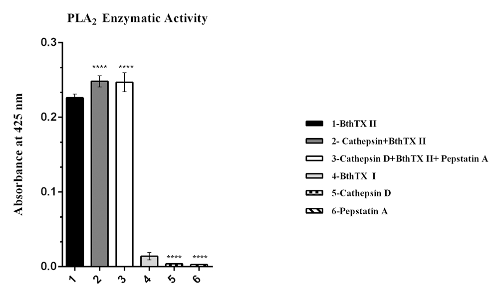
Regarding the effects of the interaction of the CatD + BthTX-II complex on BthTX-II’s catalytic function, the phospholipase activity assay (Fig. 7) shows that the complex’s formation does not interfere with nor hinder BthTX-II’s capability to cleave the artificial substrate 4N30BA. Interestingly, the presence of Pepstatin A slightly diminishes the catalytic output of the CatD + BthTX-II complex.

PLA2 enzymatic activity on artificial substrate 4N3OBA. Samples: (1) BthTX-II; (2) CatD + BthTX-II; (3) CatD + BthTX-II + PepA; (4) BthTX-I; (5) CatD; (6) PepA. Positive control: BthTX-II. Negative control: BthTX-I.
Structural analysis and molecular interaction simulations
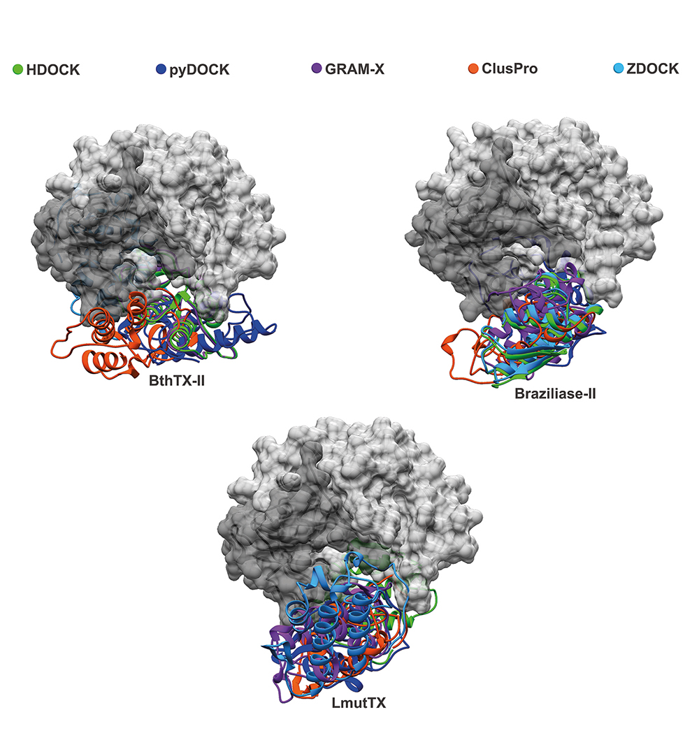
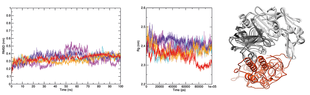
All svPLA2s showing interaction with CatD in the SPR assay and enzymatic assays were selected for further in silico investigation, seeking details about the mechanism coordinating these interactions at the atomic level and the existence of common recognition sites for svPLA2s on CatD’s surface. Thus, five molecular docking methodologies were applied, effectively employing a consensus approach, which generated sets of docking conformations (Fig. 8) for each of the svPLAs2 (BthTX-II, Braziliase-II and LmutTX). Additionally, the CatD + BthTX-II complex (Fig. 9) was subjected to a more intensive inspection due to its enzymatic activity. Molecular dynamics (MD) was used to evaluate the structural stability of this macromolecular assembly. Five independent replicas were simulated for 100 ns each. The processing and analysis of the generated trajectories included an assessment of the CatD + BthTX-II complex’s behavior in solution considering the radius of gyration (Fig. 10A) and RMSD (Fig. 10B) variations during the simulations. There were few noticeable fluctuations in the complex’s backbone and its compactness. Nevertheless, the assembly formed between these two proteins remained stable through all five replicas. The interaction between CatD and BthTX-II was evaluated, using as reference the central structures from the three most populated clusters generated in the clusterization performed with the sum of all five trajectories, exhibiting in that way an approximation of the most predominant conformation assumed by the CatD + BthTX-II complex during 500 ns of simulation (Fig. 10C). The absence of any remarkable shift in the complex’s shape suggests an overall stable and cohesive interaction.

Molecular modeling of the interaction between three snake venom PLA2s (LmuTX, Braziliase-II and BthTX-II) and human CatD using different docking tools (HDOCK, pyDOCK, GRAM-X, ClusPro, and ZDOCK). The CatD surface is represented in dark gray (light chain) and light gray (heavy chain). The svPLA2s are colored according to the docking tool used.

Molecular modeling of the CatD + BthTX-II complex. The complex formed between human CatD is shown in gray (light chain in dark gray and heavy chain in light gray) and BthTX-II is shown in orange. The interactions were enlarged to show amino acid residues in the interface and their interactions. H-bonds are highlighted by green dashed lines, and hydrophobic interactions are depicted as protrusions colored to match each amino acid residue.

CatD + BthTX-II complex molecular dynamics. CatD is shown in gray (light chain in dark gray and heavy chain in light gray) and BthTX-II is shown in orange. The radius of gyration and backbone RMSD graphics are located on the left side of the figure. All five replicas are plotted in an overlapped manner in order to highlight all minor variations and overall stability throughout the 100 ns of each replica. The structures on the right end of the figure show a superposition respective to each of the three CatD + BthTX-II complexes representing the most predominant conformations during the total 500 ns simulated. These superposed complexes are the central structures extracted from the three most populated clusters generated in the clusterization analysis performed with the trajectories of all five replicas.
Discussion
In order to proceed with the characterization of the CatD + BthTX-II complex, different methodologies were used, such as Surface Plasmon Resonance (SPR), a detection method capable of performing real-time, label-free, and high-sensitivity monitoring of molecular interactions [6464. Nguyen HH, Park J, Kang S, Kim M. Surface plasmon resonance: A versatile technique for biosensor applications. Sensors (Basel). 2015 May 5;15(5):10481-510. doi: 10.3390/s150510481.
https://doi.org/10.3390/s150510481...
], and molecular docking, a key tool in structural molecular biology and computer-aided drug design, useful to predict structural data about a potential protein-protein interaction using known three-dimensional structures [6565. Morris GM, Lim-Wilby M. Molecular docking. Methods Mol Biol. 2008;443:365-82. doi: 10.1007/978-1-59745-177-2_19.
https://doi.org/10.1007/978-1-59745-177-...
].
SPR assays carried out with immobilized human CatD showed different levels of interaction with components of all snake venoms tested, ranging from 34.1 RU mg/mL for C. d. cascavella to 2,258.3 RU mg/mL for B. jararacussu (Table 1). The interaction of venom components with human cathepsin D, especially those from bothropic venoms, strongly suggests that this could be an important and relevant new biological mechanism involving the participation of CatD and svPLA2 in snake envenomation and other physiopathological processes with the participation of homologous proteins.
The use of B. jararaca venom cation exchange chromatographic fractions for further SPR assays (Fig. 2) showed that immobilized CatD interacted only with the last fractions (10, 11, and 12), which corresponds to well-known svPLA2s, according to the monophoretic bands observed in the electrophoresis profile. This data indicated that the svPLA2s presented in the samples tested in SPR binding assays with CatD could be the respective ligands. The SPR analyses carried out with the isolated svPLA2s BthTX-II, Braziliase-II, BnuTX-I, and LmutTX revealed their ability to bind with immobilized human CatD (Fig. 3).
Two-dimensional electrophoresis showed that human CatD and BthTX-II form a stable complex of approximately 60 kDa and pI of 5.79. Initially, the apparent molecular mass and isoelectric point (pI) of the CatD + BthTX-II complex, as well as that of both enzymes separately, BthTX-II and CatD, were verified through two-dimensional electrophoresis (Fig. 4), determining a molecular mass for the CatD + BthTX-II complex. Next, the proteolytic activity of CatD and its complex with svPLA2 (BthTX-II) was evaluated using casein as a substrate at pH values from 3 to 7, and Pepstatin A as a specific inhibitor. The pH optimum of the CatD + BthTX-II complex was found to be 6, while isolated CatD shows optimal activity at pH 4 [6666. Sapolsky AI, Howell DS, Woessner, JF Jr. Neutral proteases and cathepsin D in human articular cartilage. J Clin Investig. 1974 Apr;53(4):1044-53. doi: 10.1172/JCI107641.
https://doi.org/10.1172/JCI107641...
]. Furthermore, Pepstatin A doesn’t affect the CatD + BthTX-II complex activity with the substrate (Casein) at different pH values.
Interestingly, the change in CatD pH-dependent activity, when compared to that of the CatD + BthTX-II complex, is consistent with previous CatD studies in tumoral cell lines [6767. Maynadier M, Farnoud R, Lamy P-J, Laurent-Matha V, Garcia M, Rochefort H. Cathepsin D stimulates the activities of secreted plasminogen activators in the breast cancer acidic environment. Int J Oncol. 2013 Nov;43(5):1683-90. doi: 10.3892/ijo.2013.2095.
https://doi.org/10.3892/ijo.2013.2095...
], suggesting that in the physiologic scenario, CatD’s interaction with proteins such as svPLA2 might be the factor allowing it to function in different pH ranges. Additionally, the CatD + BthTX-II complex was not inhibited by Pepstatin A, with CatD’s catalytic activity remaining steady, further corroborating the CatD + BthTX-II complex’s increased activity capacity. Moreover, the investigation of the CatD + BthTX-II complex’s impact on BthTX-II’s phospholipase activity suggests that the orientation of BthTX-II when coupled with CatD is ideal and allows BthTX-II to remain fully functional.
Computational simulations revealed a clear pattern of interaction between CatD and svPLA2s, in such a way that all svPLA2s tested in this study exhibited affinity by the concave surface formed between the heavy and light chain of CatD. This interaction profile was observed in every docking performed in this study. Furthermore, MD simulations done with the CatD + BthTX-II complex demonstrated that this may be the stable conformation assumed by CatD interacting with svPLA2s in solution. Alone, the CatD + svPLA2 complex’s interface of interaction observed in the simulations performed herein is not able to enlighten the molecular mechanisms behind the boost in CatD’s catalytic activity observed in the enzymatic assays. However, the conformation of the CatD + BthTX-II complex generated in the docking predictions and later validated in the 500 ns of simulations agrees with the phospholipase activity assays. The capability of the CatD + BthTX-II complex to retain svPLA2 makes perfect sense given BthTX-II’s orientation upon attachment to CatD (Fig. 9 and 10C), in such a way that BthTX-II’s hydrophobic channel and active site remain fully exposed to solvent.
Taking into account all these data, the in silico exploration of CatD’s complex with svPLA2 provides a clear basis for these two enzymes’ interaction in the physiologic scenario. Nevertheless, it is necessary to carry out more experimental structural studies in order to confirm the modes of interaction between these enzymes. These results also raise new questions in the investigation of pathological and inflammatory symptoms of snake envenomation, in which CatD’s interaction with svPLA2 and the complexes formed could play an important role in the cascade of systemic and local effects present in snakebite accidents.
The interaction between CatD and svPLA2 demonstrated herein will possibly have future implications for snakebite therapeutics. However, the most significant results extracted from this study may foreshadow more fundamental physiological issues involving the role of CatD in inflammatory processes, apoptosis and tumor progression. In this regard, the proteolytic process in neurons, in which CatD actively participates, is an essential maintenance step for the clearance of protein aggregates that reach the lysosomes through endocytosis and autophagy [2424. Fusek M, Vetvicka V. Dual role of cathepsin D: ligand and protease. Biomed Pap Med Fac Univ Palacky Olomouc Czech Repub. 2005 Jun;149(1):43-50. doi: 10.5507/pb.2005.003.
https://doi.org/10.5507/pb.2005.003...
].
Di Domenico and coworkers proposed that the lack of control in protein repair (proteasome and lysosomal system) is a characteristic of degenerating neurons in Alzheimer’s disease (AD), which highlights CatD’s involvement in these conditions due to its essential role in the management of lysosomal integrity [3333. Di Domenico F, Tramutola A, Perluigi M. Cathepsin D as a therapeutic target in Alzheimer’s disease. Expert Opin Ther Targets. 2016 Dec;20(12):1393-5. doi: 10.1080/14728222.2016.1252334.
https://doi.org/10.1080/14728222.2016.12...
]. Thus, the rise in PLA2 (IIA) in the cerebrospinal fluid of patients with AD indicates these enzymes as potential biomarkers in neuroinflammation [6868. Tasoulis T, Isbister GK. A review and database of snake venom proteomes. Toxins (Basel). 2017 Sep 18;9(9):290. doi: 10.3390/toxins9090290.
https://doi.org/10.3390/toxins9090290...
, 6969. Turk V, Kos J, Turk B. Cysteine cathepsins (proteases) - On the main stage of cancer? Cancer Cell. 2004 May;5(5):409-10. doi: 10.1016/s1535-6108(04)00117-5.
https://doi.org/10.1016/s1535-6108(04)00...
]. Furthermore, human brains affected by AD present a significant increase in PLA2 mRNA in the hippocampus [7070. Moses GS, Jensen MD, Lue L-F, Walker DG, Sun AY, Simonyi A, Sun GY. Secretory PLA2-IIA: a new inflammatory factor for Alzheimer’s disease. J Neuroinflammation. 2006 Oct 7;3:28. doi: 10.1186/1742-2094-3-28.
https://doi.org/10.1186/1742-2094-3-28...
]. Interestingly, reports of PLA2s’ involvement in the destabilization of lysosomal membranes have been made in different experimental systems [2929. Charlotte Johansson A, Appelqvist AJH, Nilsson C, Kagedal K, Roberg K, Ollinger K. Regulation of apoptosis-associated lysosomal membrane permeabilization. Apoptosis. 2010 May;15(5):527-40. doi: 10.1007/s10495-009-0452-5
https://doi.org/10.1007/s10495-009-0452-...
, 7171. Mirnikjoo B, Balasubramanina K, Schroit AJ. Mobilization of lysosomal calcium regulates the externalization of phosphatidylserine during Apoptosis. J Biol Chem. 2009 Mar 13;284(11):6918-23. doi: 10.1074/jbc.M805288200.
https://doi.org/10.1074/jbc.M805288200...
, 7272. Windelborn JA, Lipton P. Lysosomal release of cathepsins causes ischemic damage in the rat hippocampal slice and depends on NMDA-mediated calcium influx, arachidonic acid metabolism, and free radical production. J Neurochem. 2008 Jul;106(1):56-69. doi: 10.1111/j.1471-4159.2008.05349.x.
https://doi.org/10.1111/j.1471-4159.2008...
].
Overall, many approaches have discussed the involvement of PLA2s in inflammatory processes [7373. Murakami M, Taketomi Y, Miki Y, Sato H, Hirabayashi T, Yamamoto K. Progress in Lipid Research Recent progress in phospholipase A2 research: from cells to animals to humans. Prog Lipid Res. 2011;50:152-92. doi: 10.1016/j.plipres.2010.12.001.
https://doi.org/10.1016/j.plipres.2010.1...
-7575. Murakami M, Yamamoto K, Miki Y, Murase R, Sato H, Taketomi Y. The roles of the secreted phospholipase A2 gene family in immunology. Adv Immunol. 2016;132:91-134. doi: 10.1016/bs.ai.2016.05.001.
https://doi.org/10.1016/bs.ai.2016.05.00...
]. In addition, PLA2s also act on cell membrane metabolism and the production of arachidonic acid, a known precursor of prostaglandins, leukotrienes, and thromboxanes [7676. Ricciotti E, Fitzgerald GA. Prostaglandins and inflammation. Arterioscler Thromb Vasc Biol. 2011 May;31(5):986-1000. doi: 10.1161/ATVBAHA.110.207449.
https://doi.org/10.1161/ATVBAHA.110.2074...
-7878. Oliveira CS, Caldeira CAS, Diniz-sousa R, Romero DL, Marcussi S, Moura LA, Fuly AL, de Carvalho C, Cavalcante WLG, Gallacci M, Dal Pai M, Zuliani JP, Calderon LA, Soares AM. Pharmacological characterization of cnidarian extracts from the Caribbean Sea: evaluation of anti-snake venom and antitumor properties. J Venom Anim Toxins incl Trop Dis. 2018;1-11. doi: 10.1186/s40409-018-0161-z.
https://doi.org/10.1186/s40409-018-0161-...
]. Johansson and coworkers demonstrated that incubation of PLA2s with rat liver lysosomes resulted in the extravasation of its lysosomal constituents [2929. Charlotte Johansson A, Appelqvist AJH, Nilsson C, Kagedal K, Roberg K, Ollinger K. Regulation of apoptosis-associated lysosomal membrane permeabilization. Apoptosis. 2010 May;15(5):527-40. doi: 10.1007/s10495-009-0452-5
https://doi.org/10.1007/s10495-009-0452-...
]. Additionally, Beaujouin and coworkers demonstrated CatD’s involvement in apoptosis and showed that cancer cells that were pretreated with Pepstatin A, could not halt CatD nor hinder apoptosis, supporting the results described herein in the proteolytic activity assays. Moreover, CatD’s capability to induce cancer cell growth, even when mutated, suggests an alternative mechanism for this enzyme [7979. Beaujouin M, Baghdiguian S, Glondu-Lassis M, Berchem G, Liaudet-Coopman E. Overexpression of both catalytically active and -inactive cathepsin D by cancer cells enhances apoptosis-dependent chemo-sensitivity. Oncogene. 2006;25:1967-73. doi: 10.3390/cells9071679.
https://doi.org/10.3390/cells9071679...
].
Conclusion
For the first time, this study describes the formation of a functional muti-enzymatic complex between the human protease cathepsin D and snake venom phospholipases A2. Collectively, the in vitro assays and in silico predictions carried out in this study demonstrated interaction and the formation of a new muti-enzymatic and catalytically active complex between CatD and svPLA2. Additionally, the agreement between the data from previous studies regarding the pathways in which these enzymes are involved and the new data presented herein indicates the possibility of PLA2 and CatD acting in conjunction in the extracellular environment [4141. Burke JE, Dennis EA. Phospholipase A2 structure/function, mechanism, and signaling. J Lipid Res. 2009 Apr;50(Suppl):S237-42. doi: 10.1194/jlr.R800033-JLR200.
https://doi.org/10.1194/jlr.R800033-JLR2...
]. Nevertheless, in the face of the many possible outcomes of this new enzymatic complex, the conclusions drawn must be taken with caution and, most importantly, warrant more extensive investigation on the subject.
Acknowledgments
The authors thank the Program for Technological Development in Tools for Health-PDTIS-FIOCRUZ for the use of its facilities.
References
- 1. Yadati T, Houben T, Bitorina A, Shiri-Sverdlov R. The ins and outs of cathepsins: Physiological function and role in disease management. Cells. 2020 Jul 13;9(7):1679. doi: 10.3390/cells9071679.
» https://doi.org/10.3390/cells9071679 - 2. Turk V, Stoka V, Vasiljeva O, Renko M, Sun T, Turk B, Turk D. Cysteine cathepsins: From structure, function and regulation to new frontiers. Biochim Biophys Acta. 2012 Jan;1824(1):68-88. doi: 10.1016/j.bbapap.2011.10.002
» https://doi.org/10.1016/j.bbapap.2011.10.002 - 3. Patel S, Homaei A, El-Seedi HR, Akhtar N. Cathepsins: Proteases that are vital for survival but can also be fatal. Biomed Pharmacother. 2018 Sep;105:526-32. doi: 10.1016/j.biopha.2018.05.148.
» https://doi.org/10.1016/j.biopha.2018.05.148 - 4. Brix K, Dunkhorst A, Mayer K, Jordans S. Cysteine cathepsins: cellular roadmap to different functions. Biochimie. 2008 Feb;90(2):194-207. doi: 10.1016/j.biochi.2007.07.024.
» https://doi.org/10.1016/j.biochi.2007.07.024 - 5. Ward C, Martinez-Lopez N, Otten EG, Carroll B, Maetzel D, Singh R, Sarkar S, Korolchuk VI. Autophagy, lipophagy and lysosomal lipid storage disorders. Biochim Biophys Acta. 2016 Apr;1861(4):269-84. doi: 10.1016/j.bbalip.2016.01.006.
» https://doi.org/10.1016/j.bbalip.2016.01.006 - 6. Stoka V, Turk V, Turk B. Lysosomal cathepsins and their regulation in aging and neurodegeneration. Ageing Res Rev. 2016 Dec;32:22-37. doi: 10.1016/j.arr.2016.04.010.
» https://doi.org/10.1016/j.arr.2016.04.010 - 7. Bonnans C, Chou J, Werb Z. Remodelling the extracellular matrix in development and disease. Nat Rev Mol Cell Biol. 2014 Dec;15(12):786-801. doi: 10.1016/j.arr.2016.04.010.
» https://doi.org/10.1016/j.arr.2016.04.010 - 8. Rakash S. Role of proteases in cancer: A review. Biotechnol Mol Biol Rev. 2012 Oct;7(4):90-101. doi: 10.5897/BMBR11.027.
» https://doi.org/10.5897/BMBR11.027 - 9. Turk B, Turk V. Lysosomes as “Suicide Bags” in Cell Death: Myth or Reality? J Biol Chem. 2009 Aug 14;284(33):21783-7. doi: 10.1074/jbc.R109.023820.
» https://doi.org/10.1074/jbc.R109.023820 - 10. Repnik U, Cesen MH, Turk B, Česen MH, Turk B. The endolysosomal system in cell death and survival. Cold Spring Harb Perspect Biol. 2013 Jan 1;5(1):a008755. doi: 10.1101/cshperspect.a008755.
» https://doi.org/10.1101/cshperspect.a008755 - 11. Liaudet-Coopman E, Beaujouin M, Derocq D, Garcia M, Glondu-Lassis M, Laurent-Matha V, Prébois C, Rochefort H, Vignon F. Cathepsin D: newly discovered functions of a long-standing aspartic protease in cancer and apoptosis. Cancer Lett. 2006 Jun 18;237(2):167-79. doi: 10.1016/j.canlet.2005.06.007.
» https://doi.org/10.1016/j.canlet.2005.06.007 - 12. Conus S, Perozzo R, Reinheckel T, Peters C, Scapozza L, Yousefi S, Simon HU. Caspase-8 is activated by cathepsin D initiating neutrophil apoptosis during the resolution of inflammation. J Exp Med. 2008 Mar 17;205(3):685-98. doi: 10.1074/jbc.M111.306399.
» https://doi.org/10.1074/jbc.M111.306399 - 13. Mattson MP. Neuronal Life-and-Death Signaling, Apoptosis, and Neurodegenerative Disorders. Antioxid Redox Signal. 2006 Nov-Dec;8(11):1997-2006. doi: 10.1038/35040009.
» https://doi.org/10.1038/35040009 - 14. Sato Y, Suzuki Y, Ito E, Shimazaki S, Ishida M, Yamamoto T, Yamamoto H, Toda T, Suzuki M, Suzuki A, Endo T. Identification and characterization of an increased glycoprotein in aging: Age-associated translocation of cathepsin D. Mech Ageing Dev. 2006;127:771-8. doi: 10.1016/j.mad.2006.07.001.
» https://doi.org/10.1016/j.mad.2006.07.001 - 15. Garcia M, Platet N, Liaudet E, Laurent V, Derocq D, Brouillet J, Rochefort H. Biological and clinical significance of cathepsin D in breast cancer metastasis. Stem Cells. 1996 Nov;14(6):642-50. doi: 10.1002/stem.140642.
» https://doi.org/10.1002/stem.140642 - 16. Metcalf P, Fusek M. Two crystal structures for cathepsin D: the lysosomal targeting signal and active site. EMBO J. 1993 Apr;12(4):1293-302. doi: 10.1002/j.1460-2075.1993.tb05774.x.
» https://doi.org/10.1002/j.1460-2075.1993.tb05774.x - 17. Baldwin ET, Bhat TN, Gulnik S, Hosur M V., Sowder RC, Cachau RE, Collins J, Silva AM, Erickson JW. Crystal structures of native and inhibited forms of human cathepsin D: implications for lysosomal targeting and drug design. Proc Natl Acad Sci U S A. 1993 Jul 15;90(14):6796-800. doi: 10.1073/pnas.90.14.6796.
» https://doi.org/10.1073/pnas.90.14.6796 - 18. Zaidi N, Maurer A, Nieke S, Kalbacher H. Cathepsin D: A cellular roadmap. Biochem Biophys Res Commun. 2008 Nov 7;376(1):5-9. doi: 10.1016/j.bbrc.2008.08.099.
» https://doi.org/10.1016/j.bbrc.2008.08.099 - 19. Benes P, Vetvicka V, Fusek M. Cathepsin D - Many functions of one aspartic protease. Crit Rev Oncol Hematol. 2008 Oct;68(1):12-28. doi: 10.1016/j.critrevonc.2008.02.008.
» https://doi.org/10.1016/j.critrevonc.2008.02.008 - 20. Masson O, Bach AS, Derocq D, Prébois C, Laurent-Matha V, Pattingre S, Liaudet-Coopman E. Pathophysiological functions of cathepsin D: Targeting its catalytic activity versus its protein binding activity? Biochimie. 2010;92:1635-43. doi: 10.1016/j.biochi.2010.05.009.
» https://doi.org/10.1016/j.biochi.2010.05.009 - 21. Chaudhary H, Jena KP, Trivedi D, Purabdhar K, Vora N, Nair P, Sangtani K, Thakur S, Saini A, Singh S, Patel JSS. Cathepsin D: A novel target for apoptotic induction, as a future anti-cancer therapy: a review. J Phys Pharm Adv J Phys Pharm Adv. 2012 Feb 1;2(2):87-96.
- 22. Khalkhali-Ellis Z, Hendrix MJC. Two faces of cathepsin D: Physiological guardian angel and pathological demon. Biol Med (Aligarh). 2014 Jul;6(2):1000206. doi: 10.4172/0974-8369.1000206.
» https://doi.org/10.4172/0974-8369.1000206 - 23. Gocheva V, Joyce JA. Cysteine cathepsins and the cutting edge of cancer invasion. Cell Cycle. 2007 Jan 1;6(1):60-4. doi: 10.4161/cc.6.1.3669.
» https://doi.org/10.4161/cc.6.1.3669 - 24. Fusek M, Vetvicka V. Dual role of cathepsin D: ligand and protease. Biomed Pap Med Fac Univ Palacky Olomouc Czech Repub. 2005 Jun;149(1):43-50. doi: 10.5507/pb.2005.003.
» https://doi.org/10.5507/pb.2005.003 - 25. Johansson AC, Steen H, Öllinger K, Roberg K. Cathepsin D mediates cytochrome c release and caspase activation in human fibroblast apoptosis induced by staurosporine. Cell Death Differ. 2003 Nov;10(11):1253-9. doi: 10.1038/sj.cdd.4401290
» https://doi.org/10.1038/sj.cdd.4401290 - 26. Arnold D, Keilholz W, Schild H, Dumrese T, Stevanovic S, Rammensee HG. Substrate specificity of cathepsins D and E determined by N-terminal and C-terminal sequencing of peptide pools. Eur J Biochem. 1997 Oct 1;249(1):171-9. doi: 10.1111/j.1432-1033.1997.t01-1-00171.x.
» https://doi.org/10.1111/j.1432-1033.1997.t01-1-00171.x - 27. Pimenta DC, Oliveira A, Juliano MA, Juliano L. Substrate specificity of human cathepsin D using internally quenched fluorescent peptides derived from reactive site loop of kallistatin. Biochim Biophys Acta. 2001 Jan 12;1544(1-2):113-22. doi: 10.1016/s0167-4838(00)00209-0.
» https://doi.org/10.1016/s0167-4838(00)00209-0 - 28. Appelqvist H, Johansson A, Linderoth E, Johansson U, Antonsson B, Steinfeld R, Kagedal K, Ollinger K. Lysosome-mediated apoptosis is associated with cathepsin D-specific processing of bid at Phe24 , Trp48 , and Phe183. Ann Clin Lab Sci. 2012 Summer;42(3):231-42.
- 29. Charlotte Johansson A, Appelqvist AJH, Nilsson C, Kagedal K, Roberg K, Ollinger K. Regulation of apoptosis-associated lysosomal membrane permeabilization. Apoptosis. 2010 May;15(5):527-40. doi: 10.1007/s10495-009-0452-5
» https://doi.org/10.1007/s10495-009-0452-5 - 30. Castino R, Isidoro C, Murphy D. Autophagy-dependent cell survival and cell death in an autosomal dominant familial neurohypophyseal diabetes insipidus in vitro model. FASEB J. 2005 Jun;19(8):1024-6. doi: 10.1096/fj.04-3163fje.
» https://doi.org/10.1096/fj.04-3163fje - 31. Saftig P, Hetman M, Schmahl W, Weber K, Heine L, Mossmann H, Koster A, Hess B, Evers M, von Figura K. Mice deficient for the lysosomal proteinase cathepsin D exhibit progressive atrophy of the intestinal mucosa and profound destruction of lymphoid cells. EMBO J. 1995 Aug 1;14(15):3599-608. doi: 10.1002/j.1460-2075.1995.tb00029.x.
» https://doi.org/10.1002/j.1460-2075.1995.tb00029.x - 32. Crabtree D, Dodson M, Ouyang X, Boyer-Guittaut M, Liang Q, Ballestas ME, Fineberg N, Zhang J. Over-expression of an inactive mutant cathepsin D increases endogenous alpha-synuclein and cathepsin B activity in SH-SY5Y cells. J Neurochem. 2014 Mar;128(6):950-61. doi: 10.1111/jnc.12497.
» https://doi.org/10.1111/jnc.12497 - 33. Di Domenico F, Tramutola A, Perluigi M. Cathepsin D as a therapeutic target in Alzheimer’s disease. Expert Opin Ther Targets. 2016 Dec;20(12):1393-5. doi: 10.1080/14728222.2016.1252334.
» https://doi.org/10.1080/14728222.2016.1252334 - 34. Zhao CF, Herrington DM. The function of cathepsins B, D, and X in atherosclerosis. Am J Cardiovasc Dis. 2016;6(4):163-70.
- 35. Olson OC, Joyce JA. Cysteine cathepsin proteases: regulators of cancer progression and therapeutic response. Nat Rev Cancer. 2015 Dec;15(12):712-29. doi: 10.1038/nrc4027.
» https://doi.org/10.1038/nrc4027 - 36. Palermo C, Joyce JA. Cysteine cathepsin proteases as pharmacological targets in cancer. Trends Pharmacol Sci. 2008 Jan;29(1):22-8. doi: 10.1016/j.tips.2007.10.011.
» https://doi.org/10.1016/j.tips.2007.10.011 - 37. Almeida JR, Resende LM, Watanabe RK, Carregari VC, Huancahuire-Vega S, da S. Caldeira CA, Coutinho-Neto A, Soares AM, Vale N, Gomes PAC, Marangoni S, Calderon LA, Silva SL. Snake venom peptides and low mass proteins: Molecular tools and therapeutic agents. Curr Med Chem. 2017;24(30):3254-82. doi: 10.2174/0929867323666161028155611.
» https://doi.org/10.2174/0929867323666161028155611 - 38. da Silva GM , de Souza DHB , Waitman KB, Ebram MC, Fessel MR, Zainescu IC, Portaro FC, Heras M, Andrade SA. Design, synthesis, and evaluation of Bothrops venom serine protease peptidic inhibitors. J Venom Anim Toxins incl Trop Dis. 2021;27. doi: 10.1590/1678-9199-JVATITD-2020-0066.
» https://doi.org/10.1590/1678-9199-JVATITD-2020-0066 - 39. Murakami M, Taketomi Y, Girard C, Yamamoto K, Lambeau G. Emerging roles of secreted phospholipase A2 enzymes: Lessons from transgenic and knockout mice. Biochimie. 2010 Jun;92(6):561-82. doi: 10.1016/j.biochi.2010.03.015.
» https://doi.org/10.1016/j.biochi.2010.03.015 - 40. Fortes-Dias CL, Macedo DHF, Barbosa RP, Souza-Silva G, Ortolani PL. Identification and characterization of the first endogenous phospholipase A2 inhibitor from a non-venomous tropical snake, Boa constrictor (Serpentes: Boidae). J Venom Anim Toxins incl Trop Dis. 2020;26. doi: 10.1590/1678-9199-JVATITD-2019-0044.
» https://doi.org/10.1590/1678-9199-JVATITD-2019-0044 - 41. Burke JE, Dennis EA. Phospholipase A2 structure/function, mechanism, and signaling. J Lipid Res. 2009 Apr;50(Suppl):S237-42. doi: 10.1194/jlr.R800033-JLR200.
» https://doi.org/10.1194/jlr.R800033-JLR200 - 42. Gutiérrez JM, Lomonte B. Phospholipases A2: Unveiling the secrets of a functionally versatile group of snake venom toxins. Toxicon. 2013 Feb;62:27-39. doi: 10.1016/j.toxicon.2012.09.006.
» https://doi.org/10.1016/j.toxicon.2012.09.006 - 43. Xiong S, Huang C. Synergistic strategies of predominant toxins in snake venoms. Toxicol Lett. 2018 May 1;287:142-54. doi: 10.1016/j.toxlet.2018.02.004.
» https://doi.org/10.1016/j.toxlet.2018.02.004 - 44. Péterfi O, Boda F, Szabó Z, Ferencz E, Bába L. Hypotensive snake venom componentes -A Mini-review. Molecules. 2019 Jul 31;24(15):2778. doi: 10.3390/molecules24152778.
» https://doi.org/10.3390/molecules24152778 - 45. Harris J, Scott-Davey T. Secreted phospholipases A2 of snake venoms: Effects on the peripheral neuromuscular system with comments on the role of phospholipases A2 in disorders of the CNS and their uses in industry. Toxins (Basel). 2013 Dec 17;5(12):2533-71. doi: 10.3390/toxins5122533.
» https://doi.org/10.3390/toxins5122533 - 46. Diniz-Sousa R, Caldeira CAS, Kayano AM, Paloschi M V, Pimenta DC, Simões-Silva R, Ferreira AS, Zanchi FB, Matos NB, Grabner FP, Calderon LA, Zuliani JP, Soares AM. Identification of the molecular determinants of the antibacterial activity of LmutTX, a Lys49 phospholipase A2 homologue isolated from Lachesis muta muta snake venom (Linnaeus, 1766). Basic Clin Pharmacol Toxicol. 2018 Apr;122(4):413-23. doi: 10.1111/bcpt.12921.
» https://doi.org/10.1111/bcpt.12921 - 47. Correa EA, Kayano AM, Diniz-Sousa R, Setubal SS, Zanchi FB, Zuliani JP, Matos NB, Almeida JR, Resende LM, Marangoni S, Silva SL, Soares AM, Calderon LA. Isolation, structural and functional characterization of a new Lys49 phospholipase A2 homologue from Bothrops neuwiedi urutu with bactericidal potential. Toxicon. 2016 Jun 1;115:13-21. doi: 10.1016/j.toxicon.2016.02.021.
» https://doi.org/10.1016/j.toxicon.2016.02.021 - 48. Kayano AM, Simões-Silva R, Medeiros PSM, Maltarollo VG, Honorio KM, Oliveira E, Albericio F, da Silva SL, Aguiar ACC, Krettli AU, Fernandes CFC, Juliani JP, Calderon LA, Stábeli RG, Soares AM. BbMP-1, a new metalloproteinase isolated from Bothrops brazili snake venom with in vitro antiplasmodial properties. Toxicon. 2015 Nov;106:30-41. doi: 10.1016/j.toxicon.2015.09.005.
» https://doi.org/10.1016/j.toxicon.2015.09.005 - 49. Sobrinho JC, Kayano AM, Simões-Silva R, Alfonso JJ, Gomez AF, Gomez MCV, Zanchi FB, Moura LA, Souza VR, Fuly AL, Oliveira E, da Silva SL, Almeida JR, Zuliani JP, Soares AM. Anti-platelet aggregation activity of two novel acidic Asp49-phospholipases A2 from Bothrops brazili snake venom. Int J Biol Macromol. 2018 Feb;107(Pt A):1014-22. doi: 10.1016/j.ijbiomac.2017.09.069.
» https://doi.org/10.1016/j.ijbiomac.2017.09.069 - 50. Bradford MM. A rapid and sensitive method for the quantitation of microgram quantities of protein utilizing the principle of protein-dye binding. Anal Biochem. 1976 May 7;72:248-54. doi: 10.1006/abio.1976.9999.
» https://doi.org/10.1006/abio.1976.9999 - 51. Rodrigues VM, Soares AM, Guerra-Sá R, Rodrigues V, Fontes MRM, Giglio JR. Structural and functional characterization of neuwiedase, a nonhemorrhagic fibrin(ogen)olytic metalloprotease from Bothrops neuwiedi snake venom. Arch Biochem Biophys. 2000 Sep 15;381:213-24. doi: 10.1006/abbi.2000.1958.
» https://doi.org/10.1006/abbi.2000.1958 - 52. Petrovic N, Grove C, Langton PE, Misso NLA, Thompson PJ. A simple assay for a human serum phospholipase A2 that is associated with high-density lipoproteins. J Lipid Res. 2001 Oct;42(10):1706-13. 10.1016/S0022-2275(20)32226-4
» https://doi.org/10.1016/S0022-2275(20)32226-4 - 53. Song Y, DiMaio F, Wang RYR, Kim D, Miles C, Brunette T, Thompson J, Baker D. High resolution comparative modeling with RosettaCM. Structure. 2013 Oct 8;21(10):1735-42. doi: 10.1016/j.str.2013.08.005.
» https://doi.org/10.1016/j.str.2013.08.005 - 54. Huang J, Rauscher S, Nawrocki G, Ran T, Feig M, De Groot BL, et al. CHARMM36m: An improved force field for folded and intrinsically disordered proteins. Nat Methods. 2016 Nov 7;14:71-3. doi:10.1038/nmeth.4067.
» https://doi.org/10.1038/nmeth.4067 - 55. Bussi G, Donadio D, Parrinello M. Canonical sampling through velocity rescaling. J Chem Phys. 2007 Jan 7;126(1):014101. doi: 10.1063/1.2408420.
» https://doi.org/10.1063/1.2408420 - 56. Berendsen HJC, Postma JPM, Van Gunsteren WF, Dinola A, Haak JR. Molecular dynamics with coupling to an external bath. J Chem Phys. 1984 Jun 27;81(1984):3684-90. doi: 10.1063/1.448118
» https://doi.org/10.1063/1.448118 - 57. Hoover WG. Canonical dynamics: Equilibrium phase-space distributions. Phys Rev A. 1985 Mar;31(3):1695-7. doi: 10.1103/PhysRevA.31.1695.
» https://doi.org/10.1103/PhysRevA.31.1695 - 58. Parrinello M, Rahman A. Polymorphic transitions in single crystals: A new molecular dynamics method. J Appl Phys. 1981;52:7182-90. doi: 10.1063/1.328693.
» https://doi.org/10.1063/1.328693 - 59. Daura X, Gademann K, Jaun B, Seebach D, van Gunsteren WF, Mark AE. Peptide folding: When simulation meets experiment. Angew Chemie Int Ed. 1999;38(1-2):236-40. doi: 10.1002/(SICI)1521-3773(19990115)38:1/2%3C236::AID-ANIE236%3E3.0.CO;2-M.
» https://doi.org/10.1002/(SICI)1521-3773(19990115)38:1/2%3C236::AID-ANIE236%3E3.0.CO;2-M - 60. Pettersen EF, Goddard TD, Huang CC, Couch GS, Greenblatt DM, Meng EC, Ferrin TE. UCSF Chimera - A visualization system for exploratory research and analysis. J Comput Chem. 2004 Oct;25(13):1605-12. doi: 10.1002/jcc.20084.
» https://doi.org/10.1002/jcc.20084 - 61. Andrião-Escarso SH, Soares AM, Rodrigues VM, Angulo Y, Díaz C, Lomonte B, Gutiérrez JM, Giglio JR. Myotoxic phospholipases A2 in Bothrops snake venoms: Effect of chemical modifications on the enzymatic and pharmacological properties of bothropstoxins from Bothrops jararacussu Biochimie. 2000 Aug;82(8):755-63. doi: 10.1016/s0300-9084(00)01150-0.
» https://doi.org/10.1016/s0300-9084(00)01150-0 - 62. Dennis EA, Cao J, Hsu YH, Magrioti V, Kokotos G. Phospholipase A2 enzymes: Physical structure, biological function, disease implication, chemical inhibition, and therapeutic intervention. Chem Rev. 2011 Oct 12;111(10):6130-85. doi: 10.1021/cr200085w.
» https://doi.org/10.1021/cr200085w - 63. Conner GE, Cathepsin D. Handb proteolytic enzym. Barrett A, Rawlings N, Woessner J editors. Elsevier; 2004. p. 43-52. doi: 10.1016/B978-0-12-382219-2.00281-7.
» https://doi.org/10.1016/B978-0-12-382219-2.00281-7 - 64. Nguyen HH, Park J, Kang S, Kim M. Surface plasmon resonance: A versatile technique for biosensor applications. Sensors (Basel). 2015 May 5;15(5):10481-510. doi: 10.3390/s150510481.
» https://doi.org/10.3390/s150510481 - 65. Morris GM, Lim-Wilby M. Molecular docking. Methods Mol Biol. 2008;443:365-82. doi: 10.1007/978-1-59745-177-2_19.
» https://doi.org/10.1007/978-1-59745-177-2_19 - 66. Sapolsky AI, Howell DS, Woessner, JF Jr. Neutral proteases and cathepsin D in human articular cartilage. J Clin Investig. 1974 Apr;53(4):1044-53. doi: 10.1172/JCI107641.
» https://doi.org/10.1172/JCI107641 - 67. Maynadier M, Farnoud R, Lamy P-J, Laurent-Matha V, Garcia M, Rochefort H. Cathepsin D stimulates the activities of secreted plasminogen activators in the breast cancer acidic environment. Int J Oncol. 2013 Nov;43(5):1683-90. doi: 10.3892/ijo.2013.2095.
» https://doi.org/10.3892/ijo.2013.2095 - 68. Tasoulis T, Isbister GK. A review and database of snake venom proteomes. Toxins (Basel). 2017 Sep 18;9(9):290. doi: 10.3390/toxins9090290.
» https://doi.org/10.3390/toxins9090290 - 69. Turk V, Kos J, Turk B. Cysteine cathepsins (proteases) - On the main stage of cancer? Cancer Cell. 2004 May;5(5):409-10. doi: 10.1016/s1535-6108(04)00117-5.
» https://doi.org/10.1016/s1535-6108(04)00117-5 - 70. Moses GS, Jensen MD, Lue L-F, Walker DG, Sun AY, Simonyi A, Sun GY. Secretory PLA2-IIA: a new inflammatory factor for Alzheimer’s disease. J Neuroinflammation. 2006 Oct 7;3:28. doi: 10.1186/1742-2094-3-28.
» https://doi.org/10.1186/1742-2094-3-28 - 71. Mirnikjoo B, Balasubramanina K, Schroit AJ. Mobilization of lysosomal calcium regulates the externalization of phosphatidylserine during Apoptosis. J Biol Chem. 2009 Mar 13;284(11):6918-23. doi: 10.1074/jbc.M805288200.
» https://doi.org/10.1074/jbc.M805288200 - 72. Windelborn JA, Lipton P. Lysosomal release of cathepsins causes ischemic damage in the rat hippocampal slice and depends on NMDA-mediated calcium influx, arachidonic acid metabolism, and free radical production. J Neurochem. 2008 Jul;106(1):56-69. doi: 10.1111/j.1471-4159.2008.05349.x.
» https://doi.org/10.1111/j.1471-4159.2008.05349.x - 73. Murakami M, Taketomi Y, Miki Y, Sato H, Hirabayashi T, Yamamoto K. Progress in Lipid Research Recent progress in phospholipase A2 research: from cells to animals to humans. Prog Lipid Res. 2011;50:152-92. doi: 10.1016/j.plipres.2010.12.001.
» https://doi.org/10.1016/j.plipres.2010.12.001 - 74. Murakami M, Sato H, Miki Y, Yamamoto K, Taketomi Y. A new era of secreted phospholipase A2 J Lipid Res. 2015 Jul;56(7):1248-61. doi: 10.1194/jlr.R058123.
» https://doi.org/10.1194/jlr.R058123 - 75. Murakami M, Yamamoto K, Miki Y, Murase R, Sato H, Taketomi Y. The roles of the secreted phospholipase A2 gene family in immunology. Adv Immunol. 2016;132:91-134. doi: 10.1016/bs.ai.2016.05.001.
» https://doi.org/10.1016/bs.ai.2016.05.001 - 76. Ricciotti E, Fitzgerald GA. Prostaglandins and inflammation. Arterioscler Thromb Vasc Biol. 2011 May;31(5):986-1000. doi: 10.1161/ATVBAHA.110.207449.
» https://doi.org/10.1161/ATVBAHA.110.207449 - 77. Nicotra G, Castino R, Follo C, Peracchio C, Valente G, Isidoro C. The dilemma: Does tissue expression of cathepsin D reflect tumor malignancy? the question: Does the assay truly mirror cathepsin D mis-function in the tumor? Cancer Biomark. 2010 Oct;7(1):47-64. doi: 10.3233/CBM-2010-0143.
» https://doi.org/10.3233/CBM-2010-0143 - 78. Oliveira CS, Caldeira CAS, Diniz-sousa R, Romero DL, Marcussi S, Moura LA, Fuly AL, de Carvalho C, Cavalcante WLG, Gallacci M, Dal Pai M, Zuliani JP, Calderon LA, Soares AM. Pharmacological characterization of cnidarian extracts from the Caribbean Sea: evaluation of anti-snake venom and antitumor properties. J Venom Anim Toxins incl Trop Dis. 2018;1-11. doi: 10.1186/s40409-018-0161-z.
» https://doi.org/10.1186/s40409-018-0161-z - 79. Beaujouin M, Baghdiguian S, Glondu-Lassis M, Berchem G, Liaudet-Coopman E. Overexpression of both catalytically active and -inactive cathepsin D by cancer cells enhances apoptosis-dependent chemo-sensitivity. Oncogene. 2006;25:1967-73. doi: 10.3390/cells9071679.
» https://doi.org/10.3390/cells9071679
-
Availability of data and materials
All data generated or analyzed during this study are included in this article. -
Funding
The present study was supported by the Coordination for the Improvement of Higher Education Personnel (CAPES), National Council for Scientific and Technological Development (CNPq), Foundation for Support to the Development of Scientific and Technological Actions and the Rondônia Research (FAPERO), PDTIS/FIOCRUZ platforms, Center for the Study of Biomolecules Applied to Health-CEBio/FIOCRUZ-RO and Research Excellence Program (PROEP-FIOCRUZ), Fiocruz Rondônia. -
Ethics approval
All snake venoms used in this study were acquired from the Venom Bank at CEBio/Fiocruz Rondônia/UNIR (Center for the Study of Biomolecules Applied to Health), Porto Velho, RO, Brazil, under local government authorization license number: IBAMA nº 27131-3 and CGEN/CNPq 010627/2011-1 -
Consent for publication
Not applicable.
Publication Dates
-
Publication in this collection
04 Nov 2022 -
Date of issue
2022
History
-
Received
13 Jan 2022 -
Accepted
16 Aug 2022