Abstract
Aspidoras comprises 25 species currently considered as valid, being widely distributed in Brazil, occurring from the upper rio Paraná basin in São Paulo to coastal basins of Ceará. After Nijssen, Isbrücker’s review more than 40 years ago, no extensive work regarding the taxonomy of Aspidoras was conducted. Our paper presents a comprehensive taxonomic revision of the genus, based primarily on the extensive material that has been collected since then. Considering the new diagnosis plus the available phylogenetic data, A. pauciradiatus and A. virgulatus are transferred to Corydoras and Scleromystax, respectively. New synonymies are proposed: A. eurycephalus and A. taurus with A. albater; A. menezesi and A. spilotus with A. raimundi; and A. microgalaeus and A. marianae with A. poecilus. Additionally, a new species from the Araguaia and Paraguay river basins is described, which can be distinguished from its congeners by the morphology of its complex vertebra and infraorbital 1. Thereby, the number of valid species within Aspidoras was reduced from 25 to 18. Redescriptions for A. albater, A. belenos, A. depinnai, A. fuscoguttatus, A. lakoi, A. maculosus, A. poecilus, A. psammatides, A. raimundi, and A. velites are provided. An identification key to the species of Aspidoras is also provided.
Keywords:
Aspidoradini; Corydoradinae; Identification key; Osteology; Taxonomy
Resumo
Aspidoras compreende 25 espécies atualmente consideradas como válidas, sendo amplamente distribuído no Brasil, ocorrendo desde a bacia do alto rio Paraná em São Paulo até as bacias costeiras do Ceará. Após a revisão de Nijssen, Isbrücker há mais de 40 anos, nenhum trabalho extensivo sobre a taxonomia de Aspidoras foi conduzido. Nosso trabalho apresenta uma revisão taxonômica abrangente do gênero, baseada principalmente no extenso material que foi coletado desde então. Considerando a nova diagnose mais os dados filogenéticos disponíveis, A. pauciradiatus e A. virgulatus são transferidas para Corydoras e Scleromystax, respectivamente. Novas sinonímias são propostas: A. eurycephalus e A. taurus com A. albater; A. menezesi e A. spilotus com A. raimundi; e A. microgalaeus e A. marianae com A. poecilus. Adicionalmente, uma espécie nova é descrita das bacias dos rios Araguaia e Paraguai, podendo ser distinguida de suas congêneres pela morfologia da vértebra complexa e infraorbital 1. Assim, o número de espécies válidas em Aspidoras foi reduzido de 25 para 18. Redescrições para A. albater, A. belenos, A. depinnai, A. fuscoguttatus, A. lakoi, A. maculosus, A. poecilus, A. psammatides, A. raimundi e A. velites são fornecidas. Uma chave de identificação para as espécies de Aspidoras também é fornecida.
Palavras-chave:
Aspidoradini; Chave de identificação; Corydoradinae; Osteologia; Taxonomia
INTRODUCTION
The Callichthyidae armoured catfishes can be promptly distinguished from all other Siluriformes by the presence of two longitudinal series of dermal plates on flanks (Reis, 2003Reis RE. Family Callichthyidae (Armored catfishes). In: Reis RE, Kullander SO, Ferraris CJ, Jr., editors. Check list of the freshwater fishes of South and Central America. Porto Alegre: Edipucrs; 2003. p.291–309.). The family currently consists of around 200 valid species distributed in eight genera (Reis, 2003Reis RE. Family Callichthyidae (Armored catfishes). In: Reis RE, Kullander SO, Ferraris CJ, Jr., editors. Check list of the freshwater fishes of South and Central America. Porto Alegre: Edipucrs; 2003. p.291–309.; Fricke et al., 2021Fricke R, Eschmeyer WN, Van der Laan R. Eschmeyer’s catalog of fishes: genera, species, references [Internet]. San Francisco: California Academy of Science; 2021. Available from: http://researcharchive.calacademy.org/research/ichthyology/catalog/fishcatmain.asp
http://researcharchive.calacademy.org/re...
). Aspidoras, its second largest genus, was described by Ihering, (1907)Ihering R. Diversas espécies novas de peixes nemathognathas do Brasil. Notas preliminares. Rev Mus Paul. 1907; 1(1):13-39. as a monotypic genus harboring the then newly described species, A. rochai. According to Ihering (1907Ihering R. Diversas espécies novas de peixes nemathognathas do Brasil. Notas preliminares. Rev Mus Paul. 1907; 1(1):13-39.:30) the genus is recognized by the presence of the following unique combination of features: (I) two pairs of dorsolateral body plates between the parieto-supraoccipital and dorsal-fin base; (II) parieto-supraoccipital nearly hexagonal, with poorly developed posterior process of the parieto-supraoccipital; (III) head conspicuously deep, not depressed, rounded anteriorly and slightly laterally compressed; (IV) scapulocoracoid entirely covered by skin on ventral portion of the body; (V) barbels short, not reaching anteroventral limit of gill opening.
Gosline (1940)Gosline WA. A revision of the Neotropical catfishes of the family Callichthyidae. Stanford Ichthyol Bull. 1940; 2(1):1–36., in his revisionary study of the Callichthyidae, proposed a new diagnosis for Aspidoras based on the presence of the following combination of features: (I) head compressed; (II) lower lip reverted, forming a single pair of barbels, besides the outer mental barbel; (III) outer mental barbel reaching region of anteroventral limit of gill opening; (IV) eye slightly elevated; (V) first infraorbital naked (likely meaning exposed); (VI) fontanel small, roundish, its size nearly equal to half of the bony orbit diameter; (VII) posterior process of the parieto-supraoccipital short and broad; (VIII) dorsolateral body plates touching their counterparts between the posterior process of the parieto-supraoccipital and nuchal plate; (IX) region of pectoral girdle entirely covered by skin; (X) dorsal fin I,7, its base slightly shorter than distance between dorsal and adipose fins; and (XI) caudal fin forked (Gosline, 1940Gosline WA. A revision of the Neotropical catfishes of the family Callichthyidae. Stanford Ichthyol Bull. 1940; 2(1):1–36.:10). Although 33 years had passed between the description of the genus and Gosline’s work, Aspidoras was still considered monotypic by the author. The description of the second species within the genus, Aspidoras lakoiMiranda Ribeiro, 1949Miranda-Ribeiro P. Duas novas espécies de peixes na coleção ictiológica do Museu Nacional (Pisces, Callichthyidae et Pygidiidae). Rev Bras Biol. 1949; 9(2):143-45., was published 42 years after Ihering’s work (1907). The designation of a lectotype for A. rochai came 62 years after its description (Britski, 1969Britski HA. Lista dos tipos de peixes do Departamento de Zoologia da Secretaria da Agricultura de São Paulo. Pap Avulsos Dep Zool. 1969; 22(19):197-215.:206).
Nijssen, Isbrücker (1976)Nijssen H, Isbrücker IJH. The South American plated catfish genus Aspidoras R. von Ihering, 1907, with descriptions of nine new species from Brazil (Pisces, Siluriformes, Callichthyidae). Bijdr Dierkd. 1976; 46(1):107–31. presented the first comprehensive taxonomic review of Aspidoras, recognizing a total of 13 species. In addition to A. rochai and A. lakoi, nine species were then described, viz., A. albater, A. brunneus, A. carvalhoi, A. eurycephalus, A. fuscoguttatus, A. maculosus, A. menezesi, A. poecilus, and A. spilotus, and two species species previously assigned to Corydoras Lacepède, 1803, C. raimundiSteindachner, 1907Steindachner F. Über eine neue Coridoras-Art aus dem Rio Preto, einem sekundären Nebenflusse des Rio San Francisco, und eine Xenocara-Art aus dem Parnahyba bei Victoria und Sa. Filomena. Anz Akad Wiss. 1907; 44:290–93. and C. pauciradiatusWeitzman, Nijssen, 1970Weitzman SH, Nijssen H. Four new species and one new subspecies of the catfish genus Corydoras from Ecuador, Colombia and Brazil (Pisces, Siluriformes, Callichthyidae). Beaufortia. 1970; 18(233):119–32. Available from: https://repository.naturalis.nl/pub/504794
https://repository.naturalis.nl/pub/5047...
, were transferred to Aspidoras. The authors also provided a new diagnosis for the genus (see Nijssen, Isbrücker, 1976Nijssen H, Isbrücker IJH. The South American plated catfish genus Aspidoras R. von Ihering, 1907, with descriptions of nine new species from Brazil (Pisces, Siluriformes, Callichthyidae). Bijdr Dierkd. 1976; 46(1):107–31.:109), distinguishing Aspidoras from the other genera of Callichthyidae by the presence of two cranial fontanels, the posterior one in the parieto-supraoccipital and the anterior one between frontals (vs. a single fontanel).
Subsequent to Nijssen, Isbrücker’s (1980a) description of Aspidoras virgulatus, the taxonomy of the group was neglected for almost 20 years, until Britto, (1998)Britto MR. Two new species of the genus Aspidoras (Siluriformes: Callichthyidae) from Central Brazil. Ichthyol Explor Freshw. 1998; 8(4):359–68. proposed two new species: A. belenos, from the rio Araguaia basin, and A. microgalaeus, from the rio Xingu basin. The descriptions showed a great improvement over the previous works, furnishing external morphology data plus osteological data, which were used in the diagnosis of both new species. The diagnosis of A. belenos was the first to include the morphology of an infraorbital bone, which has proven to be extremely useful for the recognition of species within Corydoradinae (e.g., Tencatt et al., 2013Tencatt LFC, Vera-Alcaraz HS, Britto MR, Pavanelli CS. A new Corydoras Lacépède, 1803 (Siluriformes: Callichthyidae) from the rio São Francisco basin, Brazil. Neotrop Ichthyol. 2013; 11(2):257–64. https://doi.org/10.1590/S1679-62252013000200003
https://doi.org/10.1590/S1679-6225201300...
; Tencatt et al., 2014Tencatt LFC, Britto MR, Pavanelli CS. A new long-finned Corydoras Lacépède, 1803 (Siluriformes: Callichthyidae) from the lower rio Paraná basin, Brazil. Neotrop Ichthyol. 2014; 12(1):71-79. https://doi.org/10.1590/S1679-62252014000100007
https://doi.org/10.1590/S1679-6225201400...
; Britto et al., 2016Britto MR, Fukakusa CK, Malabarba LR. New species of Scleromystax Günther, 1864 (Siluriformes: Callichthyidae) - extending the meridional distribution of genera endemic to the Atlantic Forest. Neotrop Ichthyol. 2016; 14(3):493-502. https://doi.org/10.1590/1982-0224-20150158
https://doi.org/10.1590/1982-0224-201501...
; Tencatt, Britto, 2016Tencatt LFC, Britto MR. A new Corydoras Lacépède, 1803 (Siluriformes: Callichthyidae) from the rio Araguaia basin, Brazil, with comments about Corydoras araguaiaensis Sands, 1990. Neotrop Ichthyol. 2016; 14(1):53–64. https://doi.org/10.1590/1982-0224-20150062
https://doi.org/10.1590/1982-0224-201500...
; Ohara et al., 2016Ohara WM, Tencatt LFC, Britto MR. Wrapped in flames: Corydoras hephaestus, a new remarkably colored species from the rio Madeira basin (Teleostei: Callichthyidae). Zootaxa. 2016; 4170(3):539-52. https://doi.org/10.11646/zootaxa.4170.3.7
https://doi.org/10.11646/zootaxa.4170.3....
; Tencatt et al., 2019Tencatt LFC, Lima FC, Britto MR. Deconstructing an octogenarian misconception reveals the true Corydoras arcuatus Elwin 1938 (Siluriformes: Callichthyidae) and a new Corydoras species from the Amazon basin. J Fish Biol. 2019; 95(2):453–71. https://doi.org/10.1111/jfb.13980
https://doi.org/10.1111/jfb.13980...
). After Britto’s (1998) work, six species have been subsequently described in the last two decades, raising the total number of valid species of the genus to 25: A. depinnaiBritto, 2000Britto MR. Aspidoras depinnai (Siluriformes: Callichthyidae), a new species from northeastern Brazil. Copeia. 2000; 2000(4):1048–55. https://www.jstor.org/stable/1448015
https://www.jstor.org/stable/1448015...
, A. taurus Lima & Britto, 2001, A. velites Britto, Lima & Moreira, 2002, A. psammatides Britto, Lima & Santos, 2005, A. gabrieli Wosiacki, Pereira & Reis, 2014, A. marianae Leão, Britto & Wosiacki, 2015, A. kiriri Oliveira, Zanata, Tencatt & Britto, 2017, A. mephisto Tencatt & Bichuette, 2017, and A. azaghal Tencatt, Muriel-Cunha, Zuanon, Ferreira & Britto, 2020.
The phylogenetic relationships within Aspidoras are poorly known. Reis, (1998)Reis RE. Anatomy and phylogenetic analysis of the neotropical callichthyid catfishes (Ostariophysi, Siluriformes). Zool J Linn Soc. 1998; 124(2):105–68. https://doi.org/10.1111/j.1096-3642.1998.tb00571.x
https://doi.org/10.1111/j.1096-3642.1998...
provided the first phylogenetic information about Aspidoras, finding it monophyletic and as the sister group of a clade composed by Corydoras and Brochis Cope, 1871. The author also provided a diagnosis for the genus (Reis, 1998Reis RE. Anatomy and phylogenetic analysis of the neotropical callichthyid catfishes (Ostariophysi, Siluriformes). Zool J Linn Soc. 1998; 124(2):105–68. https://doi.org/10.1111/j.1096-3642.1998.tb00571.x
https://doi.org/10.1111/j.1096-3642.1998...
:161), which consisted in three features: (I) presence of fontanel on parieto-supraoccipital, (II) reduced ossified portion of pectoral- and dorsal-fin spines, and (III) absence of contact between nuchal plate and posterior process of the parieto-supraoccipital. Britto, (2003)Britto MR. Phylogeny of the subfamily Corydoradinae Hoedeman, 1952 (Siluriformes: Callichthyidae), with a definition of its genera. P Acad Nat Sci Phila. 2003; 153(1):119–54. https://doi.org/10.1635/00973157(2003)153 [0119:POTSCH]2.0.CO;2
https://doi.org/10.1635/00973157(2003)15...
, in his morphological phylogenetic analysis of the Corydoradinae, corroborated the monophyly of Aspidoras and proposed a new diagnosis for the genus based on the following synapomorphies: (I) posterior portion of mesethmoid wide, (II) frontal fontanel reduced, (III) supraoccipital fontanel present, (IV) opercle compact, and (V) ossified portion of pectoral spine strongly reduced, less than half the length of the first branched pectoral-fin ray. Additionally, Britto, (2003)Britto MR. Phylogeny of the subfamily Corydoradinae Hoedeman, 1952 (Siluriformes: Callichthyidae), with a definition of its genera. P Acad Nat Sci Phila. 2003; 153(1):119–54. https://doi.org/10.1635/00973157(2003)153 [0119:POTSCH]2.0.CO;2
https://doi.org/10.1635/00973157(2003)15...
noted that Aspidoras generally presents relatively smaller eyes in relation to the other Corydoradinae and, except for A. belenos, absence of contact between the posterior process of the parieto-supraoccipital and the nuchal plate.
Shimabukuro-Dias et al., (2004)Shimabukuro-Dias CK, Oliveira C, Reis RE, Foresti F. Molecular phylogeny of the armored catfish family Callichthyidae (Ostariophysi, Siluriformes). Mol Phylogenet Evol. 2004; 32(1):152-63. http://dx.doi.org/10.1016/j.ympev.2003.11.010
http://dx.doi.org/10.1016/j.ympev.2003.1...
published a phylogenetic study of the Callichthyidae based mainly on molecular data, but also combining their data with the morphological data provided by Reis, (1998)Reis RE. Anatomy and phylogenetic analysis of the neotropical callichthyid catfishes (Ostariophysi, Siluriformes). Zool J Linn Soc. 1998; 124(2):105–68. https://doi.org/10.1111/j.1096-3642.1998.tb00571.x
https://doi.org/10.1111/j.1096-3642.1998...
. Their results showed Aspidoras species grouped into a paraphyletic clade including Scleromystax macropterus (Regan, 1913) in three of the six consensus trees. In contrast, the maximum-parsimony trees generated by weighing morphological data five times the molecular data, in which Aspidoras species formed a monophyletic clade sister to S. macropterus (same result found in one of the two maximum-likelihood consensus trees), and by weighing morphological data ten times the molecular data, grouping Aspidoras species in a monophyletic clade sister to the remaining Corydoradinae. The most recent phylogenetic hypothesis including Aspidoras was presented by Alexandrou et al., (2011)Alexandrou MA, Oliveira C, Maillard M, McGill RAR, Newton J, Creer S, Taylor MI. Competition and phylogeny determine community structure in Müllerian co-mimics. Nature. 2011; 469(7328):84–89. https://doi.org/10.1038/nature09660
https://doi.org/10.1038/nature09660...
. In their study, the genus was paraphyletic, with A. pauciradiatus within lineage 5, the ‘Corydoras elegans group’ clade (for further comments about this group see Tencatt, Pavanelli, 2015Tencatt LFC, Pavanelli CS. Redescription of Corydoras guapore Knaack, 1961 (Siluriformes: Callichthyidae), a midwater Corydoradinae species from the rio Guaporé basin. Neotrop Ichthyol. 2015; 13(2):287–96. https://doi.org/10.1590/1982-0224-20150018
https://doi.org/10.1590/1982-0224-201500...
), and not lineage 2, the Aspidoras clade. The paraphyly of Aspidoras was also found in the unpublished phylogenetic hypothesis presented by Vera-Alcaraz, (2013)Vera-Alcaraz HS. Relações filogenéticas das espécies da família Callichthyidae (Ostariophysi, Siluriformes). [PhD Thesis]. Porto Alegre: Pontifícia Universidade Católica do Rio Grande do Sul; 2013., in which A. virgulatus appeared within the Scleromystax clade. However, despite the numerous attempts to diagnose the genus (Ihering, 1907Ihering R. Diversas espécies novas de peixes nemathognathas do Brasil. Notas preliminares. Rev Mus Paul. 1907; 1(1):13-39.; Gosline, 1940Gosline WA. A revision of the Neotropical catfishes of the family Callichthyidae. Stanford Ichthyol Bull. 1940; 2(1):1–36.; Nijssen, Isbrücker, 1976Nijssen H, Isbrücker IJH. The South American plated catfish genus Aspidoras R. von Ihering, 1907, with descriptions of nine new species from Brazil (Pisces, Siluriformes, Callichthyidae). Bijdr Dierkd. 1976; 46(1):107–31.; Reis, 1998Reis RE. Anatomy and phylogenetic analysis of the neotropical callichthyid catfishes (Ostariophysi, Siluriformes). Zool J Linn Soc. 1998; 124(2):105–68. https://doi.org/10.1111/j.1096-3642.1998.tb00571.x
https://doi.org/10.1111/j.1096-3642.1998...
; Britto, 2003Britto MR. Phylogeny of the subfamily Corydoradinae Hoedeman, 1952 (Siluriformes: Callichthyidae), with a definition of its genera. P Acad Nat Sci Phila. 2003; 153(1):119–54. https://doi.org/10.1635/00973157(2003)153 [0119:POTSCH]2.0.CO;2
https://doi.org/10.1635/00973157(2003)15...
; Vera-Alcaraz, 2013Vera-Alcaraz HS. Relações filogenéticas das espécies da família Callichthyidae (Ostariophysi, Siluriformes). [PhD Thesis]. Porto Alegre: Pontifícia Universidade Católica do Rio Grande do Sul; 2013.), the clear recognition of Aspidoras remains dubious and needs further investigation (Weitzman, Balph, 1979Weitzman SH, Balph MH. Some phylogenetic implications of a discovery of Aspidoras pauciradiatus (Pisces: Siluriformes: Callichthyidae) from the Rio Negro in Brazil. Proc Biol Soc Wash. 1979; 92(1):10-22. Available from: https://www.biodiversitylibrary.org/page/35513729#page/30/mode/1up
https://www.biodiversitylibrary.org/page...
).
Considering that the diagnosis of Aspidoras remains unclear, and the unique available taxonomic review of the genus was published over 40 years ago (Nijssen, Isbrücker, 1976Nijssen H, Isbrücker IJH. The South American plated catfish genus Aspidoras R. von Ihering, 1907, with descriptions of nine new species from Brazil (Pisces, Siluriformes, Callichthyidae). Bijdr Dierkd. 1976; 46(1):107–31.), based mainly on old and badly preserved specimens, in addition to the relatively large number of species described subsequent to that work, a new comprehensive taxonomic revision is necessary. After gathering an extensive material from many localities, it was possible to clearly delimit Aspidoras, validate most of its nominal species, and propose a new one, which is described herein. A new diagnosis for Aspidoras is proposed, as well as the reallocation of A. pauciradiatus to Corydoras and the transfer of A. virgulatus to Scleromystax. We also provide resdescriptions for A. albater, A. belenos, A. depinnai, A. fuscoguttatus, A. lakoi, A. maculosus, A. poecilus, A. psammatides, A. raimundi, and A. velites, along with an identification key to the species of Aspidoras (except for A. carvalhoi).
MATERIAL AND METHODS
Measurements were obtained using digital calipers to the nearest tenth of millimeter. Morphometric data was obtained as illustrated in Figs. 1A–D. Meristic data was taken following Reis, (1997)Reis RE. Revision of the Neotropical catfish genus Hoplosternum (Ostariophysi: Siluriformes: Callichthyidae), with the description of two new genera and three new species. Ichthyol Explor Freshw. 1997; 7:299-326.. Morphometrics are reported as proportions of standard length (SL) or head length (HL). Homology of barbels follows Britto, Lima, (2003)Britto MR, Lima FCT. Corydoras tukano, a new species of corydoradine catfish from the rio Tiquié, upper rio Negro basin, Brazil (Ostariophysi: Siluriformes: Callichthyidae). Neotrop Ichthyol. 2003; 1(2):83–91. https://doi.org/10.1590/S167962252003000200002
https://doi.org/10.1590/S167962252003000...
. For the osteological analysis, some specimens were cleared and stained (CS) according to the protocol of Taylor, Van Dyke, (1985)Taylor WR, Van Dyke GC. Revised procedures for staining and clearing small fishes and other vertebrates for bone and cartilage study. Cybium. 1985; 9(2):107–19. Available from: https://sfi-cybium.fr/en/node/2423
https://sfi-cybium.fr/en/node/2423...
. Osteological terminology was based on Reis, (1998)Reis RE. Anatomy and phylogenetic analysis of the neotropical callichthyid catfishes (Ostariophysi, Siluriformes). Zool J Linn Soc. 1998; 124(2):105–68. https://doi.org/10.1111/j.1096-3642.1998.tb00571.x
https://doi.org/10.1111/j.1096-3642.1998...
, except by using parieto-supraoccipital instead of supraoccipital (Arratia, Gayet, 1995Arratia G, Gayet M. Sensory canals and related bones of Tertiary siluriform crania from Bolivia and North America and comparison with recent forms. J Vertebr Paleontol. 1995; 15(3):482–505. https://doi.org/10.1080/02724634.1995.10011243
https://doi.org/10.1080/02724634.1995.10...
), pterotic-extrascapular instead of pterotic-supracleithrum (Slobodian, Pastana, 2018), and scapulocoracoid instead of coracoid (Lundberg, 1970Lundberg JG. The evolutionary history of North American catfishes, family Ictaluridae. [PhD Thesis]. Ann Arbor, Michigan: University of Michigan; 1970.). Nomenclature of latero-sensory canals and preopercular pores are according to Schaefer, Aquino, (2000)Schaefer SA, Aquino AE. Postotic laterosensory canal and pterotic branch homology in catfishes. J Morphol. 2000; 246(3):212–27. https://doi.org/10.1002/1097-4687(200012)246:3%3C212::aid-jmor5%3E3.0.co;2-s
https://doi.org/10.1002/1097-4687(200012...
and Schaefer, (1988)Schaefer SA. Homology and evolution of the opercular series in the loricarioid catfishes (Pisces: Siluroidei). J Zool. 1988; 214(1):81–93. https://doi.org/10.1111/j.1469-7998.1988.tb04988.x
https://doi.org/10.1111/j.1469-7998.1988...
, respectively. The supra-preopercle sensuHuysentruyt, Adriaens, (2005)Huysentruyt F, Adriaens D. Descriptive osteology of Corydoras aeneus (Siluriformes: Callichthyidae). Cybium. 2005; 29(3):261–73. https://doi.org/10.26028/cybium/2005-293-004
https://doi.org/10.26028/cybium/2005-293...
is treated here as a part of the hyomandibula according to Vera-Alcaraz, (2013)Vera-Alcaraz HS. Relações filogenéticas das espécies da família Callichthyidae (Ostariophysi, Siluriformes). [PhD Thesis]. Porto Alegre: Pontifícia Universidade Católica do Rio Grande do Sul; 2013.. Bones covered only by the thin outermost layer of skin that covers the entire body are considered as exposed. To determine the development degree of the anterior laminar expansion of infraorbital 1 in relation to the nasal capsule, the specimens were positioned to maintain the largest diameter of the nasal capsule horizontally, as illustrated in Fig. 1E. The width of frontal bone was obtained at the same point as the least interorbital width. Vertebral counts include only free centra, with the compound caudal centrum (preural 1+ ural 1) counted as a single element.
Most specimens examined herein were obtained in museums/ichthyological collections, and therefore no specific licenses were needed. Literature in which it was not possible to corroborate the species identification (through voucher specimens, drawings, or photographs) were not included in the synonymic lists. In the description, numbers in parenthesis represent the total number of specimens with those counts. Numbers with an asterisk refer to the counts of the primary types. The photographs of primary type specimens of Aspidoras albater (lateral view), A. brunneus, A. depinnai (lateral view), A. fuscoguttatus (lateral view), A. maculosus, A. microgalaeus (lateral view), A. poecilus (lateral view), A. rochai (lateral view), A. spilotus, and A. taurus (lateral view) were obtained from Morris et al., (2006)Morris PJ, Yager HM, Sabaj-Pérez MH. ACSImagebase: a digital archive of catfish images compiled by participants in the All Catfish Species Inventory [Internet]. Philadelphia: Academy of Natural Sciences; 2006. Available from: http://acsi.acnatsci.org/base/
http://acsi.acnatsci.org/base/...
. Institutional abbreviations follow Sabaj, (2020)Sabaj MH. Codes for natural history collections in ichthyology and herpetology. Copeia. 2020; 108(3):593–669. https://doi.org/10.1643/ASIHCODONS2020
https://doi.org/10.1643/ASIHCODONS2020...
, except for CITL, Coleção Ictiológica de Três Lagoas, Três Lagoas. In the list of material examined, data were arranged according to river basins; locality data include the museum acronym, catalog number, number of measured specimens of the total number of specimens in the lot, their standard-length range, State (Estado), followed by the collecting site name.

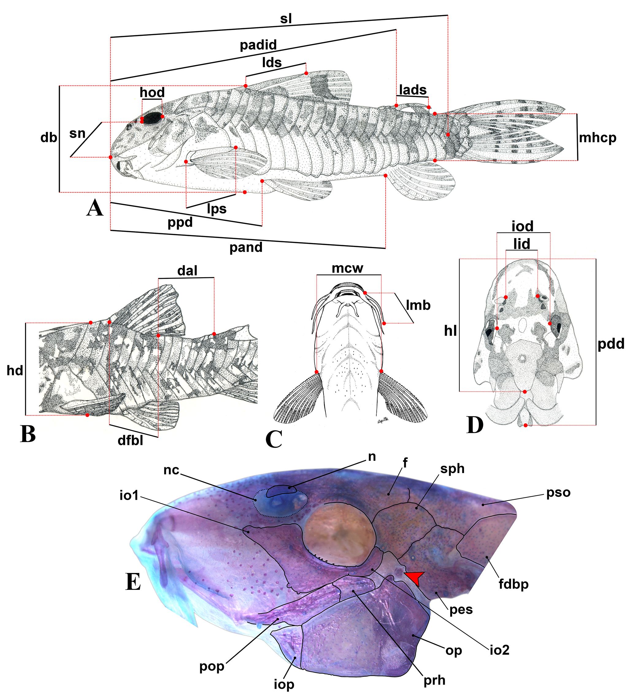
Schematic drawing illustrating applied method for obtaining measurements from Aspidoras specimens (A–D), and head of CS paratype of Aspidoras kiriri in lateral view, NUP 18246, 30.4 mm SL, positioned to maintain the largest diameter of the nasal capsule horizontally (E). Abbreviations: (A) db: depth of body, hod: horizontal orbit diameter, lads: length of adipose-fin spine, lds: length of dorsal-fin spine, lps: length of pectoral-fin spine, mhcp: minimum height of caudal peduncle, padid: preadipose distance, pand: preanal distance, ppd: prepelvic distance, sl: standard length, sn: snout length; (B) dal: distance between dorsal and adipose fins, dfbl: dorsal-fin base length, hd: head depth; (C) lmb: length of maxillary barbel, mcw: maximum cleithral width; (D) hl: head length, iod: interorbital distance, lid: least internareal distance, pdd: predorsal distance; (E) f: frontal, fdbp: first dorsolateral body plate, io1: infraorbital 1, io2: infraorbital 2, iop: interopercle, n: nasal, nc: nasal capsule (delimited by dotted lines), op: opercle, pes: pterotic-extrascapular, pop: preopercle, prh: posterodorsal ridge of hyomandibula, pso: parieto-supraoccipital, sph: sphenotic. In (E), solid black lines represent the limits of the bones; red arrow indicates the dorsal ridge of hyomandibula, modified from Oliveira et al., (2017: fig. 4).
RESULTS
Aspidoras Ihering, 1907Ihering R. Diversas espécies novas de peixes nemathognathas do Brasil. Notas preliminares. Rev Mus Paul. 1907; 1(1):13-39.
AspidorasIhering, 1907Ihering R. Diversas espécies novas de peixes nemathognathas do Brasil. Notas preliminares. Rev Mus Paul. 1907; 1(1):13-39.:30–31 (original description; type species: Aspidoras rochaiIhering, 1907Ihering R. Diversas espécies novas de peixes nemathognathas do Brasil. Notas preliminares. Rev Mus Paul. 1907; 1(1):13-39., by original designation and monotypy). —Nijssen, Isbrücker, 1976Nijssen H, Isbrücker IJH. The South American plated catfish genus Aspidoras R. von Ihering, 1907, with descriptions of nine new species from Brazil (Pisces, Siluriformes, Callichthyidae). Bijdr Dierkd. 1976; 46(1):107–31.:107‒131 (taxonomic review). —Reis, 1998Reis RE. Anatomy and phylogenetic analysis of the neotropical callichthyid catfishes (Ostariophysi, Siluriformes). Zool J Linn Soc. 1998; 124(2):105–68. https://doi.org/10.1111/j.1096-3642.1998.tb00571.x
https://doi.org/10.1111/j.1096-3642.1998...
:161 (diagnosis in identification key). —Britto, 2003Britto MR. Phylogeny of the subfamily Corydoradinae Hoedeman, 1952 (Siluriformes: Callichthyidae), with a definition of its genera. P Acad Nat Sci Phila. 2003; 153(1):119–54. https://doi.org/10.1635/00973157(2003)153 [0119:POTSCH]2.0.CO;2
https://doi.org/10.1635/00973157(2003)15...
:144, 146‒147 (phylogeny; diagnosis). —Reis, 2003Reis RE. Family Callichthyidae (Armored catfishes). In: Reis RE, Kullander SO, Ferraris CJ, Jr., editors. Check list of the freshwater fishes of South and Central America. Porto Alegre: Edipucrs; 2003. p.291–309.:291 (listed). —Ferraris, 2007Ferraris CJ, Jr. Checklist of catfishes, recent and fossil (Osteichthyes: Siluriformes), and catalogue of siluriform primary types. Zootaxa. 2007; (1418):1–628. Available from: http://www.mapress.com/zootaxa/2007f/zt01418p628.pdf
http://www.mapress.com/zootaxa/2007f/zt0...
:108 (listed).
Type species.Aspidoras rochaiIhering, 1907Ihering R. Diversas espécies novas de peixes nemathognathas do Brasil. Notas preliminares. Rev Mus Paul. 1907; 1(1):13-39., by original designation and monotypy.
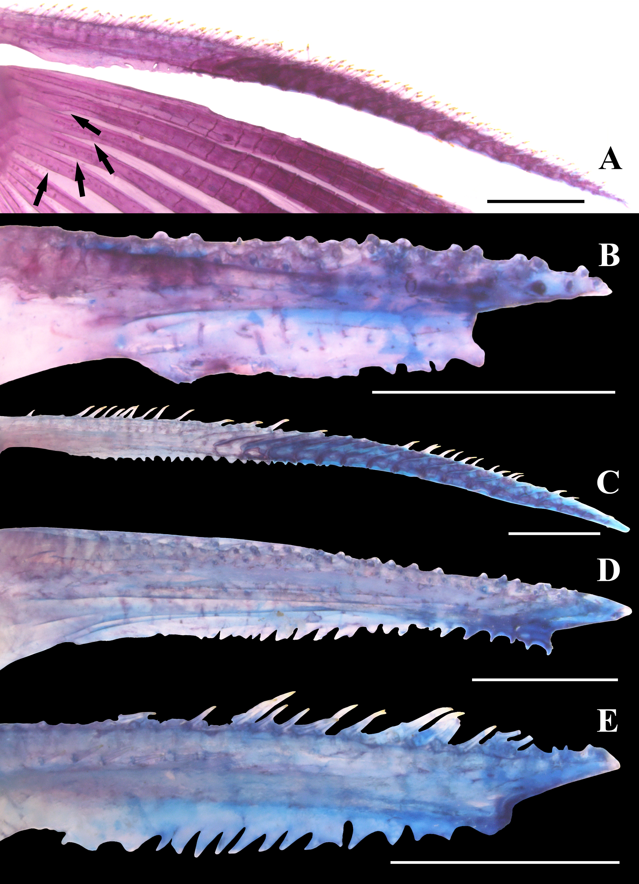
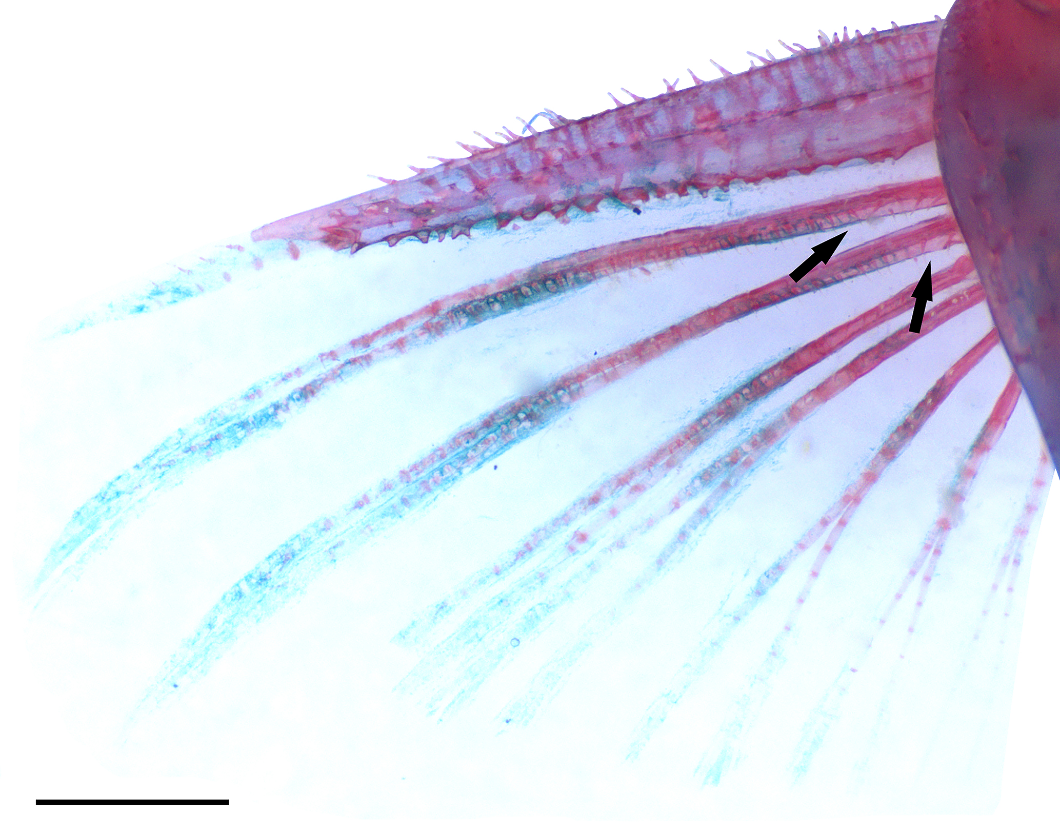
Diagnosis.Aspidoras can be distinguished from Corydoras and Scleromystax by presenting an exclusive feature among Corydoradinae: base of pectoral-fin branched rays with small laminar expansions on its inner margin, generally more evident on first rays; laminar expansions with irregular margins, forming pointed structures, in some specimens (vs. absence). Additionally, Aspidoras can be distinguished from Corydoras and Scleromystax, with exception of Corydoras pauciradiatus, C. lacerdai and S. virgulatus, by having parieto-supraoccipital fontanel (vs. fontanel absent); from C. pauciradiatus, C. lacerdai and S. virgulatus by presenting extremely reduced to moderately developed pectoral-fin spine (vs. relatively well developed).
Sexual dimorphism. Except for the presence of lanceolate genital papillae in males, presented by all Corydoradinae (see Nijssen, Isbrücker, 1980aNijssen H, Isbrücker IJH. Aspidoras virgulatus n. sp., a plated catfish from Espírito Santo, Brazil (Pisces, Siluriformes, Callichthyidae). Bull Zool Mus Univ Amst. 1980a; 7(13):133-39.; Britto, 2003Britto MR. Phylogeny of the subfamily Corydoradinae Hoedeman, 1952 (Siluriformes: Callichthyidae), with a definition of its genera. P Acad Nat Sci Phila. 2003; 153(1):119–54. https://doi.org/10.1635/00973157(2003)153 [0119:POTSCH]2.0.CO;2
https://doi.org/10.1635/00973157(2003)15...
), no other conspicuous sexually dimorphic feature was observed. In aquarium specimens, it was possible to observe that females tend to be slightly larger and more robust than males (Robert McLure, 2020, pers. comm.).
Remarks. All nominal species of Aspidoras and the summarized results of this review are presented in Tab. 1. After the examination of Aspidoras pauciradiatus from the Branco and Negro river basins, it was possible to conclude that, despite the presence of the parieto-supraoccipital fontanel, A. pauciradiatus is more closely related to the Corydoras from the lineage 5 sensuAlexandrou et al., (2011)Alexandrou MA, Oliveira C, Maillard M, McGill RAR, Newton J, Creer S, Taylor MI. Competition and phylogeny determine community structure in Müllerian co-mimics. Nature. 2011; 469(7328):84–89. https://doi.org/10.1038/nature09660
https://doi.org/10.1038/nature09660...
than to Aspidoras based on the presence of the following features: (I) pectoral-fin spine well developed (vs. spine extremely reduced to moderately developed); (II) eyes conspicuously larger (vs. conspicuously smaller); and (III) base of pectoral-fin branched rays lacking small laminar expansions on its inner margin (vs. laminar expansions present). Therefore, since both morphological and molecular (see Alexandrou et al., 2011Alexandrou MA, Oliveira C, Maillard M, McGill RAR, Newton J, Creer S, Taylor MI. Competition and phylogeny determine community structure in Müllerian co-mimics. Nature. 2011; 469(7328):84–89. https://doi.org/10.1038/nature09660
https://doi.org/10.1038/nature09660...
: suppl. fig. 2) evidence support the close relationship between A. pauciradiatus and the Corydoras species from the lineage 5, the most reasonable decision is to reallocate this species in Corydoras, as originally proposed by Weitzman, Nijssen, (1970)Weitzman SH, Nijssen H. Four new species and one new subspecies of the catfish genus Corydoras from Ecuador, Colombia and Brazil (Pisces, Siluriformes, Callichthyidae). Beaufortia. 1970; 18(233):119–32. Available from: https://repository.naturalis.nl/pub/504794
https://repository.naturalis.nl/pub/5047...
.
Calviño, Alonso (2009)Calviño PA, Alonso F. Two new species of the genus Corydoras (Ostariophysi: Siluriformes: Callichthyidae) from northwestern Argentina, and redescription of C. micracanthus Regan, 1912. Rev Mus Argent Cienc Nat. 2009; 11(2):199–214. Available from: http://www.scielo.org.ar/pdf/rmacn/v11n2/v11n2a06.pdf
http://www.scielo.org.ar/pdf/rmacn/v11n2...
provided the description of two species of Corydoras, C. gladysae and C. petracinii, and the redescription of C. micracanthusRegan, 1912Regan CT. A revision of the South-American siluroid fishes of the genus Corydoras, with a list of the specimens in the British Museum (Natural History). Ann Mag Nat Hist. 1912; 10(56):209–20., proposing the “C. micracanthus group”, which only includes these three species. The authors discussed the morphological similarities between the species from the C. micracanthus group and Aspidoras, such as short ossified portion of dorsal- and pectoral-fin spines and conspicuously slender body (see Calviño, Alonso, 2009Calviño PA, Alonso F. Two new species of the genus Corydoras (Ostariophysi: Siluriformes: Callichthyidae) from northwestern Argentina, and redescription of C. micracanthus Regan, 1912. Rev Mus Argent Cienc Nat. 2009; 11(2):199–214. Available from: http://www.scielo.org.ar/pdf/rmacn/v11n2/v11n2a06.pdf
http://www.scielo.org.ar/pdf/rmacn/v11n2...
:210). However, the authors refuted the possible allocation of these species to Aspidoras, mainly by the absence of the parieto-supraoccipital fontanel, which is present in all Aspidoras. After the examination of C. micracanthus and C. gladysae specimens, it was possible to confirm that these species do not belong to Aspidoras, not only by the absence of the parieto-supraoccipital fontanel but also by lacking the small laminar expansions on bases of first pectoral-fin branched rays, an apparently exclusive feature of the genus.
Summarized results of the present study. In the “status” column, species that were redescribed are marked with an asterisk.
Aspidoras aldebaran, new species
urn:lsid:zoobank.org:act:E3ABD342-36E5-49EA-B86A-00EC247315AF
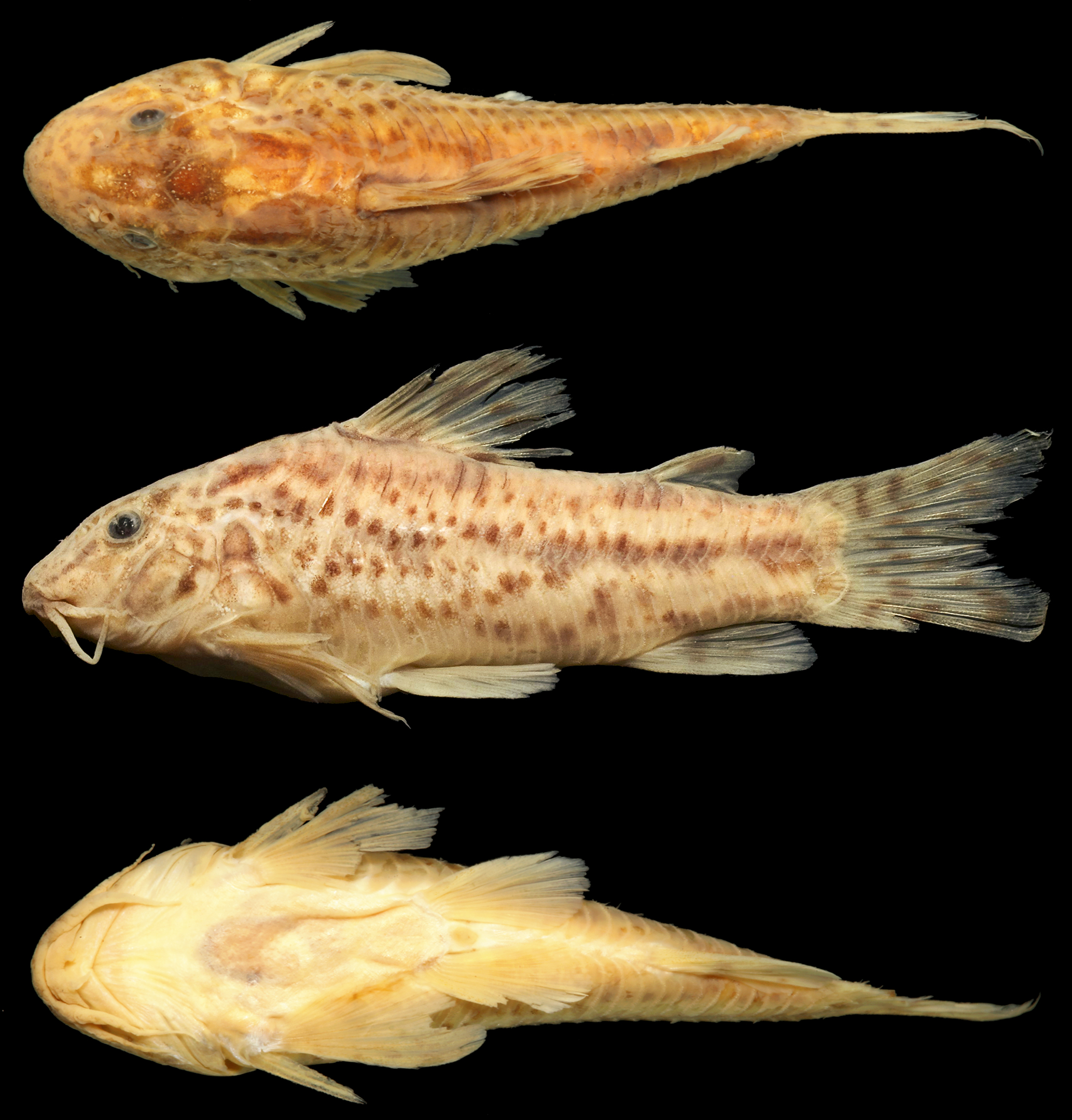
(Fig. 2)
Aspidoras taurus (non Lima, Britto, 2001Lima FCT, Britto MR. New catfish of the genus Aspidoras (Siluriformes: Callichthyidae) from the upper rio Paraguai system in Brazil. Copeia. 2001; 2001(4):1010–16. https://www.jstor.org/stable/1448389
https://www.jstor.org/stable/1448389...
): —Lima, Britto, 2001Lima FCT, Britto MR. New catfish of the genus Aspidoras (Siluriformes: Callichthyidae) from the upper rio Paraguai system in Brazil. Copeia. 2001; 2001(4):1010–16. https://www.jstor.org/stable/1448389
https://www.jstor.org/stable/1448389...
:1011 (paratypes of Aspidoras taurus; partim).
Holotype. LIRP 16933, 30.0 mm SL, Brazil, Mato Grosso State, Alto Araguaia Municipality, ribeirão do Sapo, upstream the Couto de Magalhães Waterfall, rio Araguaia basin, 17°31’10”S 53°15’33”W, 7‒8 Aug 2002, A. L. A. Melo and L. S. F. Martins.
Paratypes. All from Brazil, Mato Grosso State, Alto Araguaia Municipality, except when indicated. Rio Paraguay basin. CPUFMT 700, 11, 21.5‒28.8 mm SL, córrego Pinguelinha, 17°19’17”S 53°34’55”W, 18 Nov 2010, F. Machado and A. Ribeiro. CPUFMT 703, 3, 15.1‒29.7 mm SL, córrego Pinguela, 17°18’37”S 53°32’23”W, 18 Nov 2010, F. Machado and A. Ribeiro. CPUFMT 724, 1, 15.7 mm SL, córrego São José, 17°26’26”S 53°37’44”W, 18 Nov 2010, F. Machado and A. Ribeiro. MZUSP 41417, 2, 19.2‒27.1 mm SL, córrego da Pinguela, 17°18’37”S 53°32’22”W, 9 Mar 1989, L. P. S. Portugal and F. Langeani. MZUSP 41488, 2, 27.2‒27.4 mm SL, córrego do Mato, 9 Mar 1989, L.P.S. Portugal and F. Langeani. Rio Araguaia basin. LIRP 4477, 1, 23.7 mm SL, Goiás State, Santa Rita do Araguaia Municipality, rio Araguaia, 17°19’42”S 53°13’00”W, 5‒7 Aug 2002, A. L. A. Melo and L. S. F. Martins. MZUSP 41404, 22, 15.1‒22.6 mm SL, córrego do Rancho, approx.17°15’S 53°23’W, 8 Mar 1989, L. P. S. Portugal and F. Langeani. MZUSP 73248, 1, 16.3 mm SL, córrego Gordura, 17°18’20”S 53°16’22”W, 15 May 2001, C. L. R. Moreira and F. C. T. Lima. MZUSP 73278, 8, 10.4‒33.1 mm SL, stream tributary to the córrego Gordura, 17°17’55”S 53°16’34”W, 17 May 2001, C. L. R. Moreira and F. C. T. Lima. MZUSP 73261, 6, 15.7‒22.8 mm SL, stream tributary to the córrego Gordura, 17°19’02”S 53°15’49”W, 16 May 2001, C. L. R. Moreira and F. C. T. Lima. MZUSP 73265, 24, 10.5‒25.0 mm SL, córrego Boiadeiro, 17°20’01”S 53°14’52”W, 16 May 2001, C. L. R. Moreira and F. C. T. Lima. MZUSP 73283, 1, 12.1 mm SL, stream tributary to the córrego Gordura17°17’42”S 53°17’12”W, 17 May 2001, C. L. R. Moreira and F. C. T. Lima. MZUSP 73293, 4, 14.3‒22.7 mm SL, stream tributary to the córrego Boiadeiro, 17°20’31”S 53°14’41”W, 18 May 2001, C. L. R. Moreira and F. C. T. Lima. MZUSP 73302, 3, 22.1‒31.7 mm SL, stream tributary to the córrego Tapera, 17°21’58”S 53°14’51”W, 18 May 2001, C. L. R. Moreira and F. C. T. Lima. MZUSP 73306, 1, 19.0 mm SL, córrego Tapera, 17°22’19”S 53°14’30”W, 18 May 2001, C. L. R. Moreira and F. C. T. Lima. MZUSP 73331, 6, 12.3‒18.0 mm SL, córrego do Sapinho, 17°25’35”S 53°14’20”W, 21 May 2001, C. L. R. Moreira and F. C. T. Lima. CITL 381, 5, 24.9–29.6 mm SL; LIRP 4437, 34 of 36, 10.6‒30.5 mm SL, 2 CS of 36, 25.0‒25.6 mm SL; LIRP 4494, 26 of 28, 14.7‒27.6 mm SL, 2 CS of 28, 26.0‒27.0 mm SL; NUP 23487, 5, 22.4–26.9 mm SL, collected with the holotype.

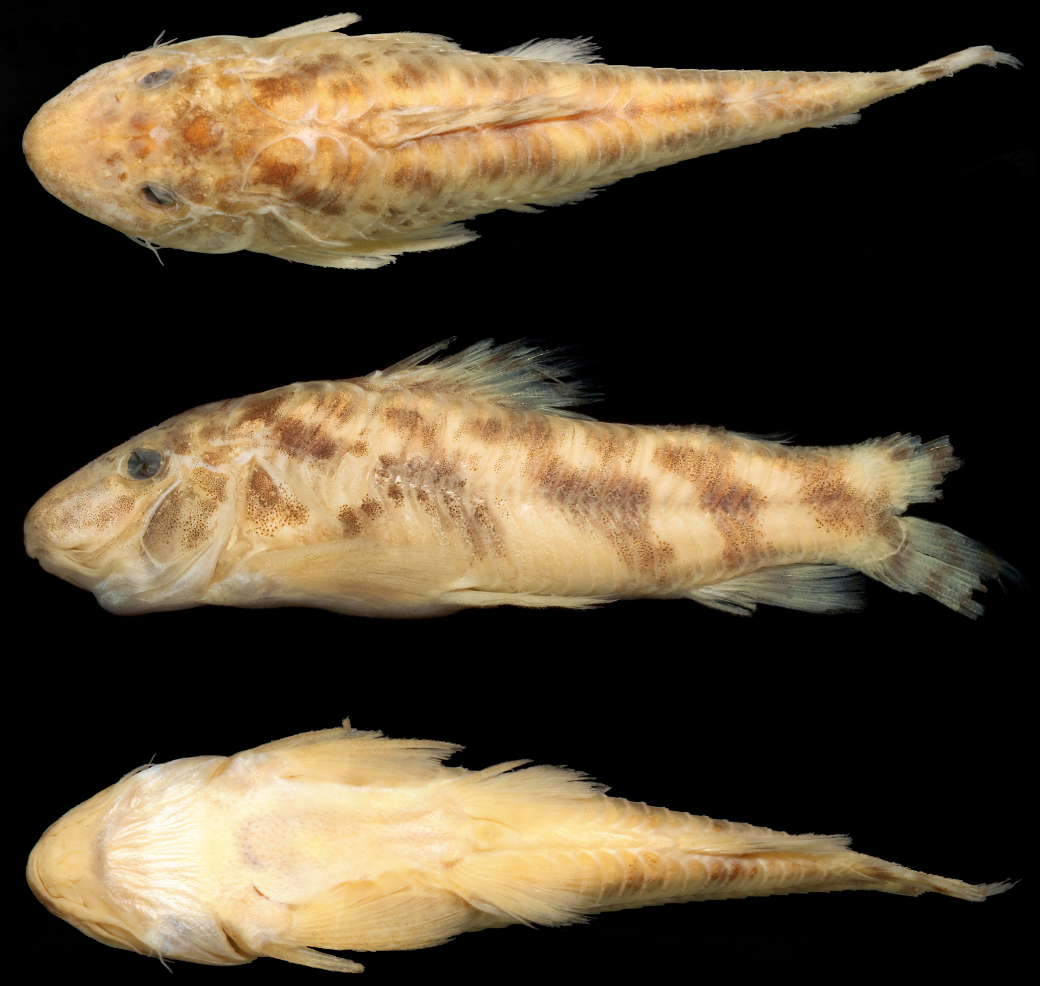
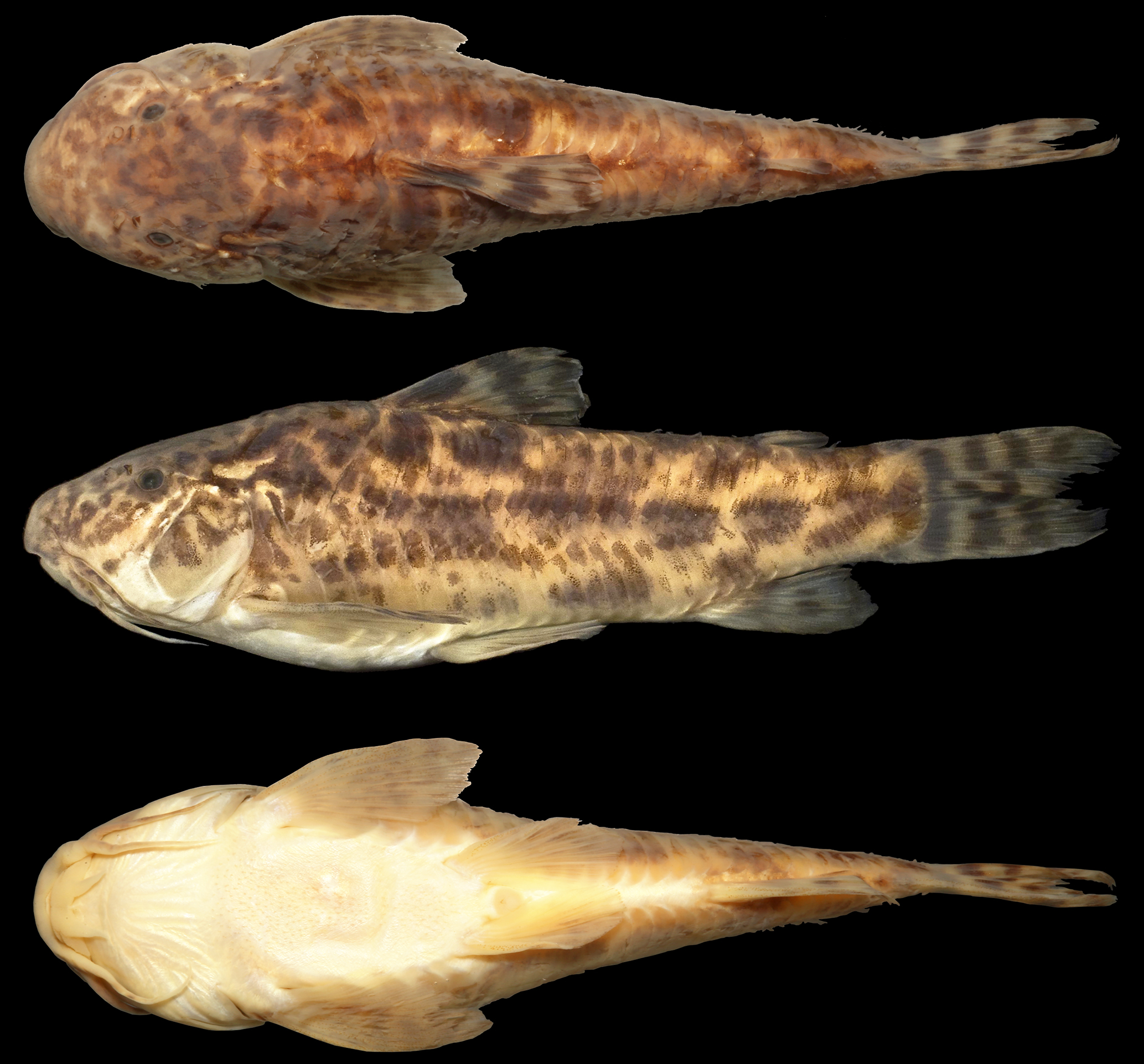
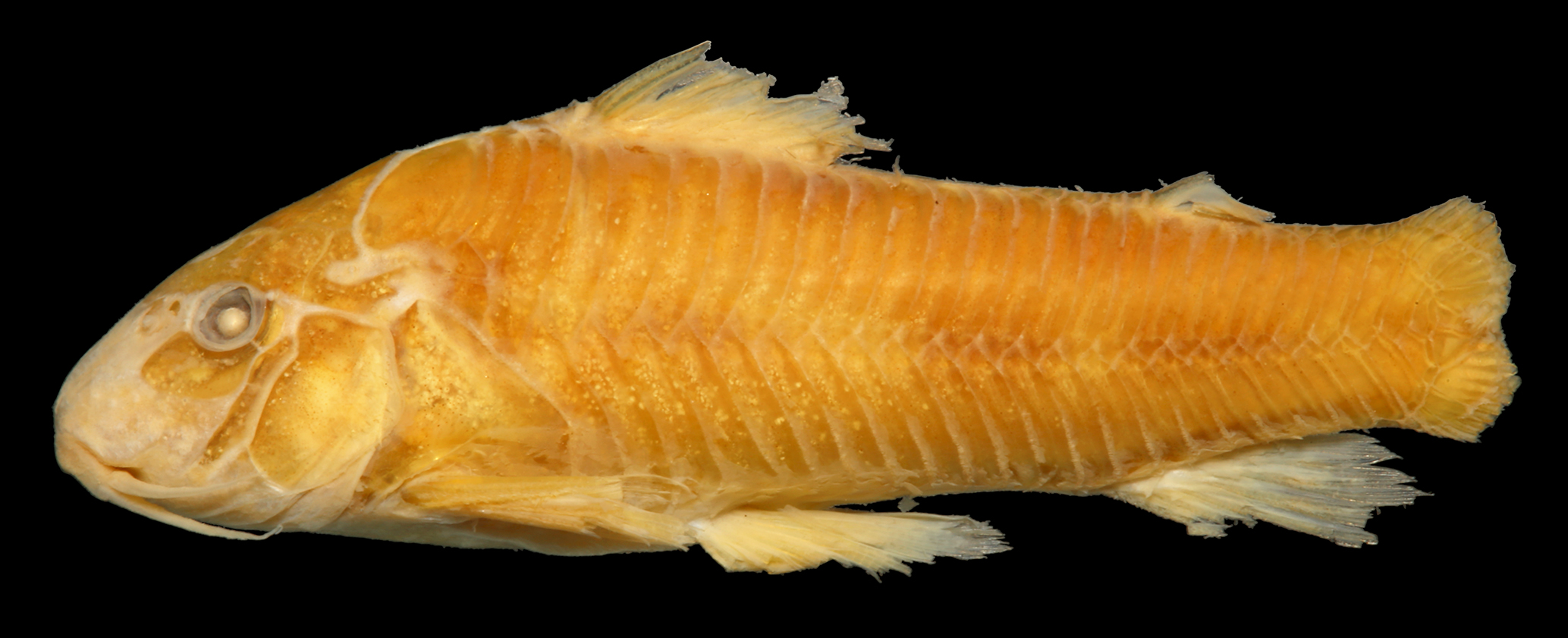
Aspidoras aldebaran, holotype, LIRP 16933,30.0 mm SL, Brazil, Mato Grosso State, Alto Araguaia Municipality, ribeirão do Sapo, upstream Couto de Magalhães Waterfall. Dorsal (top), lateral (middle) and ventral (bottom) views. Photos by Celso Ikedo.
Diagnosis.Aspidoras aldebaran can be distinguished from its congeners, with exception of A. belenos, A. kiriri, and A. raimundi, by having parapophysis of the complex vertebra well developed (vs. moderately developed in A. depinnai, A. lakoi, A. maculosus, A. mephisto, A. poecilus, A. psammatides, and A. velites; poorly or moderately developed in A. albater and A. fuscoguttatus; poorly developed in A. azaghal); it can be distinguished from A. belenos, A. kiriri, and A. raimundi by having inner laminar expansion of infraorbital 1 ranging from well developed to extremely well developed (vs. moderately developed). Additionally, it can be distinguished from A. albater, A. azaghal, A. gabrieli, A. lakoi, A. mephisto, A. psammatides, and A. rochai by having a narrow frontal bone, with width slightly smaller than half of entire length (vs. relatively wide, with width equal to or slightly larger than half of entire length in A. albater, A. azaghal, A. gabrieli, A. lakoi, A. mephisto, and A. rochai; extremely narrow, with width conspicuously smaller than half of entire length in A. psammatides); from A. azaghal, A. depinnai and A. kiriri by the presence of small black spots on dorsal fin (vs. absence).
Description. Morphometric data presented in Tab. 2. Head compressed with convex dorsal profile; somewhat trapezoid in dorsal view. Snout moderately developed and pointed; variably relatively well developed. Head profile convex from tip of snout to anterior nares; region of mesethmoid slightly concave in some specimens; ascending slightly convex from this point to dorsal-fin origin. Dorsal margin of orbit slightly elevated in some specimens. Profile nearly straight to slightly convex along dorsal-fin base. Postdorsal-fin body profile slightly concave to adipose-fin spine; slightly concave from this point to caudal-fin base. Ventral profile of body slightly convex from isthmus to pelvic-fin origin; region of gill opening slightly concave in some specimens; nearly straight from this point to anal-fin origin; slightly concave until caudal-fin base. Body roughly elliptical in cross section at pectoral girdle, gradually becoming more compressed toward caudal fin.
Eye rounded, located dorso-laterally on head; orbit delimited dorsally by lateral ethmoid, frontal and sphenotic, ventrally by infraorbitals. Anterior and posterior nares close to each other, only separated by flap of skin. Anterior naris tubular. Posterior naris close to anterodorsal margin of orbit, separated from it by distance equal to or slightly smaller than naris diameter. Mouth small, subterminal, width slightly larger than bony orbit diameter. Maxillary barbel moderate in size, not reaching anteroventral limit of gill opening. Outer mental barbel slightly larger than maxillary barbel. Inner mental barbel fleshy, with base slightly separated from its counterpart. Lower lip moderately developed, forming small semicircular or triangular fleshy flap. Small, rounded papillae covering entire surface of all barbels, upper and lower lips, snout and isthmus.
Mesethmoid short; anterior tip poorly developed, slightly smaller than 50% of bone length (see Britto, 2003Britto MR. Phylogeny of the subfamily Corydoradinae Hoedeman, 1952 (Siluriformes: Callichthyidae), with a definition of its genera. P Acad Nat Sci Phila. 2003; 153(1):119–54. https://doi.org/10.1635/00973157(2003)153 [0119:POTSCH]2.0.CO;2
https://doi.org/10.1635/00973157(2003)15...
:123, ch. 1, state 1; fig. 1B); posterior portion wide, partially exposed. Nasal slender, curved laterally, inner margin with moderately developed laminar expansion; outer margin with reduced laminar expansion; mesial border contacting frontal and mesethmoid.
Frontal elongated, narrow, with width slightly smaller than half of entire length (Fig. 3A); anterior projection ranging from short, with size smaller than nasal length, to long, with size slightly larger than nasal length; anterior margin generally exposed. Frontal fontanel relatively small, ellipsoid or somewhat rhomboid; posterior tip extension not entering anterior margin of parieto-supraoccipital. Sphenotic somewhat trapezoid, contacting parieto-supraoccipital dorsally, pterotic-extrascapular posteriorly, second infraorbital ventrally and frontal anteriorly. Pterotic-extrascapular roughly pipe-shaped, with posteriormost portion contacting first lateral-line ossicle, and ventral margin contacting opercle and cleithrum; posterior expansion almost entirely covering lateral opening of swimbladder capsule, leaving slender pseudotympanic area on dorsal margin covered only by thick layer of skin. Parieto-supraoccipital wide, posterior process strongly reduced to poorly developed, not contacting nuchal plate. Parieto-supraoccipital medial keel expanded ventrally; laminar, with posterior portion at same level as posterior process tip; expanded posteriorly in some specimens, slightly surpassing tip of posterior process. Parieto-supraoccipital fontanel small, roundish; located medially on parieto-supraoccipital.
Two laminar infraorbitals with minute odontodes; infraorbital 1 large, ventral laminar expansion poorly to moderately developed; anterior portion with laminar expansion ranging from poorly developed, slightly surpassing posterior margin of nasal capsule, to moderately developed, reaching middle of nasal capsule (Fig. 4); inner laminar expansion ranging from well developed to extremely well developed (Fig. 5A,B); small portions of external surface covered by thick layer of skin; infraorbital 2 small, slender; with posterior laminar expansion generally poorly developed; inner laminar expansion ranging from moderately to well developed; posteroventral margin close but not directly contacting posterodorsal ridge of hyomandibula; contacting in some specimens; dorsal tip contacting only sphenotic; small portions of external surface covered by thick layer of skin (Fig. 4). Posterodorsal ridge of hyomandibula, close to its articulation with opercle, oblong, relatively slender; exposed, bearing small odontodes; dorsal ridge of hyomandibula, between pterotic-extrascapular and opercle, covered by thick layer of skin. Interopercle entirely covered by thick layer of skin; with posterior portion variably exposed; somewhat triangular, anterior projection moderately developed. Preopercle relatively slender, elongated, minute odontodes sparse on external surface. Opercle compact in shape, width larger than half of its length; free margin convex; posterodorsal region variably with smoothly concave area; without serrations and covered by small odontodes; some portions of bony distal margin irregular in some specimens.
Four branchiostegal rays decreasing in size posteriorly. Hypobranchial 2 somewhat triangular, tip ossified and directed towards anterior portion, posterior margin cartilaginous; ossified portion well developed, about twice size of cartilaginous portion. Five ceratobranchials with expansions increasing posteriorly; ceratobranchial 1 with strongly reduced process on anterior margin of mesial portion; process variably absent; ceratobranchial 3 with continuous postero-lateral margin; ceratobranchial 5 toothed on postero-dorsal surface, 20 to 26 (4) teeth aligned in one row. Four epibranchials with similar size; epibranchial 2 slightly larger than others, with strongly reduced pointed process on laminar expansion of posterior margin; process variably absent; epibranchial 3 with triangular uncinate process on laminar expansion of posterior margin. Two wide pharyngobranchials (3 and 4), pharyngobranchial 3 with triangular laminar expansion on posterior margin; triangular laminar expansion with notches in some specimens. Upper tooth plate oval, with 23 to 31 (4) teeth aligned in two rows on postero-ventral surface.
Lateral-line canal entering neurocranium through pterotic-extrascapular, branching twice before entering sphenotic: pterotic branch with single pore; preoperculomandibular branch conspicuously reduced, with single pore opening close to postotic main canal; more developed, with pore opening closer to anteroventral border of pterotic-extrascapular in some specimens; postotic main canal widens just posterior to pterotic branch. Sensory canal continuing through pterotic-extrascapular, entering sphenotic as temporal canal, which splits into two branches: one branch giving rise to infraorbital canal, another branch entering frontal through supraorbital canal, both with single pore. Supraorbital canal branched, running through nasal bone. Epiphyseal branch of supraorbital canal relatively long, with pore opening close to frontal fontanel; or slightly shorter, with pore opening closer to supraorbital main canal. Nasal canal with three openings, first on posterior edge, second on posterolateral portion and third on anterior edge; second pore generally fused with first pore. Infraorbital canal running through entire second infraorbital, extending to infraorbital 1 and opening into two or three pores. Preoperculomandibular branch giving rise to preoperculo-mandibular canal, which runs through entire preopercle with three openings, leading to pores 3, 4, and 5, respectively.
Dorsal fin somewhat triangular, located just posterior to third dorsolateral body plate. Dorsal-fin rays II,7(1), II,8*(19), posterior margin of dorsal-fin spine smooth. Nuchal plate moderately developed in length; almost entirely exposed, with minute odontodes on exposed area; anterior tip covered by thick layer of skin (Fig. 6); spinelet short, partially exposed; spine relatively well developed, adpressed distal tip reaching or surpassing posterior origin of dorsal-fin base; anterior margin with small odontodes. Pectoral fin roughly triangular, its origin just posterior to gill opening. Pectoral-fin rays I,7(1), I,8*(19); posterior margin of pectoral spine with 21 to 27 moderately-developed serrations along almost its entire length; small region just posterior to origin of spine lacking serrations; some serrations directed towards origin of spine, perpendicularly directed or directed towards tip of spine; presence of bifid serrations in some specimens; base of branched rays with reduced laminar expansions on inner margin, generally more evident on first rays; laminar expansions variably with irregular margins (Fig. 7). Anteroventral portion of cleithrum partially exposed; posterolateral portion of scapulocoracoid reduced, externally visible. Pelvic fin oblong, located just below third ventrolateral body plate, and at vertical through second dorsal-fin branched ray. Pelvic-fin rays I,5*(20). Adipose fin roughly triangular, separated from posterior origin of dorsal-fin base by seven or eight dorsolateral body plates. Anal fin somewhat triangular, located just posterior to 12th or 13th ventrolateral body plates, and at vertical through region of preadipose platelets. Anal-fin rays, ii,5(3), ii,4,i(2), ii,6(2), ii,5,i*(13). Caudal-fin rays I,11,I(1), I,12,I*(19), four or five dorsal and/or ventral procurrent rays; caudal fin bilobed, dorsal and ventral lobes generally with similar size; dorsal lobe variably slightly larger than ventral lobe.
Two or three laterosensory canals on trunk; first ossicle tubular, second ossicle laminar, third lateral-line canal, if present, encased in third dorsolateral body plate. Body plates with conspicuous line of relatively large odontodes confined to posterior margins; dorsolateral body plates 24(5), 25*(12), 26(3); ventrolateral body plates 21(1), 22*(13), 23(6); dorsolateral body plates along dorsal-fin base 5(2), 6*(15), 7(3); dorsolateral body plates between adipose-fin spine and caudal-fin base 7(13), 8*(7); preadipose platelets 2(2), 3*(8), 4(9), 5(1); small platelets covering base of caudal-fin rays; small platelets disposed dorsally and ventrally between junctions of lateral plates on posterior portion of caudal peduncle. Ventral surface of trunk naked.
Vertebral count 23(4); ribs 6(3), 7(1), first pair conspicuously large; parapophysis of complex vertebra well developed (Fig. 8A).
Morphometric data of the holotype and 19 paratypes of Aspidoras aldebaran. SD = standard deviation.

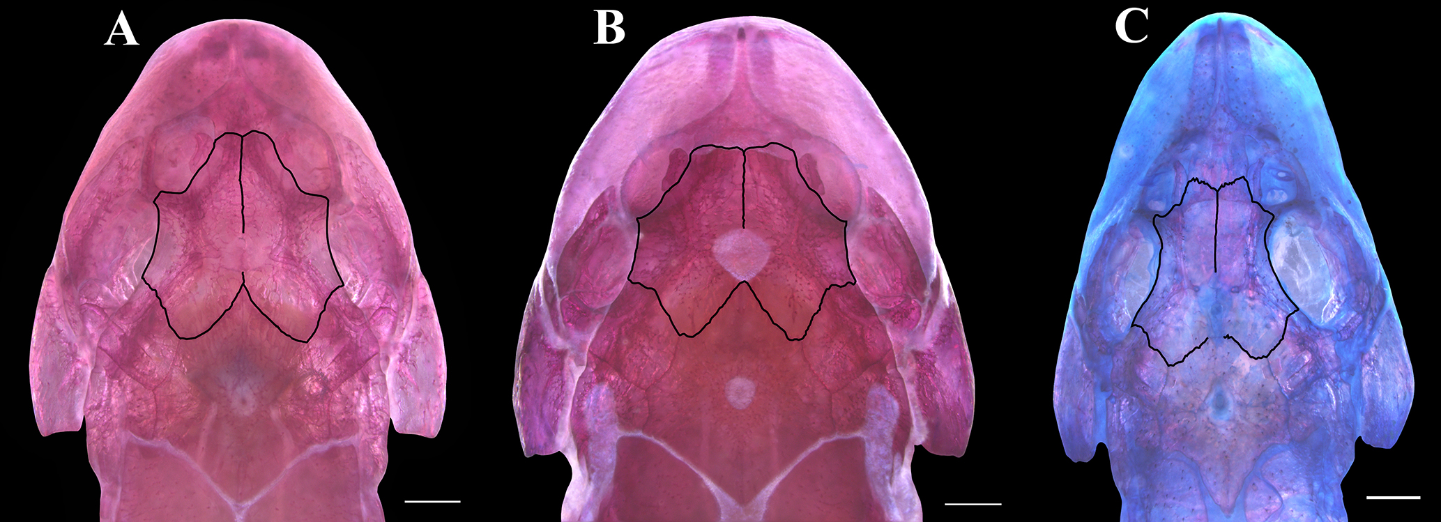
Dorsal view of the head of CS specimens showing the three patterns of paired frontal bones (outlined in black) in Aspidoras: (A) narrow, with width of each frontal slightly smaller than half of its entire length (Aspidoras maculosus, UFBA 3291, 30.7 mm SL), (B) relatively wide, with width equal to or slightly larger than half of entire length (Aspidoras albater, MZUSP 40793, 29.4 mm SL), and (C) extremely narrow, with width clearly smaller than half of entire length (Aspidoras psammatides, UNT 9604, 27.3 mm SL). Scale bars = 1 mm.

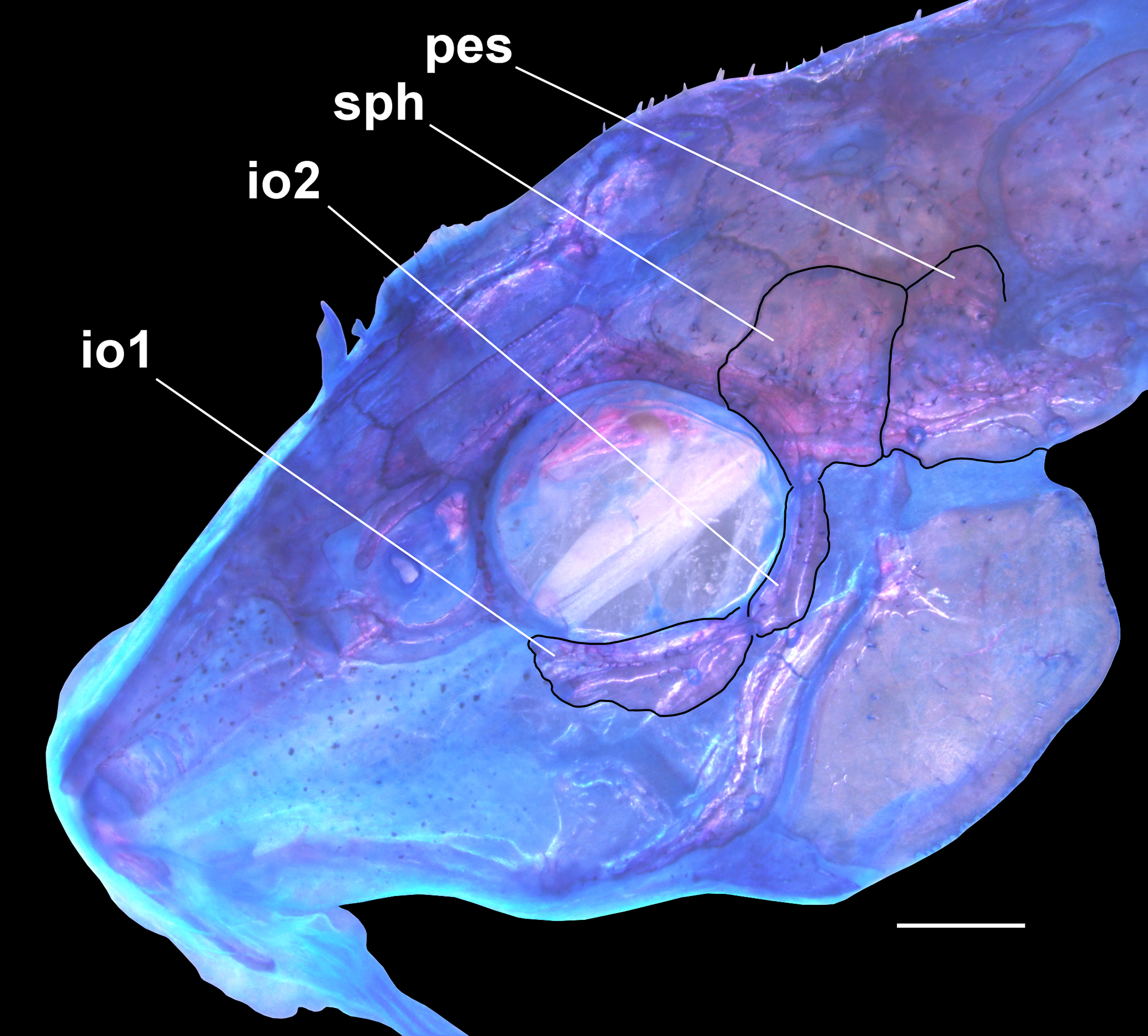
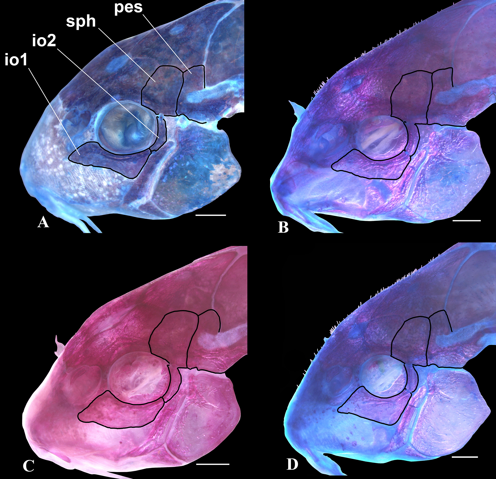
Lateral view of the head of CS paratype of Aspidoras aldebaran, LIRP 4494, 27.0 mm SL, showing general pattern of infraorbitals. Abbreviations: io1: infraorbital 1, io2: infraorbital 2, sph: sphenotic, pes: pterotic-extrascapular. Scale bar = 1 mm.

Dorsal view of the infraorbital series of CS specimens showing the four patterns of size and shape of the inner laminar expansion of infraorbital 1 (white arrows) in Aspidoras: (A) extremely well developed (Aspidoras albater, MNRJ 13080, 30.9 mm SL), (B) well developed (Aspidoras fuscoguttatus, NUP 12677, 36.1 mm SL), (C) moderately developed (Aspidoras poecilus, UNT 6249, 30.9 mm SL), and (D) poorly developed (Aspidoras psammatides, UNT 9604, 27.3 mm SL). Abbreviations: io1: infraorbital 1, io2: infraorbital 2. Scale bars = 1 mm.

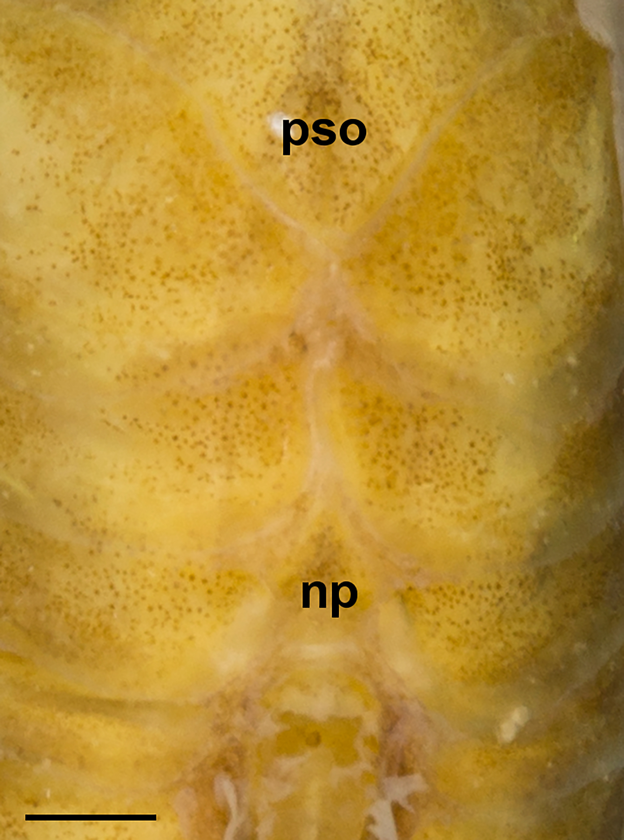
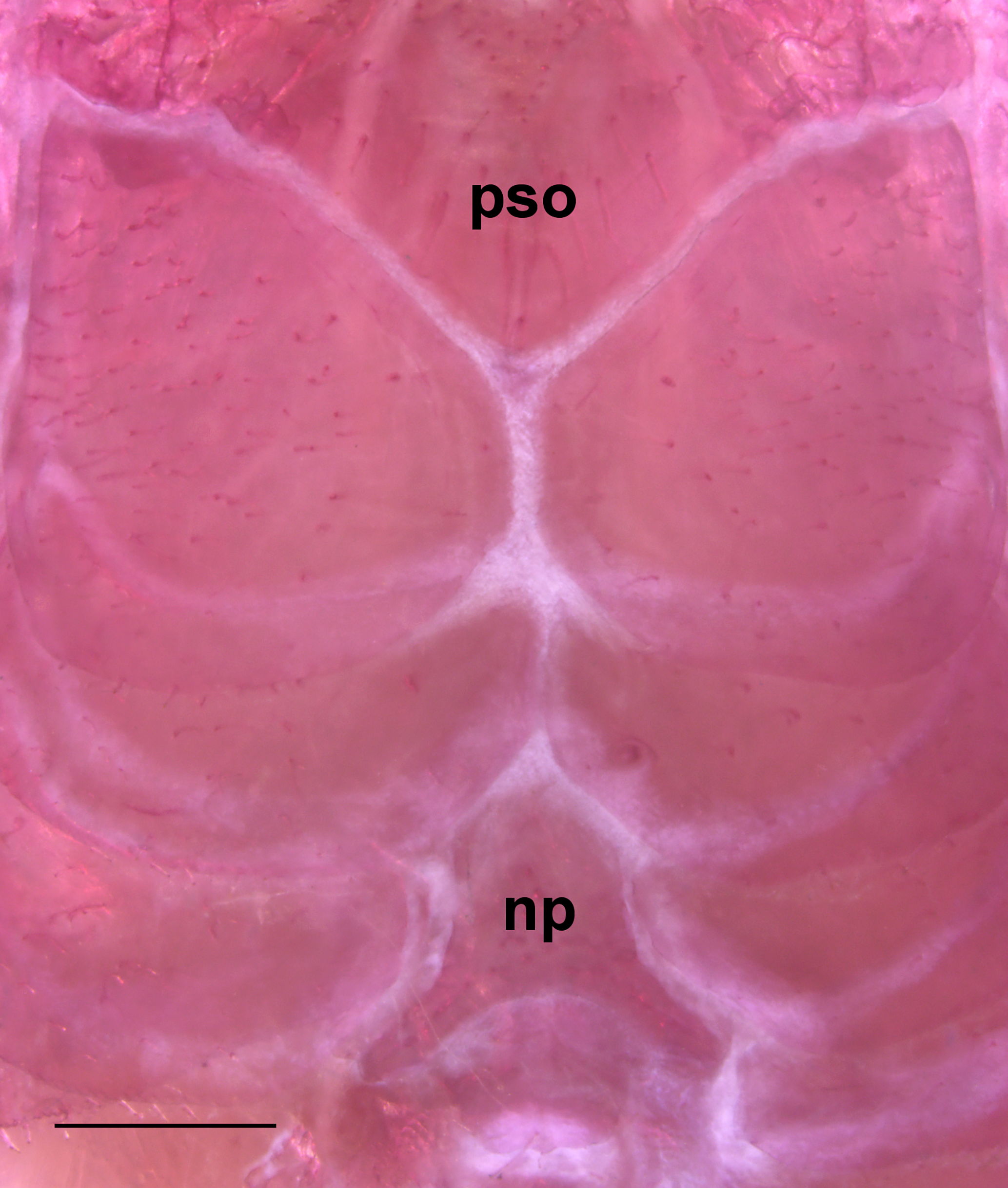
Aspidoras aldebaran, LIRP 4494, CS paratype, 27.0 mm SL, showing predorsal region of trunk in dorsal view. Dotted lines indicate the limits of the tip of the posterior process of the parieto-supraoccipital and anterior tip of the nuchal plate. Abbreviations: np: nuchal plate, pso: parieto-supraoccipital. Scale bar = 1 mm.

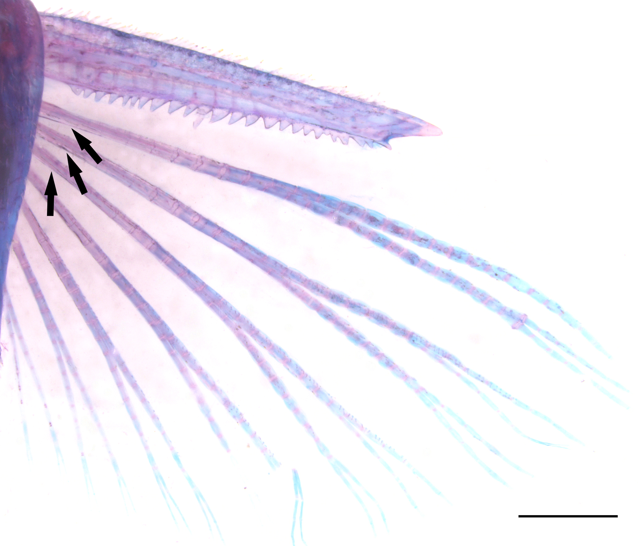
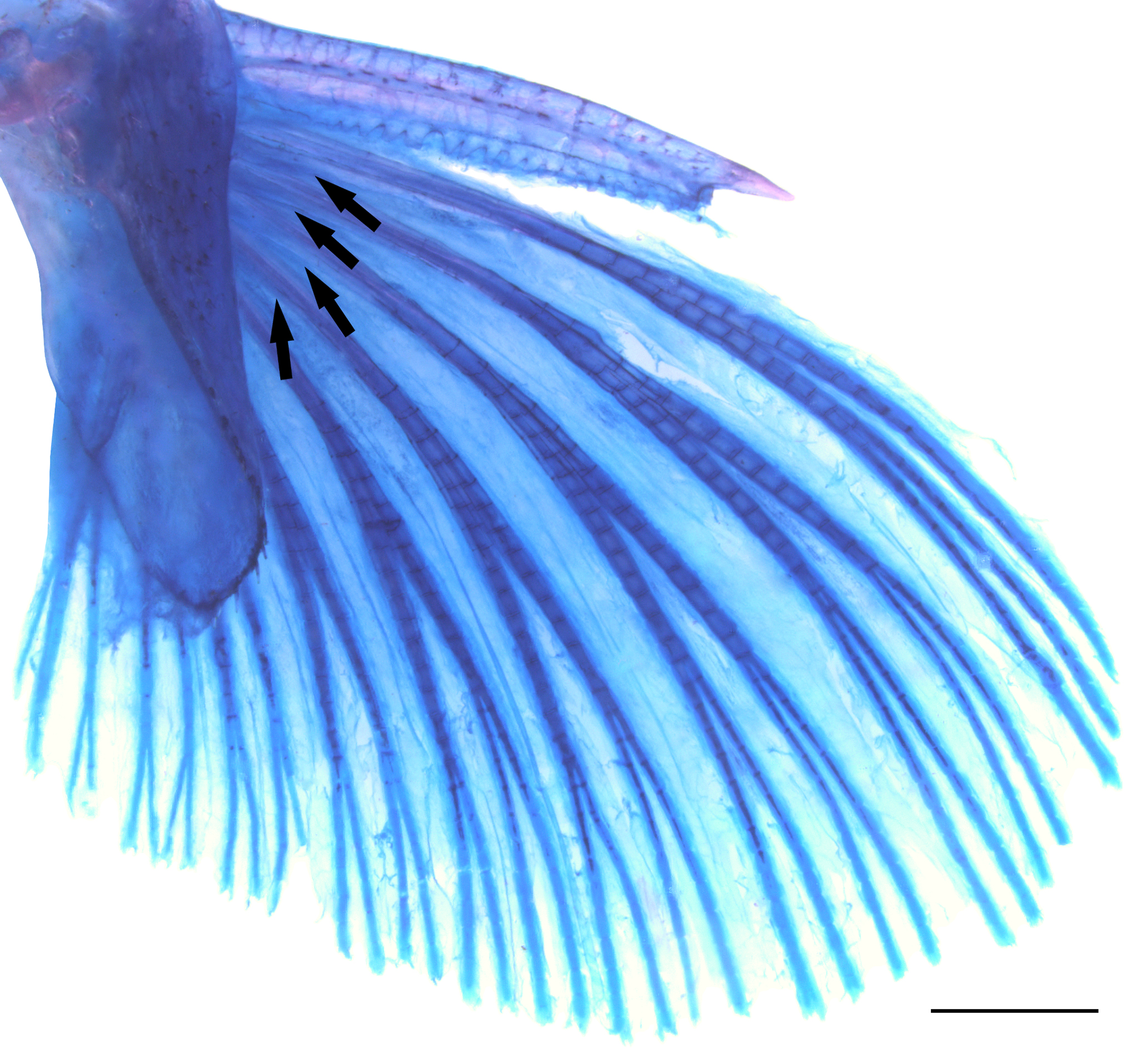
Aspidoras aldebaran, LIRP 4494, CS paratype, 27.0 mm SL, showing dorsal view of the pectoral fin. Arrows indicate the reduced laminar expansions at the base of branched rays. Scale bar = 1 mm.

Ventral view of the complex vertebra in CS specimens of (A) Aspidoras belenos, paratype, UFRJ 4419 (ex-UFRJ 3861), 22.1 mm SL, with well-developed parapophysis, (B) Aspidoras albater, MNRJ 12571, 35.3 mm SL, with poorly-developed parapophysis, and (C) Aspidoras albater, MNRJ 12581 (disarticulated, indeterminate size), with moderately-developed parapophysis. Abbreviations: ccv: centrum of the complex vertebra, pcv: parapophysis of the complex vertebra. Scale bars = 1 mm.
Coloration in alcohol. Ground color of body light or brownish yellow, with top of head dark brown. Posterodorsal portion of head, region below eye, opercle and cleithrum with scattered dark brown or black chromatophores. Snout covered dark brown or black chromatophores on its dorsal surface; chromatophores densely disposed in some specimens; generally forming dark brown or black rounded, striated or irregular relatively large spots; or forming conspicuously smaller spots; generally, with dark brown or black diffuse or conspicuous stripe from anteroventral portion of orbit to upper lip lateral area; ventrolateral portion of snout with dark brown or black chromatophores, variably forming spots, in some specimens. Upper lip and maxillary barbel with dark brown or black chromatophores; area of lateral portion of upper lip generally with conspicuous concentration of dark brown or black chromatophores; outer mental barbel with dark brown or black chromatophores, generally more evident on its proximal portion; region of isthmus around lower lip with dark brown or black chromatophores in some specimens. Dorsal series of four to five dark brown or black blotches, first on anterior portion of dorsal-fin base, second on posterior portion of dorsal-fin base, third, if present, between dorsal and adipose fins, fourth on adipose-fin base, and fifth on caudal-fin base; blotches variably diffuse. Dorsal portion of body with conspicuous concentration of dark brown or black chromatophores between counterparts of dorsolateral body plates in some specimens. Ventral surface of trunk, generally on region close to pectoral- and pelvic-fin origins, and region posterior to urogenital opening with dark brown or black chromatophores in some specimens. First dorsolateral body plate with conspicuous concentration of dark brown or black chromatophores; posterior margin of some dorso- and ventrolateral body plates, and lateral line pores with conspicuous concentration of dark brown or black chromatophores in some specimens. Midline of flank with longitudinal series of three to six medium-sized conspicuous dark brown or black blotches; blotches rounded, oblong or irregular; blotches variably fused, forming longitudinally elongated bars. Dorsal half of dorsolateral body plates with dark brown or black chromatophores; region of anterior and posterior portions of dorsal-fin base, between dorsal and adipose fins, adipose-fin base, and base of caudal peduncle with more concentrated chromatophores, forming conspicuous blotches in some specimens; blotches variably fused to flank midline blotches. Ventral half of dorsolateral body plates and dorsal half of ventrolateral body plates with concentration of dark brown or black chromatophores, forming conspicuous blotches in some specimens; blotches generally more evident on anterior portion of body and on area of flank midline blotches. Mid-ventral portion of ventrolateral body plates on area of flank midline blotches with concentration of dark brown or black chromatophores, generally forming conspicuous blotches; blotches generally more evident posteriorly to pelvic-fin origin; variably fused to flank midline blotches; ventral portion of ventrolateral body plates with concentration of dark brown or black chromatophores, generally more evident posterior to anal-fin anterior origin, in some specimens. Dorsal fin with dark brown or black spots; aligned spots, forming oblique bars in some specimens; membranes with dark brown or black chromatophores, generally more evident on region of first and second branched rays proximal portion; dorsal-fin base with conspicuous concentration of dark brown or black chromatophores, generally more concentrated on bases of first and last branched rays; spine covered by dark brown or black chromatophores. Pectoral fin with dark brown or black chromatophores on its dorsal surface, generally more evident on spine and branched rays; conspicuous concentration of dark brown and black chromatophores on proximal portion of branched rays; covered by dark brown or black spots in some specimens; spots aligned, forming oblique bars in some specimens; spots variably diffuse or more evident on first branched rays; region of body around dorsal portion of pectoral-fin origin generally with concentration of dark brown or black chromatophores. Pelvic fin with conspicuous concentration of dark brown or black chromatophores on its dorsal surface, generally forming one to three oblong dark brown or black patches; anteriormost patch generally larger and more intensely pigmented; region of body around dorsal portion of pelvic-fin origin with concentration of dark brown or black chromatophores in some specimens. Adipose-fin membrane with dark brown or black chromatophores; conspicuous concentration of dark brown or black chromatophores in some areas of membrane, generally more evident close to spine, forming isolated patches in some specimens; adipose-fin spine generally with dark brown or black chromatophores. Anal fin with conspicuous concentrations of dark brown or black chromatophores in some areas, generally more evident on its middle portion and bases of last branched rays; variably forming one to three dark brown or black blotches. Middle portion of caudal-fin base, posteriorly to last flank midline blotch, generally with small- to medium-sized dark brown or black blotch; blotch variably diffuse or fused with last midlateral blotch. Caudal fin with three to six transversal dark brown or black slender to wide bars.
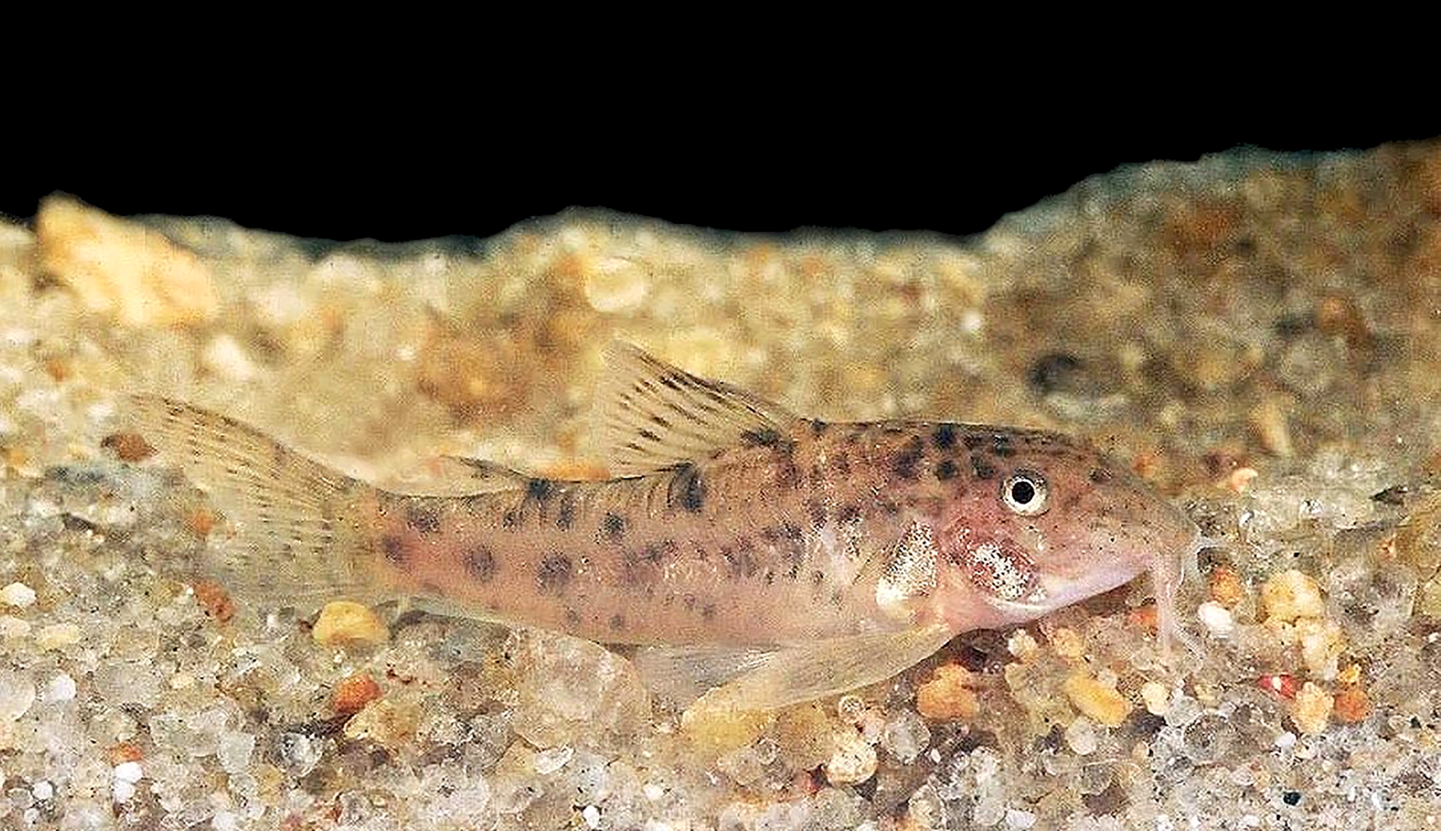
Coloration in life. Similar to that observed in preserved specimens but with clearer ground color of body. Iris orangish brown. Body covered with whitish yellow and green iridescent coloration (Fig. 9A).

Aspidoras aldebaran, uncataloged specimen photographed alive (A), and the córrego Gordura, rio Araguaia basin, showing the typical habitat of the new species (B). Photos by Hans Evers.
Geographical distribution.Aspidoras aldebaran is known from the upper rio Araguaia basin in Goiás and Mato Grosso states, and upper rio Paraguay basin in Mato Grosso State, Brazil (Fig. 10).

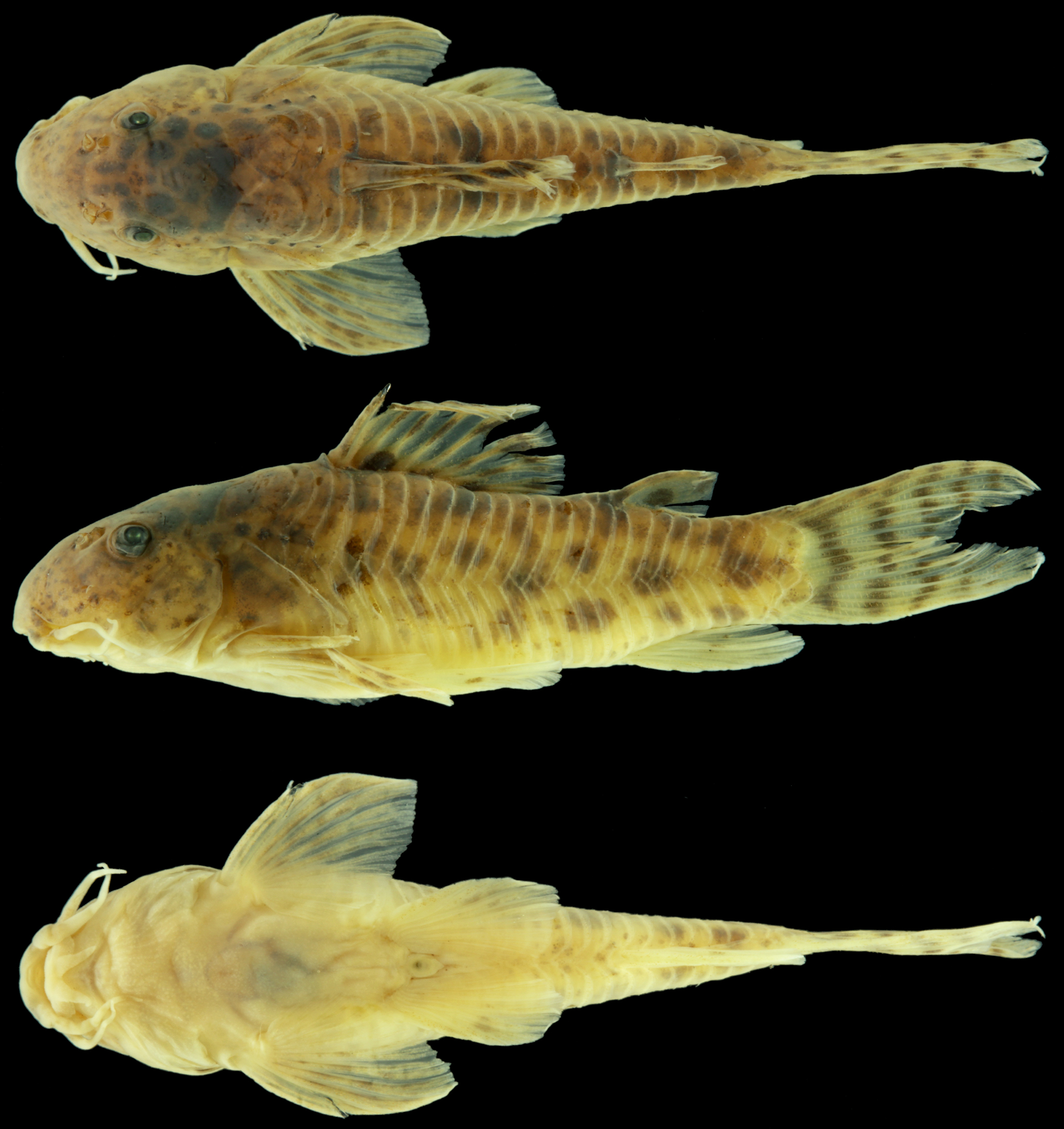
Map showing the geographical distribution of Aspidoras albater (type locality: red circle; non-type localities: black circles), A. aldebaran (type locality: red star; non-type localities: white stars), A. azaghal (purple triangle), A. belenos (type locality: purple diamond), A. brunneus (possible region of type locality: grey diamond), A. carvalhoi (type locality: purple circle), A. depinnai (type locality: red square; non-type localities: black squares), A. fuscoguttatus (type locality: red triangle; non-type localities: white triangles), A. gabrieli (type locality: red pentagon; non-type locality: white pentagon), A. kiriri (black cross), A. lakoi (type locality: red diamond; non-type locality: black diamond), A. maculosus (red cross), and A. mephisto (type locality: white cross). Each symbol may represent more than one locality.
Ecological notes. The córrego Gordura, where some of the paratypes were collected, is a medium-sized tributary to the upper rio Araguaia basin with highly transparent and clear water, width ranging from less than one meter to about 15 m, and depth ranging from about 10 cm to nearly two meters. Aspidoras aldebaran was found in shallow (up to about 20 cm) stretches in the upper portion of the córrego Gordura, with moderate current and substrate composed mainly by sand and gravel (Fig. 9B). At this site, the species was found in syntopy with Aspidoras velites, Characidium sp., and Hypostomus cf. careopinnatusMartins, Marinho, Langeani & Serra, 2012Martins FO, Marinho MM, Langeani F, Serra JP. A new species of Hypostomus (Siluriformes: Loricariidae) from the upper rio Paraguay basin, Brazil. Copeia. 2012(3); 2012(3):494-500. http://dx.doi.org/10.1643/CI-11-011
http://dx.doi.org/10.1643/CI-11-011...
. Aspidoras aldebaran and A. velites were placed in a small biotope aquarium (i.e., one that recreates the natural habitat) to record their color pattern in life. During this event, a specimen of A. velites displayed an aggressive behavior towards an A. aldebaran specimen, chasing and pushing it throughout the aquarium. The A. aldebaran specimen did not react aggressively but tried to evade the assaults by the A. velites specimen. Interestingly, A. aldebaran is generally larger and a clearly more robust species than A. velites. In nature, such confrontations seem unlikely considering that the new species was generally found associated with the sandy substrate of small shores whereas A. velites was most commonly captured in areas associated with marginal and/or submerged vegetation.
Etymology. The epithet “aldebaran” refers to the red giant Aldebaran or Alpha Tauri (α Tauri), the brightest star of the Taurus constellation, deriving from the Arabic al Dabarān, which means “the follower”. The star presents a bright orange glow and it is positioned at the left eye of the mythological bull. The name alludes to the fact that A. aldebaran was firstly found among Aspidoras taurus type series (see Remarks below), being promptly recognized as a different and new species by its peculiar morphology and color pattern. A noun in apposition.
Conservation status. The Extent of Occurrence of A. aldebaran was estimated to be 864 km2. Additionally, the species is known only from two relatively small subareas, one of them with streams draining to the rio Araguaia basin, and the other one with streams draining to the rio Paraguay basin. Although the number of specimens in fish collections is relatively large, the region where the new species was found, the Southern portion of the border between Mato Grosso and Goiás states, close to Mato Grosso Sul State limits, is severely impacted by anthropic action, especially for agricultural and cattle raising purposes. Therefore, it is expected that such a relatively restricted species, essentially inhabiting headwater streams, will be negatively affected in the near future. According to the International Union for Conservation of Nature (IUCN) categories and criteria (IUCN Standards and Petitions Subcommittee, 2019International Union for Conservation of Nature (IUCN). Standards and petitions subcommittee. Guidelines for using the IUCN Red List categories and criteria. Version 14 [Internet]. Gland; 2019. Available from: https://www.iucnredlist.org/resources/redlistguidelines
https://www.iucnredlist.org/resources/re...
), Aspidoras aldebaran can be classified as Near Threatened (NT), approximating the Endangered (EN) category by criterion B1b(iii).
Remarks. The analysis of the Aspidoras taurus type series revealed the presence of two species, one that included the holotype of A. taurus, and a second one under the vouchers MZUSP 41404, MZUSP 41417 and MZUSP 41488, which is clearly distinct from all congeners (see Diagnosis). Although Lima, Britto (2001Lima FCT, Britto MR. New catfish of the genus Aspidoras (Siluriformes: Callichthyidae) from the upper rio Paraguai system in Brazil. Copeia. 2001; 2001(4):1010–16. https://www.jstor.org/stable/1448389
https://www.jstor.org/stable/1448389...
:1011) stated that A. taurus only occurs in the upper rio Paraguay basin, the data available for MZUSP 41404 showed that its collecting site, the do Rancho Stream, is in fact a tributary to the upper rio Araguaia basin, and not upper rio Paraguay basin, as stated by the authors.
As explained by Tencatt, Evers (2016Tencatt LFC, Evers H-G. A new species of Corydoras Lacépède, 1803 (Siluriformes: Callichthyidae) from the río Madre de Dios basin, Peru. Neotrop Ichthyol. 2016; 14(1):13–26. https://doi.org/10.1590/1982-0224-20150019
https://doi.org/10.1590/1982-0224-201500...
:20), the C- and CW-number coding system was implemented by the fishkeeping hobby for putative new species to avoid the creation of nomina nuda in taxonomy. The species coded as CW141 is said to be from the same area as A. aldebaran, the rio Araguaia basin at Alto Araguaia, Mato Grosso, and strikingly resembles the new species. Therefore, considering the clear compatibility between locality data and general morphology and color pattern, we attribute CW141 to A. aldebaran.
Aspidoras albater Nijssen & Isbrücker, 1976
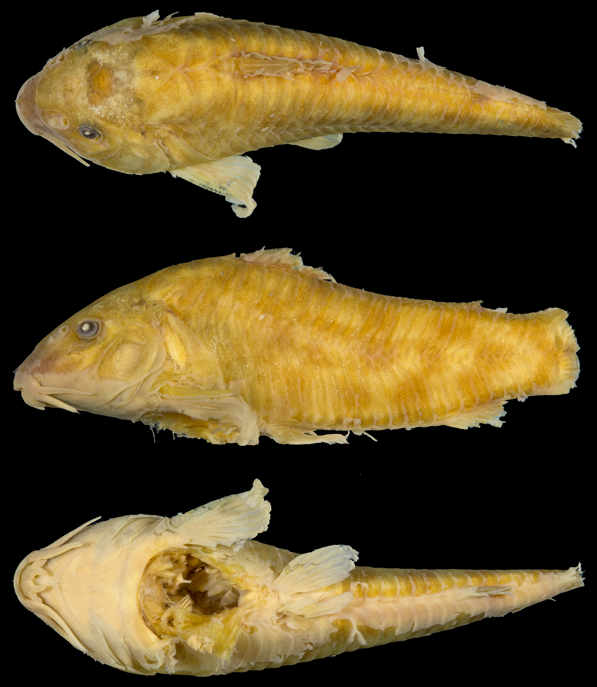
(Fig. 11)
Aspidoras albater Nijssen & Isbrücker, 1976:115 (original description; type locality: rio Tocantinzinha [sic, = Tocantinzinho] near São João da Aliança, Goiás State, Brazil). —Britto, 1998Britto MR. Two new species of the genus Aspidoras (Siluriformes: Callichthyidae) from Central Brazil. Ichthyol Explor Freshw. 1998; 8(4):359–68.:360 (listed as comparative material; partim). —Britto, 2000Britto MR. Aspidoras depinnai (Siluriformes: Callichthyidae), a new species from northeastern Brazil. Copeia. 2000; 2000(4):1048–55. https://www.jstor.org/stable/1448015
https://www.jstor.org/stable/1448015...
:1054 (listed as comparative material; partim). —Lima, Britto, 2001Lima FCT, Britto MR. New catfish of the genus Aspidoras (Siluriformes: Callichthyidae) from the upper rio Paraguai system in Brazil. Copeia. 2001; 2001(4):1010–16. https://www.jstor.org/stable/1448389
https://www.jstor.org/stable/1448389...
:1015 (listed as comparative material; partim). —Reis, 2003Reis RE. Family Callichthyidae (Armored catfishes). In: Reis RE, Kullander SO, Ferraris CJ, Jr., editors. Check list of the freshwater fishes of South and Central America. Porto Alegre: Edipucrs; 2003. p.291–309.:292 (listed). —Ferraris, 2007Ferraris CJ, Jr. Checklist of catfishes, recent and fossil (Osteichthyes: Siluriformes), and catalogue of siluriform primary types. Zootaxa. 2007; (1418):1–628. Available from: http://www.mapress.com/zootaxa/2007f/zt01418p628.pdf
http://www.mapress.com/zootaxa/2007f/zt0...
:108 (listed). —Wosiacki et al., 2014Wosiacki WB, Pereira TG, Reis RE. Description of a new species of Aspidoras (Siluriformes, Callichthyidae) from the Serra dos Carajás, lower Tocantins River basin, Brazil. Copeia. 2014; 2014(2):309–16. https://doi.org/10.1643/CI-13-091
https://doi.org/10.1643/CI-13-091...
:311 (morphological comparison, listed as comparative material). —Tencatt, Bichuette, 2017Tencatt LFC, Bichuette ME. Aspidoras mephisto, new species: The first troglobitic Callichthyidae (Teleostei: Siluriformes) from South America. PLoS ONE. 2017; 12(3):1-24. https://doi.org/10.1371/journal.pone.0171309
https://doi.org/10.1371/journal.pone.017...
:8 (morphological comparison).
Aspidoras eurycephalus Nijssen & Isbrücker, 1976:118 (original description; type locality: Vermelho Stream into rio das Almas, Goiás State, Brazil; new synonym). —Reis, 2003Reis RE. Family Callichthyidae (Armored catfishes). In: Reis RE, Kullander SO, Ferraris CJ, Jr., editors. Check list of the freshwater fishes of South and Central America. Porto Alegre: Edipucrs; 2003. p.291–309.:292 (listed). —Ferraris, 2007Ferraris CJ, Jr. Checklist of catfishes, recent and fossil (Osteichthyes: Siluriformes), and catalogue of siluriform primary types. Zootaxa. 2007; (1418):1–628. Available from: http://www.mapress.com/zootaxa/2007f/zt01418p628.pdf
http://www.mapress.com/zootaxa/2007f/zt0...
:109 (listed). —Tencatt, Bichuette, 2017Tencatt LFC, Bichuette ME. Aspidoras mephisto, new species: The first troglobitic Callichthyidae (Teleostei: Siluriformes) from South America. PLoS ONE. 2017; 12(3):1-24. https://doi.org/10.1371/journal.pone.0171309
https://doi.org/10.1371/journal.pone.017...
:8 (morphological comparison).
Aspidoras poecilus (non Nijssen, Isbrücker, 1976Nijssen H, Isbrücker IJH. The South American plated catfish genus Aspidoras R. von Ihering, 1907, with descriptions of nine new species from Brazil (Pisces, Siluriformes, Callichthyidae). Bijdr Dierkd. 1976; 46(1):107–31.): —Oliveira et al., 2017Oliveira LMA, Zanata AM, Tencatt LFC, Britto MR. A new species of Aspidoras (Siluriformes: Callichthyidae) from a small coastal drainage in northeastern Brazil. Neotrop Ichthyol. 2017; 15(1):e160118. https://doi.org/10.1590/1982-0224-20160118
https://doi.org/10.1590/1982-0224-201601...
:e160118[7] (listed as comparative material; partim).
Aspidoras aff. poecilus (non Nijssen, Isbrücker, 1976Nijssen H, Isbrücker IJH. The South American plated catfish genus Aspidoras R. von Ihering, 1907, with descriptions of nine new species from Brazil (Pisces, Siluriformes, Callichthyidae). Bijdr Dierkd. 1976; 46(1):107–31.): —Britto, 1998Britto MR. Two new species of the genus Aspidoras (Siluriformes: Callichthyidae) from Central Brazil. Ichthyol Explor Freshw. 1998; 8(4):359–68.:361 (listed as comparative material; partim). —Britto, 2000Britto MR. Aspidoras depinnai (Siluriformes: Callichthyidae), a new species from northeastern Brazil. Copeia. 2000; 2000(4):1048–55. https://www.jstor.org/stable/1448015
https://www.jstor.org/stable/1448015...
:1054 (listed as comparative material; partim). —Lima, Britto, 2001Lima FCT, Britto MR. New catfish of the genus Aspidoras (Siluriformes: Callichthyidae) from the upper rio Paraguai system in Brazil. Copeia. 2001; 2001(4):1010–16. https://www.jstor.org/stable/1448389
https://www.jstor.org/stable/1448389...
:1015 (listed as comparative material; partim).
Aspidoras taurus Lima & Britto, 2001:1011 (original description; type locality: rio Itiquira, Mato Grosso State, Brazil; partim; new synonym). —Ferraris, 2007Ferraris CJ, Jr. Checklist of catfishes, recent and fossil (Osteichthyes: Siluriformes), and catalogue of siluriform primary types. Zootaxa. 2007; (1418):1–628. Available from: http://www.mapress.com/zootaxa/2007f/zt01418p628.pdf
http://www.mapress.com/zootaxa/2007f/zt0...
:110 (listed). —Wosiacki et al., 2014Wosiacki WB, Pereira TG, Reis RE. Description of a new species of Aspidoras (Siluriformes, Callichthyidae) from the Serra dos Carajás, lower Tocantins River basin, Brazil. Copeia. 2014; 2014(2):309–16. https://doi.org/10.1643/CI-13-091
https://doi.org/10.1643/CI-13-091...
:311 (morphological comparison, listed as comparative material). —Tencatt, Bichuette, 2017Tencatt LFC, Bichuette ME. Aspidoras mephisto, new species: The first troglobitic Callichthyidae (Teleostei: Siluriformes) from South America. PLoS ONE. 2017; 12(3):1-24. https://doi.org/10.1371/journal.pone.0171309
https://doi.org/10.1371/journal.pone.017...
:21 (listed as comparative material).

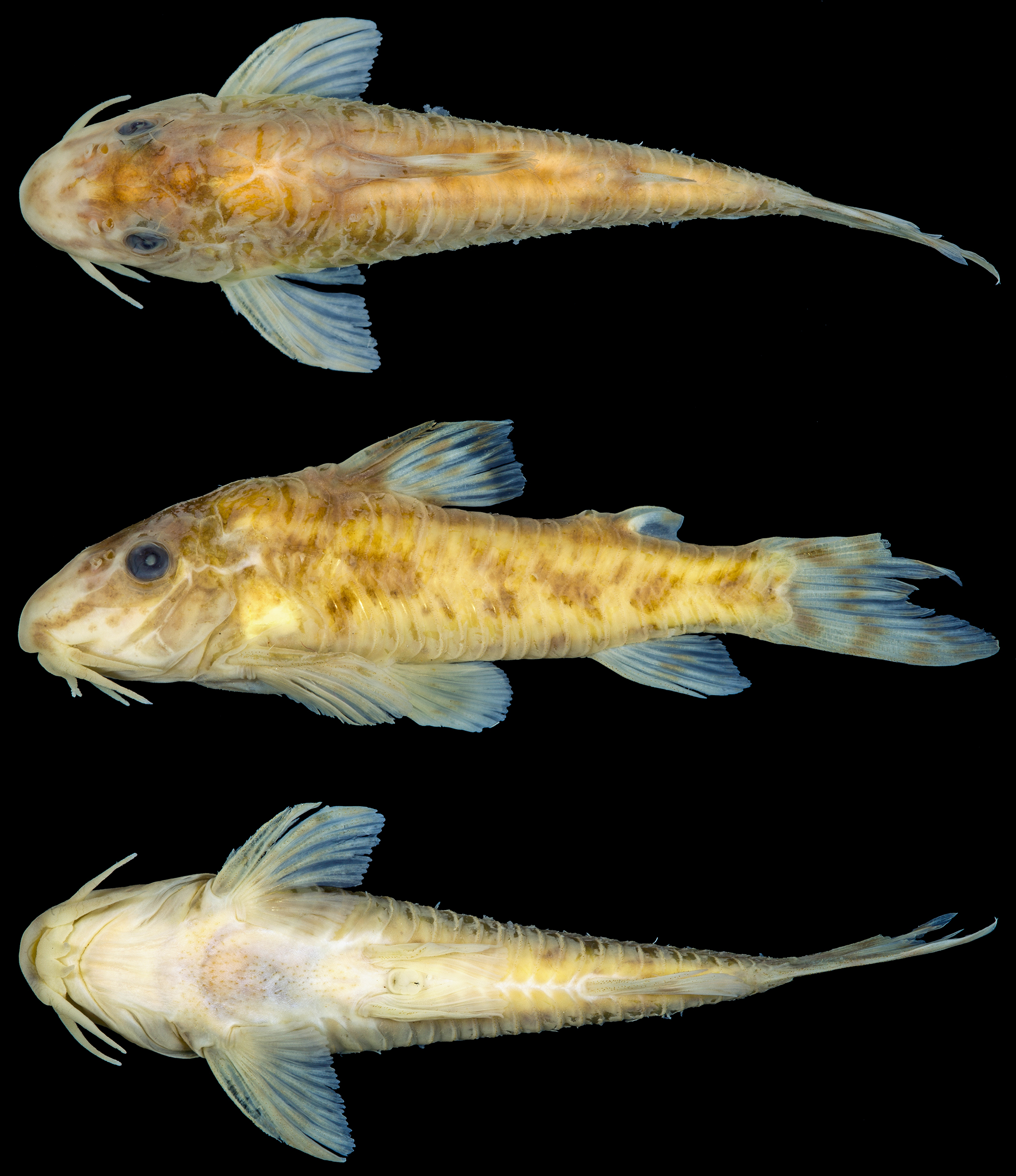
Aspidoras albater, holotype, MZUSP 12991, 34.2 mm SL, rio Tocantinzinho near São João da Aliança, Goiás State, Brazil. Dorsal (top), lateral (middle) and ventral (bottom) views. Photo in lateral view by Eduardo Baena.
Diagnosis.Aspidoras albater can be distinguished from its congeners, with exception of A. aldebaran, A. azaghal, A. depinnai, A. fuscoguttatus, A. gabrieli, A. lakoi, and A. poecilus, by having inner laminar expansion of infraorbital 1 ranging from well developed to extremely well developed (vs. moderately developed in A. belenos, A. maculosus, A. mephisto, and A. raimundi; poorly developed in A. psammatides and A. velites); it differs from A. fuscoguttatus by having anterior portion of infraorbital 1 with laminar expansion ranging from strongly reduced, at same level as posterior margin of nasal capsule, to moderately-developed expansion, reaching middle of nasal capsule (vs. expansion ranging from well developed, surpassing middle of nasal capsule, to extremely well developed, reaching anterior margin of nasal capsule); from A. gabrieli by the presence of acutely furcate caudal fin (vs. smoothly furcate), and dorsolateral body plates on predorsal region touching or closer to their counterparts (vs. dorsolateral body plates more distant from their counterparts); from A. lakoi it can be distinguished by lacking a pointed process on anterodorsal portion of infraorbital 1 (vs. presence); from A. depinnai and A. poecilus plus A. aldebaran, A. belenos, A. kiriri, A. psammatides, A. raimundi, and A. velites by having relatively wide frontal bone, with width equal to or slightly larger than half of entire length (vs. narrow, with width slightly smaller than half of entire length in A. aldebaran, A. belenos, A. depinnai, A. kiriri, A. poecilus, A. raimundi and A. velites; extremely narrow, with width conspicuously smaller than half of entire length in A. psammatides); from A. azaghal it differs by the presence of the first dorsal-fin element, the spinelet (vs. absence). It can be further distinguished from A. aldebaran, A. belenos, and A. kiriri by having parapophysis of the complex vertebra poorly or moderately developed (vs. well developed); from A. rochai by having preadipose azygous plates generally with similar sizes; anteriormost plates smaller than remaining plates in some specimens (vs. preadipose azygous plates with variable sizes, alternating between smaller and larger plates).
Description. Morphometric data presented in Tab. 3; additional morphometric data available in Lima, Britto (2001Lima FCT, Britto MR. New catfish of the genus Aspidoras (Siluriformes: Callichthyidae) from the upper rio Paraguai system in Brazil. Copeia. 2001; 2001(4):1010–16. https://www.jstor.org/stable/1448389
https://www.jstor.org/stable/1448389...
:1012, tab. 1). Head compressed with convex dorsal profile, somewhat triangular or trapezoid in dorsal view. Snout relatively well developed and pointed; or moderately developed and more rounded. Head profile convex from tip of snout to anterior nares; region of mesethmoid slightly concave in some specimens; ascending slightly convex to nearly straight from this point to dorsal-fin origin. Profile slightly convex along dorsal-fin base. Postdorsal-fin body profile slightly concave or nearly straight to adipose-fin spine; slightly concave from this point to caudal-fin base. Ventral profile of body slightly convex from isthmus to pelvic-fin origin; lump on isthmus region in some specimens (apparently by malformation); region of gill opening slightly concave in some specimens; nearly straight from this point to anal-fin origin; slightly concave until caudal-fin base. Body roughly elliptical in cross section at pectoral girdle, gradually becoming more compressed toward caudal fin.
Eye rounded, located dorso-laterally on head; orbit delimited dorsally by lateral ethmoid, frontal and sphenotic, ventrally by infraorbitals. Anterior and posterior nares close to each other, only separated by flap of skin. Anterior naris tubular. Posterior naris close to anterodorsal margin of orbit, separated from it by distance equal to or slightly smaller than naris diameter. Mouth small, subterminal, width larger than bony orbit diameter. Maxillary barbel moderate to large in size, ranging from not reaching to surpassing anteroventral limit of gill opening. Outer mental barbel slightly larger than maxillary barbel. Inner mental barbel fleshy, with base close to its counterpart. Lower lip moderately developed, forming small semicircular or triangular fleshy flap; with two triangular fleshy flaps in some specimens. Small rounded papillae covering entire surface of all barbels, upper and lower lips, snout and isthmus.
Mesethmoid short; anterior tip long, slightly larger than 50% of entire bone length (see Britto, 2003Britto MR. Phylogeny of the subfamily Corydoradinae Hoedeman, 1952 (Siluriformes: Callichthyidae), with a definition of its genera. P Acad Nat Sci Phila. 2003; 153(1):119–54. https://doi.org/10.1635/00973157(2003)153 [0119:POTSCH]2.0.CO;2
https://doi.org/10.1635/00973157(2003)15...
:123, ch. 1, state 0; fig. 1A); posterior portion wide, entirely, or almost entirely covered by thick layer of skin. Nasal slender, curved laterally, inner margin generally with moderately developed laminar expansion; variably poorly developed; outer margin with reduced laminar expansion; absent in some specimens; mesial border generally contacting only frontal; or contacting frontal and mesethmoid.
Frontal elongated, relatively wide, with width equal to or slightly larger than half of entire length (Fig. 3B); anterior projection ranging from short, with size smaller than nasal length, to long, with size larger than nasal length; anterior margin generally covered by thick layer of skin. Frontal fontanel relatively small, ellipsoid or somewhat rhomboid; posterior tip extension not entering anterior margin of parieto-supraoccipital. Sphenotic somewhat trapezoid, contacting parieto-supraoccipital dorsally, pterotic-extrascapular posteriorly, second infraorbital ventrally and frontal anteriorly. Pterotic-extrascapular roughly pipe-shaped, with posteriormost portion contacting first lateral-line ossicle, and ventral margin contacting opercle and cleithrum; posterior expansion almost entirely covering lateral opening of swimbladder capsule, leaving slender pseudotympanic area on its dorsal margin covered only by thick layer of skin. Parieto-supraoccipital wide, posterior process strongly reduced to poorly developed; not contacting nuchal plate. Parieto-supraoccipital medial keel expanded ventrally; laminar, with posterior portion at same level as posterior process tip; with posterior portion variably not reaching tip of posterior process; or expanded posteriorly, surpassing tip of posterior process. Parieto-supraoccipital fontanel small, roundish; located medially on parieto-supraoccipital; or slightly displaced towards posterior portion of parieto-supraoccipital; fontanel occluded, reduced to a small fossa, in some specimens.
Two laminar infraorbitals with minute odontodes; infraorbital 1 large, ventral laminar expansion generally ranging from poorly developed to well developed; almost reduced to latero-sensory canal in some specimens; single specimen with extremely well-developed ventral laminar expansion (MZUSP 49853, 35.4 mm SL); anterior portion with laminar expansion ranging from strongly reduced, at same level as posterior margin of nasal capsule, to moderately developed, reaching middle of nasal capsule (Fig. 12); inner laminar expansion ranging from well developed to extremely well developed (Figs. 5A,B); small portions of external surface covered by thick layer of skin; infraorbital 2 small, generally slender; slightly more compact in some specimens; generally with posterior laminar expansion moderately developed; variably poorly or relatively well developed; inner laminar expansion ranging from moderately to well developed; posteroventral margin close but not directly contacting posterodorsal ridge of hyomandibula; contacting in some specimens; dorsal tip contacting only sphenotic; external surface partially covered by thick layer of skin; or entirely covered by thick layer of skin (Fig. 12). Posterodorsal ridge of hyomandibula, close to its articulation with opercle, oblong, relatively slender; generally exposed; entirely or almost entirely covered by thick layer of skin in some specimens; dorsal ridge of hyomandibula, between pterotic-extrascapular and opercle, covered by thick layer of skin; variably covered by thin layer of skin, exposed; exposed areas generally bearing small odontodes. Interopercle with posterior portion exposed; or entirely covered by thick layer of skin; somewhat triangular, anterior projection moderately developed; variably well developed. Preopercle relatively slender, elongated, minute odontodes sparse on external surface; entirely or almost entirely covered by thick layer of skin in some specimens. Opercle compact in shape, width larger than half of its length; free margin convex; posterodorsal region variably with smoothly concave area; without serrations and covered by small odontodes; some portions of bony distal margin irregular in some specimens.
Four branchiostegal rays decreasing in size posteriorly. Hypobranchial 2 somewhat triangular, tip ossified and directed towards anterior portion, posterior margin cartilaginous; ossified portion well developed, about twice size of cartilaginous portion; or conspicuously well developed, about triple size of cartilaginous portion. Five ceratobranchials with expansions increasing posteriorly; ceratobranchial 1 with small process on anterior margin of mesial portion; process strongly reduced in some specimens; ceratobranchial 3 generally with continuous postero-lateral margin; variably notched; ceratobranchial 5 toothed on postero-dorsal surface, 25 to 34 (22) teeth aligned in one row. Four epibranchials with similar size; epibranchial 2 slightly larger than others, with small-pointed process on laminar expansion of posterior margin; epibranchial 3 with triangular uncinate process on laminar expansion of posterior margin. Two wide pharyngobranchials (3 and 4), pharyngobranchial 3 with triangular laminar expansion on posterior margin; triangular laminar expansion variably with notches. Upper tooth plate oval, with 29 to 42 (22) teeth aligned in two rows on postero-ventral surface.
Lateral-line canal entering neurocranium through pterotic-extrascapular, branching twice before entering sphenotic: pterotic branch with single pore; preoperculomandibular branch conspicuously reduced, with single pore opening close to postotic main canal; more developed, with pore opening closer to anteroventral border of pterotic-extrascapular in some specimens; postotic main canal widens just posterior to pterotic branch. Sensory canal continuing through pterotic-extrascapular, entering sphenotic as temporal canal, which splits into two branches: one branch giving rise to infraorbital canal, another branch entering frontal through supraorbital canal, both with single pore. Supraorbital canal branched, running through nasal bone. Epiphyseal branch of supraorbital canal relatively long, with pore opening close to frontal fontanel; or slightly shorter, with pore opening closer to supraorbital main canal. Nasal canal with three openings, first on posterior edge, second on posterolateral portion and third on anterior edge; second pore generally fused with first pore. Infraorbital canal running through entire second infraorbital, extending to infraorbital 1 and opening into two or three pores. Preoperculomandibular branch giving rise to preoperculo-mandibular canal, which runs through entire preopercle with three openings, leading to pores 3, 4, and 5, respectively; pore 3 opening at posterodorsal ridge of hyomandibula in some specimens.
Dorsal fin somewhat triangular, located just posterior to third or fourth dorsolateral body plate. Dorsal-fin rays II,7(1), II,8*(23), II,9(1), posterior margin of dorsal-fin spine smooth. Nuchal plate poorly developed in length; strongly reduced in some specimens; generally, entirely covered by thick layer of skin; posterior portion variably exposed and with minute odontodes (Fig. 13); spinelet extremely short or short; partially exposed or entirely covered by thick layer of skin; spine poorly developed, adpressed distal tip reaching middle portion of dorsal-fin base; or moderately developed, adpressed distal tip slightly surpassing middle portion of dorsal-fin base; anterior margin with small odontodes. Pectoral fin roughly triangular, its origin just posterior to gill opening. Pectoral-fin rays I,8(1), I,9*(24); posterior margin of pectoral spine with eight to 24 poorly- or moderately-developed serrations along almost its entire length; small region just posterior to origin of spine lacking serrations; some serrations directed towards origin of spine, perpendicularly directed or directed towards tip of spine; presence of bifid serrations in some specimens; base of branched rays with small laminar expansions on inner margin, generally more evident on first rays; laminar expansions variably with irregular margins (Fig. 14). Anteroventral portion of cleithrum partially exposed; posterolateral portion of scapulocoracoid reduced, generally externally visible, or covered by thick layer of skin, not externally visible. Pelvic fin oblong, located just below fourth or fifth ventrolateral body plate, and at vertical through third dorsal-fin branched ray. Pelvic-fin rays i,5*(25). Adipose fin roughly triangular, separated from posterior origin of dorsal-fin base by nine to 11 dorsolateral body plates. Anal fin somewhat triangular, located just posterior to 13th, 14th, or 15th ventrolateral body plates, and at vertical through region of preadipose platelets; or anterior to region of preadipose platelets. Anal-fin rays ii,6*(25). Caudal-fin rays i,12,i*(24), i,13,i(1), generally five or six dorsal and/or ventral procurrent rays; caudal fin bilobed, dorsal lobe slightly larger than ventral lobe.
Two to four laterosensory canals on trunk; first ossicle tubular, second ossicle laminar, third and fourth lateral-line canals, if present, encased in third and fourth dorsolateral body plates, respectively. Body plates with conspicuous line of relatively large odontodes confined to posterior margins; dorsolateral body plates 26*(4), 27(10), 28(10), 29(1); ventrolateral body plates 23*(2), 24(6), 25(17); dorsolateral body plates along dorsal-fin base 5(2), 6*(16), 7(7); dorsolateral body plates between adipose-fin spine and caudal-fin base 7(2), 8*(14), 9(8), 10(1); preadipose platelets 4(3), 5*(12), 6(7), 7(3), 8(1); small platelets covering base of caudal-fin rays; small platelets disposed dorsally and ventrally between junctions of lateral plates on posterior portion of caudal peduncle. Anterior margin of orbit, above region of junction of frontal with lateral ethmoid, with small platelets; platelets variably absent. Ventrolateral portion of snout, region of nasal capsule and dorsal portion of snout, above mesethmoid, with small platelets in some specimens. Region between nuchal plate and posterior process of parieto-supraoccipital variably with relatively large platelets. Ventral surface of trunk covered by small platelets, generally more abundant and/or concentrated on anterior half of trunk; absence of platelets in some specimens.
Vertebral count 24(1), 25(11), 26(8), 27(2); ribs 6(2), 7(18), 8(2), first pair conspicuously large; parapophysis of complex vertebra poorly or moderately developed (Figs. 8B,C).
Morphometric data of the holotype and four paratypes (MZUSP 12992) plus 20 non-type specimens of Aspidoras albater. SD = standard deviation.

Lateral view of the head of CS specimens of Aspidoras albater, showing the patterns of infraorbitals within the species. Ventral laminar expansion of infraorbital 1 strongly reduced in (A) and (B) (LBP 1427, 36.7 mm SL and LIRP 4447, 29.4 mm SL, respectively), poorly developed in (C) (MNRJ 11716, 27.8 mm SL), moderately developed in (D) (MZUSP 40793, 29.4 mm SL) and well developed in (E) (MNRJ 13050, 31.3 mm SL). Abbreviations: io1: infraorbital 1, io2: infraorbital 2, sph: sphenotic, pes: pterotic-extrascapular. Scale bars = 1 mm.

Aspidoras albater, MZUSP 40793, CS, 29.4 mm SL, showing predorsal region of trunk in dorsal view. Dotted lines indicate the limits of the tip of the posterior process of the parieto-supraoccipital and anterior tip of the nuchal plate. Abbreviations: np: nuchal plate, pso: parieto-supraoccipital. Scale bar = 1 mm.

Dorsal view of pectoral spines in CS specimens of Aspidoras albater, showing variation in the morphology of pectoral-fin spine: presence of laminar expansion and simple and/or bifid poorly- and/or moderately-developed serrations on its posterior margin in (A) and (B) (MZUSP 114401, 30.1 mm SL and MNRJ 13080, 30.9 mm SL, respectively), (C) with only simple poorly-developed serrations on its posterior margin (LIRP 4472, 29.4 mm SL), (D) with only simple and/or bifid poorly-developed serrations on its posterior margin (MNRJ 12779, 31.3 mm SL), or (E) with only simple and/or bifid moderately-developed serrations on its posterior margin (MNRJ 12581, 31.1 mm SL). Arrows in (A) indicate the small laminar expansions at the base of branched rays. Scale bars = 1 mm.
Coloration in alcohol. Color pattern highly variable. Ground color of body light or brownish yellow, with top of head dark brown. Posterodorsal portion of head, region below eye, opercle and cleithrum with scattered dark brown or black chromatophores. Snout covered by dark brown or black chromatophores on its dorsal surface; chromatophores densely disposed in some specimens; variably forming dark brown or black rounded, striated or irregular spots; with dark brown or black diffuse stripe from anteroventral portion of orbit to upper lip lateral area in some specimens; ventrolateral portion of snout variably with dark brown or black chromatophores. Upper lip and maxillary barbel with dark brown or black chromatophores; area of lateral portion of upper lip with conspicuous concentration of dark brown or black chromatophores in some specimens; outer mental barbel with dark brown or black chromatophores, generally more evident on its proximal portion, in some specimens; region of isthmus around lower lip variably with dark brown or black chromatophores. Dorsal series of four to five dark brown or black blotches, first on anterior portion of dorsal-fin base, second, if present, on middle portion of dorsal-fin base, third on posterior portion of dorsal-fin base, fourth on adipose-fin base, fifth on caudal-fin base; blotches variably diffuse. Dorsal portion of body with conspicuous concentration of dark brown or black chromatophores between counterparts of dorsolateral body plates in some specimens. Ventral surface of trunk and region posterior to urogenital opening with dark brown or black chromatophores in some specimens. First dorsolateral body plate with conspicuous concentration of dark brown or black chromatophores; posterior margin of some dorso- and ventrolateral plates, and lateral line pores with conspicuous concentration of dark brown or black chromatophores in some specimens. Midline of flank with longitudinal series of three to six medium- to large-sized conspicuous dark brown or black blotches; blotches rounded, oblong or irregular; blotches fused with each other, forming longitudinal stripe in single specimen (DZSJRP 12585, 29.9 mm SL). Dorsal half of dorsolateral body plates with dark brown or black chromatophores; region of anterior and posterior portions of dorsal-fin base, between dorsal and adipose fins, adipose-fin base, between adipose and base of caudal peduncle and base of caudal peduncle with more concentrated chromatophores, forming conspicuous blotches in some specimens; blotches variably fused to flank midline blotches. Ventral half of dorsolateral body plates and dorsal half of ventrolateral body plates with concentration of dark brown or black chromatophores, forming conspicuous blotches in some specimens; blotches generally more evident on anterior portion of body and on area of flank midline blotches. Mid-ventral portion of ventrolateral body plates on area of flank midline blotches with concentration of dark brown or black chromatophores; forming conspicuous blotches in some specimens; blotches generally more evident posteriorly to pelvic-fin origin; variably fused to flank midline blotches; ventral portion of ventrolateral body plates with concentration of dark brown or black chromatophores, generally more evident posterior to anal-fin anterior origin, in some specimens. Dorsal fin with dark brown or black spots; aligned spots, forming longitudinal or oblique bars in some specimens; conspicuous concentration of dark brown or black chromatophores on some areas of membranes, forming larger dark brown or black patches in some specimens; almost entirely solid black or covered by dark brown or black chromatophores in some specimens; dorsal-fin base with conspicuous concentration of dark brown or black chromatophores, generally more concentrated on bases of first and last branched rays; spine covered by dark brown or black chromatophores. Pectoral fin with dark brown or black chromatophores on its dorsal surface, generally more evident on spine and first branched rays; covered by dark brown or black spots in some specimens; spots generally diffuse and more evident on first branched rays; or entirely hyaline; region of body around dorsal portion of pectoral-fin origin generally with concentration of dark brown or black chromatophores. Pelvic fin generally hyaline or with sparse dark brown or black chromatophores on its dorsal surface; with conspicuous concentration of dark brown or black chromatophores on its proximal two thirds or forming up to two oblong dark brown or black patches in some specimens; region of body around dorsal portion of pelvic-fin origin with concentration of dark brown or black chromatophores in some specimens. Adipose-fin membrane with dark brown or black chromatophores; conspicuous concentration of dark brown or black chromatophores in some areas of membrane, generally more evident close to spine, forming isolated patches in some specimens; adipose-fin spine generally with dark brown or black chromatophores. Anal fin with conspicuous concentrations of dark brown or black chromatophores in some areas, generally more evident on its middle portion and bases of last branched rays; with one to three dark brown or black blotches in some specimens; entirely or almost entirely covered by dark brown or black chromatophores in some specimens. Middle portion of caudal-fin base, posteriorly to last flank midline blotch, generally with small- to medium-sized dark brown or black blotch; blotch diffuse or fused with last midlateral blotch in some specimens. Caudal fin with three to six transversal dark brown or black slender to wide bars.
Coloration in life. Similar to that observed in preserved specimens but with ground color of body grayish yellow in some specimens. Body covered by whitish yellow and green iridescent coloration (Fig. 15).

Aspidoras albater specimens photographed alive. (A) uncataloged aquarium specimen from the rio Araguaia basin, (B) specimen from the rio Muquém, rio Tocantins basin (MZUSP 114357, not measured), (C) uncatalogued aquarium specimens from the rio Araguaia basin, (D) uncatalogued aquarium specimen from Mato Grosso, possibly from the rio Paraguay basin, and (E) specimen from a stream with unknown name tributary to the rio Tocantins basin (LBP 19312, 27.6 mm SL). Photos by Ingo Seidel (A), José Birindelli (B), Hans Evers (C, D), and Martin Taylor (E).
Geographical distribution.Aspidoras albater is known from the upper portions of the Tocantins and Paraná River basins in Goiás State, upper rio Araguaia basin in Goiás and Mato Grosso states, and upper rio Paraguay basin in Mato Grosso State, Brazil (Fig. 10).
Conservation status.Aspidoras albater is widespread in central Brazil, occurring in the Tocantins-Araguaia, Paraná, and Paraguay river basins of Goiás and Mato Grosso states. No threat to the species is currently known. According to the International Union for Conservation of Nature (IUCN) categories and criteria (IUCN Standards and Petitions Subcommittee, 2019International Union for Conservation of Nature (IUCN). Standards and petitions subcommittee. Guidelines for using the IUCN Red List categories and criteria. Version 14 [Internet]. Gland; 2019. Available from: https://www.iucnredlist.org/resources/redlistguidelines
https://www.iucnredlist.org/resources/re...
), Aspidoras albater can be classified as Least Concern (LC).
Remarks. According to Nijssen, Isbrücker, (1976)Nijssen H, Isbrücker IJH. The South American plated catfish genus Aspidoras R. von Ihering, 1907, with descriptions of nine new species from Brazil (Pisces, Siluriformes, Callichthyidae). Bijdr Dierkd. 1976; 46(1):107–31., A. albater can be recognized by the presence of four large oblique black blotches on body. As aforementioned (see Color in alcohol), the dark brown or black blotches on dorsal and ventral portions of flanks can be fused to flank midline blotches, resulting in these somewhat oblique, irregular, large blotches. However, this pattern is not present in all type specimens. Among the 10 type specimens re-examined herein for this feature (MZUSP 12991, MZUSP 12992 and USNM 213540, and considering the left side of the body), only the holotype and four paratypes display such a pattern for all midline blotches (Figs. 11, 16A,E,F,H). In the other five paratypes, it was possible to observe that some midline blotches can be variably fused with the blotches on dorsal and/or ventral portions of flanks (Figs. 16B,C,D), or even present no fusion with any of them (Figs. 16C,D,G). Also, the fusion between midline and ventral blotches often results in large transversally elongated blotches (Figs. 16B‒D), whereas the fusion between dorsal, midline and ventral blotches may result in chevron-shaped large blotches instead of the oblique pattern (Fig. 16). Such variable patterns were also observed in several non-type specimens. Therefore, since this feature is highly variable and may be present in other Aspidoras species, such as A. depinnai and A. kiriri, it should not be considered as diagnostic for A. albater.

Paratypes of Aspidoras albater, (A–D) MZUSP 12992, 4, 25.9–31.2 mm SL, and (E–I) USNM 213540, 5, 26.1–31.2 mm SL, showing general morphological and color patterns in lateral view. Scale bars = 5 mm.
Aspidoras eurycephalus (Fig. 17) was described from the rio Maranhão basin, also part of the upper rio Tocantins drainage. Comparing only the holotypes, A. albater can be distinguished from A. eurycephalus by presenting the following combination of features: (I) absence of contact between counterparts of dorsolateral body plates on predorsal region of body (vs. presence); (II) posterior process of the parieto-supraoccipital poorly developed (vs. strongly reduced); (III) pectoral spine poorly developed but clearly larger (vs. poorly developed but clearly smaller); (IV) smaller interorbital width (vs. larger); and (V) ventral laminar expansion of infraorbital 1 moderately developed (vs. well developed). Despite the holotypes presenting these remarkable differences, most of the examined specimens from the upper rio Tocantins basin, including even some paratypes of A. albater, present an intermediate morphology or a mix of features found in either the holotype of A. albater or the holotype of A. eurycephalus. Intermediate or mixed morphotypes were also found in the upper portions of the rivers Araguaia, Paraguay, and Paraná basins.

Holotype of Aspidoras eurycephalus, junior synonym of Aspidoras albater, CAS 16010, 29.5 mm SL, córrego Vermelho into rio das Almas, Goiás State, Brazil. Dorsal (top), lateral (middle) and ventral (bottom) views. Photo by the California Academy of Sciences.
Lima, Britto (2001)Lima FCT, Britto MR. New catfish of the genus Aspidoras (Siluriformes: Callichthyidae) from the upper rio Paraguai system in Brazil. Copeia. 2001; 2001(4):1010–16. https://www.jstor.org/stable/1448389
https://www.jstor.org/stable/1448389...
described A. taurus (Fig. 18) based on the following diagnostic features: (I) infraorbitals and preopercle covered by thick skin and not visible externally; (II) nuchal plate reduced, covered by thick skin and not visible externally; and (III) absence of lateral line posterior to two small lateral line ossicles. Regarding feature (I), the examination of several specimens from the A. taurus type series, including the holotype, and additional non-type specimens (LBP 1427), revealed that only the infraorbital 2 and preopercle can be entirely covered by thick layer of skin in some specimens, contrary to the infraorbital 1, that is at least partially externally visible in all examined specimens. In some A. taurus specimens, as well as in some A. albater specimens from the rivers Araguaia, Tocantins and Paraná basins, the ventral laminar expansion of infraorbital 1 is almost reduced to the latero-sensory canal, resulting in an extremely small exposed area in those specimens, which is difficult to observe with naked eyes and may cause the wrong perception that the infraorbital 1 is not exposed. Despite the fact that it was possible to confirm the presence of the features (II) and (III) in A. taurus, they can also be observed in A. albater specimens from the rivers Araguaia, Tocantins and Paraná basins.

Holotype of Aspidoras taurus, junior synonym of Aspidoras albater, MZUSP 57154, 52.1 mm SL, rio Itiquira, Mato Grosso State, Brazil. Dorsal (top), lateral (middle) and ventral (bottom) views. Photo in lateral view by Eduardo Baena.
One of the most remarkable features of A. taurus is the relatively large size of some specimens, reaching more than 50 mm SL, though most specimens present the expected size for the genus, which is generally inferior to 40 mm SL. However, considering general morphological pattern, even these unusually large specimens are notably similar to the holotype of A. eurycephalus and some paratypes of A. albater. Additionally, it is also possible to find relatively large-sized specimens in populations from the rivers Araguaia (LIRP 4472, up to 40.1 mm SL, Fig. 19A; MZUSP 41409, up to 44.6 mm SL), Tocantins (MZUSP 114391, up to 40.0 mm SL) and Paraná (DZSJRP 9963, up to 41.6 mm SL) basins. Considering each examined feature (e.g., infraorbital 1, predorsal, pectoral spine, interorbital, and color patterns), the holotypes of A. albater A. eurycephalus and A. taurus alternately represented two extreme forms of a morphological range, with one of them, as well as most additional examined specimens, falling in between the other two, and, because no conspicuous diagnostic features were observed, the most reasonable decision is to place both A. eurycephalus and A. taurus in the synonymy of A. albater.

Relatively large specimen of (A) Aspidoras albater, LIRP 4472, 40.1 mm SL, and darker specimen of (B) Aspidoras albater, MZUSP 114401, 35.9 mm SL, showing general morphological and color patterns in lateral view. Photo (A) by Celso Ikedo.
Specimens clearly compatible with A. albater are scarce in the aquarium hobby. However, the species coded as Aspidoras sp. C35, which is known as “the black phantom” among hobbyists due to its intensely pigmented pattern, especially on dorsal fin, likely represents A. albater. According to Hans Evers (2020, pers. comm.), who captured the specimens originally used to assign the code C35 (Fig. 15C), this species was found in a place called “Bacalhau”, a small, shallow, and crystal clear-water stream, while traveling from Aruanã to Goiânia, in Goiás State. Around the middle of the shortest route from Aruanã to Goiânia, on the outskirts of the Goiás Velho City, a small stream called córrego Bacalhau crosses the road GO-070 (15°57’01”S 50°08’52”W). This stream is a tributary to the rio Vermelho, rio Araguaia basin. Considering the presence of A. albater specimens with intensely black pigmentation on dorsal fin, including specimens from the rio Tocantins basin in Goiás (Fig. 19B), allied to the compatible geographical distribution, it seems reasonable to conclude that C35 corresponds to A. albater. On the other hand, the species coded as CW52, often attributed to A. albater, presents unknown origin and intermediate morphological and color patterns between A. albater and A. poecilus. It is known that many Aspidoras species that were introduced into the hobby were kept mixed, easily interbreeding with fertile F1 offspring (Robert McLure, 2020, pers. comm.). This may explain C52’s simultaneous similarity to two completely different species such as A. albater and A. poecilus. Therefore, CW52 is considered herein as a possible aquarium strain.
Material examined. In addition to the material examined by Nijssen, Isbrücker, (1976)Nijssen H, Isbrücker IJH. The South American plated catfish genus Aspidoras R. von Ihering, 1907, with descriptions of nine new species from Brazil (Pisces, Siluriformes, Callichthyidae). Bijdr Dierkd. 1976; 46(1):107–31., the following material was examined. All from Brazil. Rio Paraguay basin. DZSJRP 12585, 20 of 33, 14.5‒29.9 mm SL, Mato Grosso, stream tributary to the rio Itiquira. LBP 1427, 29, 17.2‒39.5 mm SL, 2 CS, 23.8‒36.7 mm SL, Mato Grosso, rio Itiquira. MZUSP 57154, 52.1 mm SL, holotype of Aspidoras taurusLima, Britto, 2001Lima FCT, Britto MR. New catfish of the genus Aspidoras (Siluriformes: Callichthyidae) from the upper rio Paraguai system in Brazil. Copeia. 2001; 2001(4):1010–16. https://www.jstor.org/stable/1448389
https://www.jstor.org/stable/1448389...
, Mato Grosso, Fazenda Cabeceira do Itiquira, close to highway BR-364, rio Itiquira. MNRJ 19951, 5, 30.9–40.3 mm SL; MZUSP 41455, 228 (5 CS not measured), 14.1‒54.1 mm SL; USNM 363174, 5, 31.5–40.9 mm SL, paratypes of Aspidoras taurusLima, Britto, 2001Lima FCT, Britto MR. New catfish of the genus Aspidoras (Siluriformes: Callichthyidae) from the upper rio Paraguai system in Brazil. Copeia. 2001; 2001(4):1010–16. https://www.jstor.org/stable/1448389
https://www.jstor.org/stable/1448389...
, all collected with holotype. Rio Paraná basin. DZSJRP 9963, 8, 24.3‒41.6 mm SL, Distrito Federal, córrego Vicente Pires. MNRJ 19984, 1, 35.6 mm SL, Distrito Federal, stream tributary to the lago Paranoá. MNRJ 20258, 3, 14.3‒36.8 mm SL, Goiás, córrego Coqueiro. MNRJ 20332, 2, 26.9‒29.7 mm SL, Goiás, córrego Águas Emendadas. MZUSP 41434, 8, 9.8‒21.1 mm SL, Goiás, ribeirão Capivara. NUP 1098, 5, 20.2‒39.7 mm SL, 1 CS, 33.8 mm SL, Goiás, Corumbá Reservoir. NUP 1286, 2, 19.7‒27.9 mm SL, Goiás, ribeirão Cachoeira. Rio Araguaia basin. DZSJRP 19858, 23, 21.9‒37.9 mm SL, Mato Grosso, córrego Fundo II. LBP 15657, 1, 22.2 mm SL, Mato Grosso, tributary to the rio das Mortes. LIRP 4447, 5, 14.9‒25.8 mm SL, 1 CS, 29.4 mm SL, Mato Grosso, córrego Rico. LIRP 4472, 85 of 162, 20.4‒40.1 mm SL, 3 CS of 162, 29.4‒31.9 mm SL, Mato Grosso, córrego Carneiro. MNRJ 21075, 17, 14.7‒28.2 mm SL, 2 CS, 25.0‒25.9 mm SL, Mato Grosso, stream tributary to the rio das Garças. MZUSP 28599, 29, 13.8‒36.4 mm SL, Mato Grosso, rio Noidore. MZUSP 41409, 131, 12.1–44.6 mm SL, Goiás, grotto on the road to Couto de Magalhães Falls. MZUSP 41424, 7, 14.5‒33.9 mm SL, Goiás, córrego do Jacaré. MZUSP 88144, 2, 23.0‒34.4 mm SL, Mato Grosso, tributary to the rio das Garças. MZUSP 89424, 2, 25.2‒27.3 mm SL, Goiás, ribeirão d’Anta. MZUSP 97858, 10 of 23, 13.3‒27.3 mm SL, Mato Grosso, unnamed stream. UFRJ 6434, 13, 16.8‒28.5 mm SL, Goiás, stream tributary to the rio Caiapó. Rio Tocantins basin. INPA 39901, 10 of 91, 17.4‒19.3 mm SL, Goiás, rio Areia. LBP 15302, 2, 28.3‒33.4 mm SL, Goiás, rio da Lapa. LBP 19277, 8, 24.8‒35.4 mm SL, Goiás, córrego Kavanca. LBP 19312, 1, 27.6 mm SL, Goiás, unnamed stream. LESCI 334, 1, 32.7 mm SL, Goiás, Angélica Cave. LISDEBE 6606, 24.7 mm SL, Goiás, unnamed stream. MCP 45556, 6, 18.7‒24.2 mm SL, Goiás, córrego Galheiros. MNRJ 11716, 66 of 69, 16.0‒29.6 mm SL, 3 CS of 69, 26.7‒29.9 mm SL, Goiás, córrego Pontilhão. MNRJ 12564, 6, 24.3‒34.7 mm SL, Goiás State, ribeirão do Padre. MNRJ 12571, 10 of 26, 22.2–33.4 mm SL, 1 CS of 26, 35.3 mm SL, Goiás, stream tributary to the rio Tocantins. MNRJ 12581, 39 of 44, 14.6‒29.7 mm SL, 5 CS of 44, 22.5‒31.1 mm SL, Goiás, stream tributary to the rio Tocantins. MNRJ 12725, 3, 25.0‒27.3 mm SL, Goiás, córrego Batéias. MNRJ 12766, 1, 23.4 mm SL, Goiás, rio da Mula. MNRJ 12775, 3, 14.5‒25.5 mm SL, Goiás, stream tributary to the rio Tocantins. MNRJ 12779, 8 of 12, 27.4‒28.8 mm SL, 4 CS of 12, 26.2–31.3 mm SL, Goiás, córrego Bateinha. MNRJ 13044, 2, 18.5‒32.9 mm SL, Goiás, córrego Quineira or Mangueira. MNRJ 13045, 34 of 36, 16.6‒25.6 mm SL, 2 CS of 36, 24.0‒29.3 mm SL, Goiás, córrego Batéias. MNRJ 13050, 25 of 28, 19.0‒29.9 mm SL, 3 CS of 28, 17.6‒32.5 mm SL, Goiás, córrego Pontilhão. MNRJ 13051, 1, 20.9 mm SL, Goiás, córrego Pirapitinga. MNRJ 13052, 5, 27.7‒32.2 mm SL, 1 CS, 36.5 mm SL, Goiás, córrego Grota Seca. MNRJ 13053, 3, 28.1‒37.4 mm SL, Goiás, tributary to the córrego Lageado. MNRJ 13054, 20, 14.0‒26.6 mm SL, Goiás, córrego Batéias. MNRJ 13055, 4, 20.2‒36.0 mm SL, 1 CS, 27.2 mm SL, Goiás, Matula Stream. MNRJ 13056, 9, 19.3‒29.8 mm SL, Goiás, stream tributary to the córrego Lageado. MNRJ 13080, 8 of 9, 10.4‒34.7 mm SL, 1 CS of 9, 30.9 mm SL, Goiás, córrego São Bento. MNRJ 13081, 2, 28.4‒33.0 mm SL, Goiás, stream tributary to the córrego São Bento. MNRJ 13082, 6, 15.2‒29.5 mm SL, Goiás, córrego Quineira or Mangueira. MNRJ 28880, 8, 22.7‒32.9 mm SL, Goiás, córrego Água Boa. MNRJ 28881, 3, 25.7‒26.7 mm SL, Goiás, córrego Batéia. MNRJ 36166, 9, 26.5‒33.4 mm SL, Goiás, córrego Água Boa. MZUSP 40624, 4, 23.7‒30.1 mm SL, Goiás, rio Angélica. MZUSP 40656, 2, 19.4‒24.8 mm SL, Goiás, rio Preto. MZUSP 40683, 1, 28.8 mm SL, Goiás, rio Bezerra. MZUSP 40793, 45 of 46, 14.8‒33.5 mm SL, 1 CS of 46, 29.4 mm SL, Goiás, stream tributary to the rio das Almas. MZUSP 40799, 7, 23.3‒28.5 mm SL, Goiás, swamp tributary to the córrego Ave Maria. MZUSP 40806, 3, 21.8‒27.2 mm SL, Goiás, stream tributary to the córrego Ave Maria. MZUSP 40811, 74, 21.2‒32.9 mm SL, Goiás, stream tributary to the córrego Ave Maria. MZUSP 40815, 2, 21.3‒28.3 mm SL, Goiás, stream tributary to the rio das Pedras. MZUSP 40886, 5, 21.6‒35.9 mm SL, Goiás, córrego Poço Danta. MZUSP 49853, 5, 17.0‒35.4 mm SL, Goiás, stream in the Serra da Mesa. MZUSP 50157, 35, 11.3‒31.3 mm SL, Goiás, Serra da Mesa Lake. MZUSP 54468, 100, 21.1‒35.4 mm SL, Goiás, rio Bagagem. MZUSP 55221, 56, 14.7‒31.8 mm SL, Goiás, rio da Lapa. MZUSP 55238, 6, 16.8‒23.4 mm SL, Goiás, rio da Lapa. MZUSP 55244, 1, 25.2 mm SL, Goiás, rio Angélica. MZUSP 55250, 1, 26.0 mm SL, Goiás, rio Angélica. MZUSP 55967, 9, 18.7‒28.0 mm SL, Goiás, rio da Lapa sinkhole. MZUSP 55983, 2, 26.6‒27.1 mm SL, Goiás, córrego Quilombo. MZUSP 55985, 1, 25.3 mm SL, Goiás, lapa Angélica. MZUSP 57407, 16, 15.9‒23.1 mm SL, Goiás, stream tributary to the rio São Domingos. MZUSP 81661, 1, 33.0 mm SL, Goiás, rio Angélica. MZUSP 81662, 1, 27.1 mm SL, Goiás, lapa Angélica. MZUSP 85771, 5, 18.2‒27.9 mm SL, Goiás, rio da Lapa. MZUSP 114074, 3, 15.6‒23.2 mm SL, Goiás, ribeirão dos Bois. MZUSP 114080, 2, 25.9–30.2 mm SL, Goiás, stream tributary to the rio dos Bois. MZUSP 114082, 12, 20.1‒32.7 mm SL, Goiás, stream tributary to the rio dos Bois. MZUSP 114089, 4, 16.2‒29.5 mm SL, Goiás, rio São João. MZUSP 114128, 5, 19.6‒26.2 mm SL, Tocantins, córrego Bezerra. MZUSP 114300, 15, 18.0‒30.6 mm SL, Goiás, stream tributary to the rio Galheiros. MZUSP 114357, 7, 22.2‒29.4 mm SL, Goiás, rio Muquém. MZUSP 114372, 4, 22.4‒30.8 mm SL, Goiás, stream tributary to the rio Claro. MZUSP 114391, 64, 13.2‒40.0 mm SL, Goiás, tributary to the rio das Almas. MZUSP 114398, 18, 16.2‒28.7 mm SL, Goiás, stream tributary to the rio Paranã. MZUSP 114401, 20 of 21, 20.3‒35.9 mm SL, 1 CS of 21, 30.1 mm SL, Goiás, stream tributary to the rio Paranã. UNT 10233, 5, 14.6‒21.5 mm SL, Goiás, rio do Peixe. UNT 14686, 14, 12.6‒33.2 mm SL, Goiás, córrego Grotão or Maria Rita. UFBA 8077, 6 of 11, 19.4‒30.1 mm SL, Goiás, stream tributary to the rio das Almas. UNT 10225, 6, 8.8‒24.4 mm SL, Goiás, rio Buritizal.
Aspidoras azaghal Tencatt, Muriel-Cunha, Zuanon, Ferreira & Britto, 2020
Aspidoras azaghal Tencatt, Muriel-Cunha, Zuanon, Ferreira & Britto, 2020:3 (original description; type locality: unnamed Igarapé I, tributary to the Igarapé do Pontal, itself a tributary to the lower rio Xingu basin).
Diagnosis.Aspidoras azaghal can be distinguished from all its congeners by the absence of the median foramen at the dorsal-fin spine base (vs. presence). Additionally, the species can be distinguished from its congeners, with exception of A. velites, by the absence of the first dorsal-fin element, the spinelet (vs. presence); from A. velites it differs by having the parieto-supraoccipital fontanel located medially on bone (vs. close to origin of posterior process), absence of a longitudinal dark brown or black stripe along flank midline (vs. presence) and ventral surface of trunk covered by relatively small irregular and/or roundish platelets (vs. relatively large, irregular and/or elongated platelets). See Tencatt et al., (2020Tencatt LFC, Muriel-Cunha J, Zuanon J, Ferreira MFC, Britto MR. A journey through the Amazon Middle Earth reveals Aspidoras azaghal (Siluriformes: Callichthyidae), a new species of armoured catfish from the rio Xingu basin, Brazil. J Fish Biol. 2020; 97(4):1072–86. https://doi.org/10.1111/jfb.14467
https://doi.org/10.1111/jfb.14467...
:3–4) for a more detailed diagnosis.
Conservation status.Aspidoras azaghal is currently known only from its type locality, the Igarapé do Pontal basin within the limits of the Parque Nacional da Serra do Pardo, a Conservation Unit. Despite the relatively scarce available material and restricted geographical distribution, the species occurs in a protected area and no threat to the species is currently known. According to the International Union for Conservation of Nature (IUCN) categories and criteria (IUCN Standards and Petitions Subcommittee, 2019International Union for Conservation of Nature (IUCN). Standards and petitions subcommittee. Guidelines for using the IUCN Red List categories and criteria. Version 14 [Internet]. Gland; 2019. Available from: https://www.iucnredlist.org/resources/redlistguidelines
https://www.iucnredlist.org/resources/re...
), Aspidoras azaghal can be classified as Least Concern (LC).
Material examined. Same as listed by Tencatt et al., (2020Tencatt LFC, Muriel-Cunha J, Zuanon J, Ferreira MFC, Britto MR. A journey through the Amazon Middle Earth reveals Aspidoras azaghal (Siluriformes: Callichthyidae), a new species of armoured catfish from the rio Xingu basin, Brazil. J Fish Biol. 2020; 97(4):1072–86. https://doi.org/10.1111/jfb.14467
https://doi.org/10.1111/jfb.14467...
:3).
Aspidoras belenos Britto, 1998Britto MR. Two new species of the genus Aspidoras (Siluriformes: Callichthyidae) from Central Brazil. Ichthyol Explor Freshw. 1998; 8(4):359–68.
(Fig. 20)
Aspidoras belenosBritto, 1998Britto MR. Two new species of the genus Aspidoras (Siluriformes: Callichthyidae) from Central Brazil. Ichthyol Explor Freshw. 1998; 8(4):359–68.:361 (original description; type locality: creek at Primavera do Leste – Paranatinga road, 82 km north from Primavera do Leste, Mato Grosso State, Brazil). —Britto, 2000Britto MR. Aspidoras depinnai (Siluriformes: Callichthyidae), a new species from northeastern Brazil. Copeia. 2000; 2000(4):1048–55. https://www.jstor.org/stable/1448015
https://www.jstor.org/stable/1448015...
:1054 (listed as comparative material). —Lima, Britto, 2001Lima FCT, Britto MR. New catfish of the genus Aspidoras (Siluriformes: Callichthyidae) from the upper rio Paraguai system in Brazil. Copeia. 2001; 2001(4):1010–16. https://www.jstor.org/stable/1448389
https://www.jstor.org/stable/1448389...
:1015 (listed as comparative material). —Britto, 2003Britto MR. Phylogeny of the subfamily Corydoradinae Hoedeman, 1952 (Siluriformes: Callichthyidae), with a definition of its genera. P Acad Nat Sci Phila. 2003; 153(1):119–54. https://doi.org/10.1635/00973157(2003)153 [0119:POTSCH]2.0.CO;2
https://doi.org/10.1635/00973157(2003)15...
:144 (phylogeny). —Reis, 2003Reis RE. Family Callichthyidae (Armored catfishes). In: Reis RE, Kullander SO, Ferraris CJ, Jr., editors. Check list of the freshwater fishes of South and Central America. Porto Alegre: Edipucrs; 2003. p.291–309.:292 (listed). —Ferraris, 2007Ferraris CJ, Jr. Checklist of catfishes, recent and fossil (Osteichthyes: Siluriformes), and catalogue of siluriform primary types. Zootaxa. 2007; (1418):1–628. Available from: http://www.mapress.com/zootaxa/2007f/zt01418p628.pdf
http://www.mapress.com/zootaxa/2007f/zt0...
:108 (listed). —Wosiacki et al., 2014Wosiacki WB, Pereira TG, Reis RE. Description of a new species of Aspidoras (Siluriformes, Callichthyidae) from the Serra dos Carajás, lower Tocantins River basin, Brazil. Copeia. 2014; 2014(2):309–16. https://doi.org/10.1643/CI-13-091
https://doi.org/10.1643/CI-13-091...
:315 (listed as comparative material). —Leão et al., 2015Leão MDV, Britto MR, Wosiacki WB. A new species of Aspidoras Ihering (Siluriformes: Callichthyidae: Corydoradinae) from the rio Xingu basin, Pará, Brazil. Zootaxa. 2015; 3986(5):577-87. https://doi.org/10.11646/zootaxa.3986.5.5
https://doi.org/10.11646/zootaxa.3986.5....
:578 (morphological comparision). —Oliveira et al., 2017Oliveira LMA, Zanata AM, Tencatt LFC, Britto MR. A new species of Aspidoras (Siluriformes: Callichthyidae) from a small coastal drainage in northeastern Brazil. Neotrop Ichthyol. 2017; 15(1):e160118. https://doi.org/10.1590/1982-0224-20160118
https://doi.org/10.1590/1982-0224-201601...
:e160118[7] (listed as comparative material with wrong catalog number, MNRJ 12933 instead of MNRJ 12433). —Tencatt, Bichuette, 2017Tencatt LFC, Bichuette ME. Aspidoras mephisto, new species: The first troglobitic Callichthyidae (Teleostei: Siluriformes) from South America. PLoS ONE. 2017; 12(3):1-24. https://doi.org/10.1371/journal.pone.0171309
https://doi.org/10.1371/journal.pone.017...
:8 (morphological comparison).

Aspidoras belenos, holotype, MNRJ 12433, 27.3 mm SL, stream at the Primavera do Leste – Paranatinga Road, 82 km north from Primavera do Leste, Mato Grosso State, Brazil. Dorsal (top), lateral (middle) and ventral (bottom) views. Photos by Victor de Brito.
Diagnosis.Aspidoras belenos can be distinguished from its congeners, with exception of A. aldebaran, A. kiriri, and A. raimundi, by having parapophysis of the complex vertebra well developed (vs. moderately developed in A. depinnai, A. lakoi, A. maculosus, A. mephisto, A. poecilus, A. psammatides, and A. velites; poorly to moderately developed in A. albater and A. fuscoguttatus; poorly developed in A. azaghal); it differs from A. kiriri by the absence of a large and intensely pigmented black blotch on dorsal fin (vs. presence); from A. aldebaran by having anterior portion of infraorbital 1 with extremely well-developed laminar expansion, reaching or surpassing anterior margin of nasal capsule (vs. ranging from poorly developed, slightly surpassing posterior margin of nasal capsule, to moderately developed, reaching middle of nasal capsule); it can be distinguished from A. raimundi by having the following combination of features: absence of large and intensely pigmented dark brown or black blotch on dorsal fin (vs. blotch generally present), and markings on ventrolateral body plates generally present and clearly more numerous, not considering the number of blotches in the series along flank midline (vs. markings, when present, clearly scarcer). It can be further distinguished from A. albater, A. azaghal, A. gabrieli, A. lakoi, A. mephisto, A. psammatides, and A. rochai by having a narrow frontal bone, with width slightly smaller than half of entire length (vs. relatively wide, with width equal to or slightly larger than half of entire length in A. albater, A. azaghal, A. gabrieli, A. lakoi, A. mephisto, and A. rochai; extremely narrow, with width conspicuously smaller than half of entire length in A. psammatides).
Description. Head compressed with convex dorsal profile; triangular or somewhat trapezoid in dorsal view. Snout moderately developed and pointed. Head profile convex from tip of snout to anterior nares; ascending slightly convex from this point to dorsal-fin origin; region of parieto-supraoccipital process slightly concave in some specimens. Profile slightly convex along dorsal-fin base. Postdorsal-fin body profile slightly concave to adipose-fin spine; slightly concave from this point to caudal-fin base. Ventral profile of body slightly convex from isthmus to pelvic-fin origin; region of gill opening slightly concave in some specimens; nearly straight from this point to anal-fin origin; slightly concave until caudal-fin base. Body roughly elliptical in cross section at pectoral girdle, gradually becoming more compressed toward caudal fin.
Eye rounded, located dorso-laterally on head; orbit delimited dorsally by lateral ethmoid, frontal and sphenotic, ventrally by infraorbitals. Anterior and posterior nares close to each other, only separated by flap of skin. Anterior naris tubular. Posterior naris close to anterodorsal margin of orbit, separated from it by distance equal to or slightly smaller than naris diameter. Mouth small, subterminal, width larger than bony orbit diameter. Maxillary barbel moderate in size, not reaching anteroventral limit of gill opening. Outer mental barbel slightly larger than maxillary barbel. Inner mental barbel fleshy, with base close to its counterpart. Lower lip moderately developed, forming one triangular or semicircular fleshy flap, forming structure similar to barbel in some specimens. Small rounded papillae covering entire surface of all barbels, upper and lower lips, snout and isthmus.
Mesethmoid short; anterior tip relatively well developed, equal to or slightly larger than 50% of bone length (see Britto, 2003Britto MR. Phylogeny of the subfamily Corydoradinae Hoedeman, 1952 (Siluriformes: Callichthyidae), with a definition of its genera. P Acad Nat Sci Phila. 2003; 153(1):119–54. https://doi.org/10.1635/00973157(2003)153 [0119:POTSCH]2.0.CO;2
https://doi.org/10.1635/00973157(2003)15...
:123, ch. 1, state 0; fig. 1A); posterior portion wide, entirely covered by thick layer of skin; exposed in some specimens. Nasal slender, curved laterally, inner margin with moderately-developed laminar expansion; outer margin with reduced laminar expansion; mesial border generally contacting only frontal.
Frontal elongated, narrow, with width slightly smaller than half of entire length (Fig. 3A); anterior projection short, size smaller than nasal length. Frontal fontanel relatively small, ellipsoid or somewhat rhomboid; posterior tip extension not entering anterior margin of parieto-supraoccipital. Sphenotic somewhat trapezoid, contacting parieto-supraoccipital dorsally, compound pterotic posteriorly, second infraorbital ventrally and frontal anteriorly. Pterotic-extrascapular roughly pipe-shaped, with posteriormost portion contacting first lateral-line ossicle, and ventral margin contacting opercle and cleithrum; posterior expansion almost entirely covering lateral opening of swimbladder capsule, leaving slender pseudotympanic area on its dorsal margin covered only by thick layer of skin. Parieto-supraoccipital wide, posterior process moderately developed, close but not directly contacting nuchal plate; directly in contact in some specimens. Parieto-supraoccipital medial keel expanded ventrally; laminar, with posterior portion generally expanded posteriorly, slightly surpassing tip of posterior process. Parieto-supraoccipital fontanel small, roundish; located medially on parieto-supraoccipital.
Two laminar infraorbitals with minute odontodes; infraorbital 1 large, ventral laminar expansion from well to extremely well developed; anterior portion generally with extremely well-developed laminar expansion, reaching or surpassing anterior margin of nasal capsule (Fig. 21); inner laminar expansion generally moderately developed (Fig. 5C); small portions of external surface covered by thick layer of skin; infraorbital 2 small, slender; with posterior laminar expansion poorly developed; inner laminar expansion moderately developed; posteroventral margin contacting posterodorsal ridge of hyomandibula, dorsal tip contacting only sphenotic; small portions of external surface covered by thick layer of skin (Fig. 21). Posterodorsal ridge of hyomandibula, close to its articulation with opercle, oblong, relatively slender; exposed; dorsal ridge of hyomandibula, between pterotic-extrascapular and opercle, exposed; exposed areas generally bearing small odontodes. Interopercle covered by thick layer of skin on its anterior portion; somewhat triangular, anterior projection moderately developed. Preopercle relatively slender, elongated, minute odontodes sparse on external surface. Opercle more elongated dorso-ventrally, width equal to or slightly larger than half of its length; free margin convex; posterodorsal region with smoothly concave area in some specimens; without serrations and covered by small odontodes; some portions of bony distal margin irregular in some specimens.
Four branchiostegal rays decreasing in size posteriorly. Hypobranchial 2 somewhat triangular, tip ossified and directed towards anterior portion, posterior margin cartilaginous; ossified portion moderately developed, about same size of cartilaginous portion. Five ceratobranchials with expansions increasing posteriorly; ceratobranchial 1 with small process on anterior margin of mesial portion; ceratobranchial 3 with continuous postero-lateral margin; ceratobranchial 5 toothed on postero-dorsal surface, 19 to 22 (2) teeth aligned in one row. Four epibranchials with similar size; epibranchial 2 slightly larger than others, lacking small pointed process on laminar expansion of posterior margin; epibranchial 3 with triangular uncinate process on laminar expansion of posterior margin. Two wide pharyngobranchials (3 and 4), pharyngobranchial 3 with triangular laminar expansion on posterior margin; triangular laminar expansion with rounded notch in some specimens. Upper tooth plate oval, with 24 to 28 (2) teeth aligned in two rows on postero-ventral surface.
Lateral-line canal entering neurocranium through pterotic-extrascapular, branching twice before entering sphenotic: pterotic branch with single pore; preoperculomandibular branch conspicuously reduced, with single pore opening close to postotic main canal; more developed, with pore opening closer to anteroventral border of pterotic-extrascapular in some specimens; postotic main canal widens just posterior to pterotic branch. Sensory canal continuing through pterotic-extrascapular, entering sphenotic as temporal canal, which splits into two branches: one branch giving rise to infraorbital canal, another branch entering frontal through supraorbital canal, both with single pore. Supraorbital canal branched, running through nasal bone. Epiphyseal branch of supraorbital canal relatively long, with pore opening close to frontal fontanel. Nasal canal with three openings, first on posterior edge, second on posterolateral portion, generally fused with the first pore, and third on anterior edge. Infraorbital canal running through entire second infraorbital, extending to infraorbital 1 and generally opening into two pores. Preoperculomandibular branch giving rise to preoperculo-mandibular canal, which runs through entire preopercle with three openings, leading to pores 3, 4, and 5, respectively; pore 3 opening at posterodorsal ridge of hyomandibula in some specimens.
Dorsal fin somewhat triangular, located just posterior to second or third dorsolateral body plate. Dorsal-fin rays II,7,i*(2), II,8(1); posterior margin of dorsal-fin spine smooth. Nuchal plate moderately developed in length; almost entirely exposed; anterior tip covered by thick layer of skin (Fig. 22); spinelet short, partially exposed; spine relatively well developed, adpressed distal tip reaching or slightly surpassing posterior origin of dorsal-fin base; anterior margin with small odontodes. Pectoral fin roughly triangular, its origin just posterior to gill opening. Pectoral-fin rays I,8*(3); posterior margin of pectoral spine with 14 to 23 poorly- or moderately-developed serrations along almost its entire length; small region just posterior to origin of spine lacking serrations; some serrations directed towards origin of spine, perpendicularly directed or directed towards tip of spine; bifid serrations generally present; base of branched rays with small laminar expansions on inner margin, generally more evident on first rays; laminar expansions with irregular margins in some specimens (Fig. 23). Anteroventral portion of cleithrum partially exposed; posterolateral portion of scapulocoracoid reduced, externally visible. Pelvic fin oblong, located just below third ventrolateral body plate, and at vertical through second dorsal-fin branched ray. Pelvic-fin rays I,5*(3). Adipose fin somewhat triangular, separated from posterior origin of dorsal-fin base by seven or eight dorsolateral body plates. Anal fin somewhat triangular, located just posterior to 12th or 13th ventrolateral body plates, and at vertical through adipose-fin spine origin or region of preadipose platelets. Anal-fin rays, ii,5(1), ii,5,i*(2). Caudal-fin rays i,12,i*(3), generally four or five dorsal and/or ventral procurrent rays; caudal fin bilobed, dorsal and ventral lobes with similar size; dorsal lobe slightly larger or smaller than ventral lobe in some specimens.
Three to five laterosensory canals on trunk; first ossicle tubular, second ossicle laminar, third, fourth, and fifth lateral-line canals, if present, encased in third, fourth, fifth dorsolateral body plates, respectively; sixth plate on left side of body with isolated pore in one paratype (UFRJ 1206, 21.4 mm SL). Body plates with conspicuous line of relatively large odontodes confined to posterior margins; dorsolateral body plates 23(1), 25*(2); ventrolateral body plates 21(1), 22*(2); dorsolateral body plates along dorsal-fin base 5*(3); dorsolateral body plates between adipose-fin spine and caudal-fin base 7(1), 9*(2); preadipose platelets 2(1), 3*(2); small platelets covering base of caudal-fin rays; small platelets disposed dorsally and ventrally between junctions of lateral plates on posterior portion of caudal peduncle. Anterior margin of orbit, above junction of frontal and lateral ethmoid, with small platelets. Ventrolateral portion of snout with relatively large platelets in some specimens. Ventral surface of trunk with small irregular platelets.
Vertebral count 23(2); ribs 5(1), 6(1), first pair conspicuously large; parapophysis of complex vertebra well developed (Fig. 8A).

Lateral view of the head of CS paratype of Aspidoras belenos, UFRJ 4419 (ex-UFRJ 3861), 22.1 mm SL, showing its general infraorbitals pattern. Abbreviations: io1: infraorbital 1, io2: infraorbital 2, sph: sphenotic, pes: pterotic-extrascapular. Scale bar = 1 mm.

Aspidoras belenos, UFRJ 1206, paratype, 21.4 mm SL, showing predorsal region of trunk in dorsal view. Abbreviations: np: nuchal plate, pso: parieto-supraoccipital. Scale bar = 1 mm.

Aspidoras belenos, UFRJ 4419 (ex-UFRJ 3861), CS paratype, 22.1 mm SL, showing dorsal view of the pectoral fin. Arrows indicate the small laminar expansions at the base of branched rays. Scale bar = 1 mm.
Coloration in alcohol. Ground color of body light or brownish yellow, with top of head dark brown. Posterodorsal portion of head, region below eye, opercle and cleithrum with scattered dark brown or black chromatophores. Snout with dark brown or black chromatophores on its dorsal surface, generally forming dark brown or black rounded, irregular or striated and relatively large spots in some specimens; generally, with dark brown or black diffuse or conspicuous stripe from anteroventral portion of orbit to upper lip lateral area. Upper lip and maxillary barbel with dark brown or black chromatophores; area of lateral portion of upper lip with conspicuous concentration of dark brown or black chromatophores in some specimens; outer mental barbel with dark brown or black chromatophores, generally more evident on its proximal portion, in some specimens. Dorsal series of four to six dark brown or black blotches, first on anterior portion of dorsal-fin base, second on posterior portion of dorsal-fin base, third, if present, between dorsal and adipose fins, fourth on adipose-fin base, fifth, if present, on middle portion of caudal peduncle, and sixth on caudal-fin base; blotches variably diffuse. Dorsal portion of body with conspicuous concentration of dark brown or black chromatophores between counterparts of dorsolateral body plates in some specimens. First dorsolateral body plate with conspicuous concentration of dark brown or black chromatophores; posterior margin of some dorsolateral plates and lateral line pores with conspicuous concentration of dark brown or black chromatophores in some specimens. Midline of flank with longitudinal series of four to five medium-sized conspicuous dark brown or black blotches; blotches rounded, oblong or irregular. Dorsal half of dorsolateral body plates with dark brown or black chromatophores; region of anterior and posterior portions of dorsal-fin base and adipose-fin base with more concentrated chromatophores, forming conspicuous blotches in some specimens. Ventral half of dorsolateral body plates and dorsal half of ventrolateral body plates with concentration of dark brown or black chromatophores, forming conspicuous blotches in some specimens; blotches generally more evident on anterior portion of body and on area of flank midline blotches. Mid-ventral portion of ventrolateral body plates on area of flank midline blotches with concentration of dark brown or black chromatophores, forming conspicuous blotches in some specimens; blotches generally more evident posteriorly to pelvic-fin origin; ventral portion of ventrolateral body plates with concentration of dark brown or black chromatophores, generally more evident posterior to anal-fin anterior origin, in some specimens. Dorsal fin with dark brown or black spots; aligned spots, forming oblique bars in some specimens; membranes with dark brown or black chromatophores, generally more evident on region of first and second branched rays proximal portion; dorsal-fin base with conspicuous concentration of dark brown or black chromatophores, generally more concentrated on bases of first and last branched rays; spine covered by dark brown or black chromatophores. Pectoral fin with dark brown or black chromatophores on its dorsal surface; chromatophores more evident on spine and first branched rays in some specimens; covered by dark brown or black spots in some specimens; spots generally diffuse and more evident on first branched rays. Pelvic fin generally with sparse dark brown or black chromatophores on its dorsal surface; almost entirely hyaline in some specimens. Adipose-fin membrane with dark brown or black chromatophores, generally more concentrated on its posterior portion; spine generally with dark brown or black chromatophores. Anal fin with conspicuous concentrations of dark brown or black chromatophores in some areas, generally more evident on its middle portion and bases of last branched rays; generally, forming one dark brown or black blotch. Middle portion of caudal-fin base, posteriorly to last flank midline blotch, generally with small- to medium-sized dark brown or black blotch; blotch diffuse or fused with last midlateral blotch in some specimens. Caudal fin with three to four transversal dark brown or black slender bars.
Coloration in life. Similar to that observed in preserved specimens but with ground color of body grayish yellow in some specimens. Body covered by whitish yellow iridescent coloration (Fig. 24A).

Uncatalogued topotype of Aspidoras belenos photographed alive (A), and its typical habitat at the type locality, a stream with unknown name tributary to the rio Suspiro, rio das Mortes basin (B). Photos by Hans Evers.
Geographical distribution.Aspidoras belenos is known only from the rio Suspiro basin, its type locality, a tributary to the rio das Mortes, Mato Grosso State, Brazil (Fig. 10).
Ecological notes. The type locality of A. belenos, a small stream tributary to the rio Suspiro, rio das Mortes basin, presents width ranging from less than one meter to about five meters, and depth ranging from about 10 cm to nearly two meters (Fig. 24B). The species was found in shallow stretches (up to about 20 cm) with moderate current and substrate composed by sand, fine gravel, and larger rocks, where the specimens were observed burying themselves on sandy substrate through undulatory movements of the body. Aspidoras belenos seems to form small shoals of less than 10 individuals and was often found in syntopy with an unidentified species of Characidium.
Conservation status.Aspidoras belenos is currently known only from its type locality, a small stream tributary to the rio Suspiro, rio das Mortes basin, Mato Grosso. A new attempt to capture additional material of this species was made in 2017. The collecting trip led by LFCT to the type locality and the rio Suspiro itself resulted in the capture of additional specimens only at the type locality, where it seems to occur in low abundance. Considering the apparent rarity of the species and the fact that the region is still poorly known and presents severe evidence of anthropic impact, especially for agricultural purposes, it is not possible to adequately assess the risk of extinction of the population. According to the International Union for Conservation of Nature (IUCN) categories and criteria (IUCN Standards and Petitions Subcommittee, 2019International Union for Conservation of Nature (IUCN). Standards and petitions subcommittee. Guidelines for using the IUCN Red List categories and criteria. Version 14 [Internet]. Gland; 2019. Available from: https://www.iucnredlist.org/resources/redlistguidelines
https://www.iucnredlist.org/resources/re...
), Aspidoras belenos can be classified as Data Deficient (DD).
Material examined. In addition to the material examined by Britto (1998Britto MR. Two new species of the genus Aspidoras (Siluriformes: Callichthyidae) from Central Brazil. Ichthyol Explor Freshw. 1998; 8(4):359–68.:361), the following material was examined. All from Brazil, Mato Grosso, rio das Mortes basin, rio Araguaia basin. MZUSP 97703, 1, 22.3 mm SL, stream tributary to the rio Suspiro. CITL 388, 4, 21.8–28.0 mm SL, same locality as holotype.
Aspidoras brunneus Nijssen & Isbrücker, 1976
Aspidoras brunneus Nijssen & Isbrücker, 1976:116 (original description; type locality: Serra do Roncador, km 125 of the road Chavantina-Casximba [sic, = Xavantina-Cachimbo], Mato Grosso State, Brazil). —Britto, 2000Britto MR. Aspidoras depinnai (Siluriformes: Callichthyidae), a new species from northeastern Brazil. Copeia. 2000; 2000(4):1048–55. https://www.jstor.org/stable/1448015
https://www.jstor.org/stable/1448015...
:1054 (listed as comparative material). —Lima, Britto, 2001Lima FCT, Britto MR. New catfish of the genus Aspidoras (Siluriformes: Callichthyidae) from the upper rio Paraguai system in Brazil. Copeia. 2001; 2001(4):1010–16. https://www.jstor.org/stable/1448389
https://www.jstor.org/stable/1448389...
:1015 (listed as comparative material). —Reis, 2003Reis RE. Family Callichthyidae (Armored catfishes). In: Reis RE, Kullander SO, Ferraris CJ, Jr., editors. Check list of the freshwater fishes of South and Central America. Porto Alegre: Edipucrs; 2003. p.291–309.:292 (listed). —Ferraris, 2007Ferraris CJ, Jr. Checklist of catfishes, recent and fossil (Osteichthyes: Siluriformes), and catalogue of siluriform primary types. Zootaxa. 2007; (1418):1–628. Available from: http://www.mapress.com/zootaxa/2007f/zt01418p628.pdf
http://www.mapress.com/zootaxa/2007f/zt0...
:109 (listed). —Leão et al., 2015Leão MDV, Britto MR, Wosiacki WB. A new species of Aspidoras Ihering (Siluriformes: Callichthyidae: Corydoradinae) from the rio Xingu basin, Pará, Brazil. Zootaxa. 2015; 3986(5):577-87. https://doi.org/10.11646/zootaxa.3986.5.5
https://doi.org/10.11646/zootaxa.3986.5....
:578 (morphological comparison). —Oliveira et al., 2017Oliveira LMA, Zanata AM, Tencatt LFC, Britto MR. A new species of Aspidoras (Siluriformes: Callichthyidae) from a small coastal drainage in northeastern Brazil. Neotrop Ichthyol. 2017; 15(1):e160118. https://doi.org/10.1590/1982-0224-20160118
https://doi.org/10.1590/1982-0224-201601...
:e160118[7] (morphological comparison).

Aspidoras brunneus, holotype, ZMA 113588, 20.9 mm SL, Serra do Roncador, km 125 of the road Xavantina-Cachimbo, Mato Grosso, Brazil. Dorsal (top), lateral (middle) and ventral (bottom) views. Photos by Mark Allen.

Aspidoras brunneus, paratype, USNM 213569, 17.9 mm SL, Serra do Roncador, km 125 of the road Xavantina-Cachimbo, Mato Grosso, Brazil. Photo by Sandra Raredon.
Diagnosis.Aspidoras brunneus can be distinguished from all of its congeners by having a wide, longitudinal, conspicuous dark brown stripe along dorsal portion of flank (vs. absence of such stripe). Additionally, it can be distinguished from A. aldebaran, A. belenos, A. depinnai, A. fuscoguttatus, A. maculosus, A. poecilus, A. psammatides, A. raimundi, and A. rochai by having nuchal plate entirely covered by a thick layer of skin (vs. nuchal plate with at least posterior portion exposed).
Conservation status.Aspidoras brunneus is currently known only from its type locality, which is dubious regarding drainage (see Remarks below), and the sole available material is composed only by its type series. Two attempts to capture this species in the Serra do Roncador region led by LFCT in 2016 (September and November) were unsuccessful. Additionally, a visit to the Universidade Federal do Mato Grosso in Barra do Garças/Pontal do Araguaia, where the ichthyologist Dilermando Lima holds a small fish collection from the Serra do Roncador area, also did not reveal any specimens of A. brunneus. Based on the material gathered for our study, the only Aspidoras species recently found in the region are A. albater and A. poecilus. Considering that this species is known only from its unclear type locality, and the fact that the region presents severe signs of anthropic impact, especially for agricultural and cattle raising purposes, it is not possible to adequately assess the risk of extinction of the population. According to the International Union for Conservation of Nature (IUCN) categories and criteria (IUCN Standards and Petitions Subcommittee, 2019International Union for Conservation of Nature (IUCN). Standards and petitions subcommittee. Guidelines for using the IUCN Red List categories and criteria. Version 14 [Internet]. Gland; 2019. Available from: https://www.iucnredlist.org/resources/redlistguidelines
https://www.iucnredlist.org/resources/re...
), Aspidoras brunneus can be classified as Data Deficient (DD).
Remarks.Aspidoras brunneus was described based on six specimens collected in “Mato Grosso, Serra do Roncador, km 125 of the road Chavantina-Casximba” (see Nijssen, Isbrücker, 1976Nijssen H, Isbrücker IJH. The South American plated catfish genus Aspidoras R. von Ihering, 1907, with descriptions of nine new species from Brazil (Pisces, Siluriformes, Callichthyidae). Bijdr Dierkd. 1976; 46(1):107–31.:116). The road mentioned by the authors must be the Xavantina-Cachimbo road, depicted by Lowe-McConnell (1991:65, fig. 1), which seems to be, at least partially, equivalent to the current BR-158 road. Streams of the Serra do Roncador region is drain into the rivers Araguaia and Xingu basins (see Lowe-McConnell, 1991Lowe-McConnell RH. Natural history of fishes in Araguaia and Xingu Amazonian tributaries, Serra do Roncador, Mato Grosso, Brazil. Ichthyol Explor Freshw. 1991; 2(1):63-82.). Following both BR-158 and Xavantina-Cachimbo roads, it is possible to see tributaries from both basins crossing or close to them at several points. Therefore, even with the kilometer reference, it is not possible to confidently state the basin in which A. brunneus was captured, though some studies explicitly pointed to it as being from the rio Araguaia basin (Wosiacki et al., 2014Wosiacki WB, Pereira TG, Reis RE. Description of a new species of Aspidoras (Siluriformes, Callichthyidae) from the Serra dos Carajás, lower Tocantins River basin, Brazil. Copeia. 2014; 2014(2):309–16. https://doi.org/10.1643/CI-13-091
https://doi.org/10.1643/CI-13-091...
:314) or the rio Xingu basin (Britto, 2000Britto MR. Aspidoras depinnai (Siluriformes: Callichthyidae), a new species from northeastern Brazil. Copeia. 2000; 2000(4):1048–55. https://www.jstor.org/stable/1448015
https://www.jstor.org/stable/1448015...
:1048; Leão et al., 2015Leão MDV, Britto MR, Wosiacki WB. A new species of Aspidoras Ihering (Siluriformes: Callichthyidae: Corydoradinae) from the rio Xingu basin, Pará, Brazil. Zootaxa. 2015; 3986(5):577-87. https://doi.org/10.11646/zootaxa.3986.5.5
https://doi.org/10.11646/zootaxa.3986.5....
:583). Contrary to its unclear type locality, A. brunneus is one of the most peculiar species of the genus, being promptly diagnosed from all its congeners by the presence of a wide, longitudinal conspicuous dark brown stripe along dorsal portion of flank (Figs. 25, 26). However, even with the relatively large number of specimens examined herein from both the Araguaia and Xingu basins in the Serra do Roncador region, no specimen with the unusual color pattern of A. brunneus was observed, and only A. albater and A. poecilus were recorded from there. Considering that the only available material of this species is restricted to the six poorly preserved type specimens, a redescription of A. brunneus is not provided herein.
Material examined. Same as listed by Nijssen, Isbrücker (1976Nijssen H, Isbrücker IJH. The South American plated catfish genus Aspidoras R. von Ihering, 1907, with descriptions of nine new species from Brazil (Pisces, Siluriformes, Callichthyidae). Bijdr Dierkd. 1976; 46(1):107–31.:116).
Aspidoras carvalhoi Nijssen & Isbrücker, 1976
(Fig. 27)
Aspidoras carvalhoi Nijssen & Isbrücker, 1976:117 (original description; type locality: Açude Canabrava, Guaramiranga, Ceará State, Brazil). —Britto, 2000Britto MR. Aspidoras depinnai (Siluriformes: Callichthyidae), a new species from northeastern Brazil. Copeia. 2000; 2000(4):1048–55. https://www.jstor.org/stable/1448015
https://www.jstor.org/stable/1448015...
:1054 (listed as comparative material). —Lima, Britto, 2001Lima FCT, Britto MR. New catfish of the genus Aspidoras (Siluriformes: Callichthyidae) from the upper rio Paraguai system in Brazil. Copeia. 2001; 2001(4):1010–16. https://www.jstor.org/stable/1448389
https://www.jstor.org/stable/1448389...
:1015 (listed as comparative material). —Reis, 2003Reis RE. Family Callichthyidae (Armored catfishes). In: Reis RE, Kullander SO, Ferraris CJ, Jr., editors. Check list of the freshwater fishes of South and Central America. Porto Alegre: Edipucrs; 2003. p.291–309.:292 (listed). —Ferraris, 2007Ferraris CJ, Jr. Checklist of catfishes, recent and fossil (Osteichthyes: Siluriformes), and catalogue of siluriform primary types. Zootaxa. 2007; (1418):1–628. Available from: http://www.mapress.com/zootaxa/2007f/zt01418p628.pdf
http://www.mapress.com/zootaxa/2007f/zt0...
:109 (listed). ‒Leão et al., 2015Leão MDV, Britto MR, Wosiacki WB. A new species of Aspidoras Ihering (Siluriformes: Callichthyidae: Corydoradinae) from the rio Xingu basin, Pará, Brazil. Zootaxa. 2015; 3986(5):577-87. https://doi.org/10.11646/zootaxa.3986.5.5
https://doi.org/10.11646/zootaxa.3986.5....
:585 (listed as comparative material). —Oliveira et al., 2017Oliveira LMA, Zanata AM, Tencatt LFC, Britto MR. A new species of Aspidoras (Siluriformes: Callichthyidae) from a small coastal drainage in northeastern Brazil. Neotrop Ichthyol. 2017; 15(1):e160118. https://doi.org/10.1590/1982-0224-20160118
https://doi.org/10.1590/1982-0224-201601...
:e160118[3], e160118[7] (morphological comparison; listed as comparative material). —Tencatt, Bichuette, 2017Tencatt LFC, Bichuette ME. Aspidoras mephisto, new species: The first troglobitic Callichthyidae (Teleostei: Siluriformes) from South America. PLoS ONE. 2017; 12(3):1-24. https://doi.org/10.1371/journal.pone.0171309
https://doi.org/10.1371/journal.pone.017...
:8‒9, 21 (morphological comparison; listed as comparative material).

Aspidoras carvalhoi, holotype, MNRJ 5230, 25.4 mm SL, açude Canabrava (or Cana Brava), Guaramiranga, Ceará State, Brazil. Lateral view. Photo by Victor de Brito.
Conservation status.Aspidoras carvalhoi is currently known only from its type locality, and the only available material of this species are the two type specimens, of which the holotype is in a very poor state of preservation (Fig. 27). As a result, the recognition of this taxon is virtually impossible at the present moment. Considering that there is no information on distribution, population trends, or threats, it is not possible to adequately assess the risk of extinction of the population. According to the International Union for Conservation of Nature (IUCN) categories and criteria (IUCN Standards and Petitions Subcommittee, 2019International Union for Conservation of Nature (IUCN). Standards and petitions subcommittee. Guidelines for using the IUCN Red List categories and criteria. Version 14 [Internet]. Gland; 2019. Available from: https://www.iucnredlist.org/resources/redlistguidelines
https://www.iucnredlist.org/resources/re...
), Aspidoras carvalhoi can be classified as Data Deficient (DD).
Remarks.Aspidoras carvalhoi was described based on two specimens from the Guaramiranga region, Ceará State, Brazil. Contrary to A. rochai, also described from Guaramiranga (see the “Remarks” section for this species), there is further available data regarding the type locality of A. carvalhoi: Açude Canabrava (or Cana Brava), apparently drained by the Aracoiaba River system. According to Nijssen, Isbrücker (1976Nijssen H, Isbrücker IJH. The South American plated catfish genus Aspidoras R. von Ihering, 1907, with descriptions of nine new species from Brazil (Pisces, Siluriformes, Callichthyidae). Bijdr Dierkd. 1976; 46(1):107–31.:117), this species presents a very reduced pigmentation on body, being described as “almost completely devoid of chromatophores; only a few dark brown isolated minute pigment spots on the lateral scutes, concentrated along or near the posterior edge of the scutes.”. Apparently, the authors considered this to be the undamaged color pattern of A. carvalhoi, using “Body without color pattern” as a diagnostic feature in their identification key (see p.110). However, considering that these specimens were captured in 1947, nearly 30 years before they were examined by Nijssen, Isbrüecker (1976)Nijssen H, Isbrücker IJH. The South American plated catfish genus Aspidoras R. von Ihering, 1907, with descriptions of nine new species from Brazil (Pisces, Siluriformes, Callichthyidae). Bijdr Dierkd. 1976; 46(1):107–31., it seems more plausible to presume that the color pattern faded after preservation. Since the holotype of A. carvalhoi is severely damaged (Fig. 27) and the original description is insufficiently informative, allied to the fact that no additional specimen from the rio Aracoiaba basin was found, it was not possible to clearly recognize A. carvalhoi. Therefore, we were not able to provide a new diagnosis and redescription of this species, or even include it in the identification key. Despite that, the presence of two cranial fontanels (Nijssen, Isbrüecker, 1976Nijssen H, Isbrücker IJH. The South American plated catfish genus Aspidoras R. von Ihering, 1907, with descriptions of nine new species from Brazil (Pisces, Siluriformes, Callichthyidae). Bijdr Dierkd. 1976; 46(1):107–31.:126, fig. 16g), absence of contact between nuchal plate and posterior process of the parieto-supraoccipital (p.126, fig. 16g), and pectoral-fin spine serration pattern (p.125, fig. 15h) undoubtedly place this species within Aspidoras.
Material examined. Same as listed by Nijssen, Isbrücker (1976Nijssen H, Isbrücker IJH. The South American plated catfish genus Aspidoras R. von Ihering, 1907, with descriptions of nine new species from Brazil (Pisces, Siluriformes, Callichthyidae). Bijdr Dierkd. 1976; 46(1):107–31.:117).
Aspidoras
depinnai
Britto, 2000Britto MR. Aspidoras depinnai (Siluriformes: Callichthyidae), a new species from northeastern Brazil. Copeia. 2000; 2000(4):1048–55. https://www.jstor.org/stable/1448015
https://www.jstor.org/stable/1448015...
(Fig. 28)
Aspidoras depinnaiBritto, 2000Britto MR. Aspidoras depinnai (Siluriformes: Callichthyidae), a new species from northeastern Brazil. Copeia. 2000; 2000(4):1048–55. https://www.jstor.org/stable/1448015
https://www.jstor.org/stable/1448015...
:1049 (original description; type locality: creek at Amaraji-Primavera road). —Reis, 2003Reis RE. Family Callichthyidae (Armored catfishes). In: Reis RE, Kullander SO, Ferraris CJ, Jr., editors. Check list of the freshwater fishes of South and Central America. Porto Alegre: Edipucrs; 2003. p.291–309.:292 (listed). —Ferraris, 2007Ferraris CJ, Jr. Checklist of catfishes, recent and fossil (Osteichthyes: Siluriformes), and catalogue of siluriform primary types. Zootaxa. 2007; (1418):1–628. Available from: http://www.mapress.com/zootaxa/2007f/zt01418p628.pdf
http://www.mapress.com/zootaxa/2007f/zt0...
:109 (listed). —Wosiacki et al., 2014Wosiacki WB, Pereira TG, Reis RE. Description of a new species of Aspidoras (Siluriformes, Callichthyidae) from the Serra dos Carajás, lower Tocantins River basin, Brazil. Copeia. 2014; 2014(2):309–16. https://doi.org/10.1643/CI-13-091
https://doi.org/10.1643/CI-13-091...
:311 (morphological comparison). —Tencatt, Bichuette, 2017Tencatt LFC, Bichuette ME. Aspidoras mephisto, new species: The first troglobitic Callichthyidae (Teleostei: Siluriformes) from South America. PLoS ONE. 2017; 12(3):1-24. https://doi.org/10.1371/journal.pone.0171309
https://doi.org/10.1371/journal.pone.017...
:21 (listed as comparative material).

Aspidoras depinnai, holotype, MZUSP 56214, 32.5 mm SL, stream at the Amaraji – Primavera Road, Pernambuco State, Brazil. Dorsal (top), lateral (middle) and ventral (bottom) views. Photo in lateral view by Eduardo Baena.
Diagnosis.Aspidoras depinnai can be distinguished from its congeners, with exception of A. albater, A. azaghal, A. fuscoguttatus, A. gabrieli, A. kiriri, A. poecilus, and A. raimundi by having dorsal fin with conspicuous concentration of dark brown or black chromatophores on its middle portion, forming large dark brown or black blotch (vs. blotch absent); it can be distinguished from A. albater, A. azaghal, and A. gabrieli by having narrow frontal bone, with width slightly smaller than half of its entire length (vs. relatively wide, with width equal to or slightly larger than half of entire length in A. albater and A. gabrieli; relatively wide, with width equal to half of entire length in A. azaghal); from A. fuscoguttatus by lacking small spots on dorsal fin (vs. spots present); from A. kiriri by the presence of parapophysis of complex vertebra moderately developed (vs. well developed); from A. poecilus it differs by the presence of the following combination of features: dark brown or black stripe from anteroventral portion of orbit to upper lip lateral area, when present, less evident (vs. generally present and more evident), and flanks generally with fewer and/or less evident dark brown or black markings, not considering the number of blotches in the series along flank midline (vs. markings generally more numerous and/or more evident); and from A. raimundi it can be distinguished by the dorsal-fin color pattern (dark brown or black blotch on middle portion of dorsal fin less pigmented vs. dark brown or black blotch on middle portion of dorsal fin generally present and intensely pigmented; variably with obliquely fragmented blotch, forming two smaller blotches; region of blotch(es) with almost entirely hyaline membranes in some specimens, forming series of dark brown or black spots; presence of non-aligned dark brown or black small spots in some specimens), and by having inner laminar expansion of infraorbital 1 well developed (vs. moderately developed).
Description. Morphometric data of type specimens in Britto, (2000)Britto MR. Aspidoras depinnai (Siluriformes: Callichthyidae), a new species from northeastern Brazil. Copeia. 2000; 2000(4):1048–55. https://www.jstor.org/stable/1448015
https://www.jstor.org/stable/1448015...
, data on additional examined specimens presented in Tab. 4. Head compressed with convex dorsal profile; triangular or somewhat trapezoid in dorsal view. Snout moderately developed and rounded. Head profile convex from tip of snout to anterior nares; ascending slightly convex from this point to dorsal-fin origin; region of parieto-supraoccipital process slightly concave in some specimens. Profile slightly convex along dorsal-fin base. Postdorsal-fin body profile slightly concave to adipose-fin spine; slightly concave from this point to caudal-fin base. Ventral profile of body slightly convex from isthmus to pelvic-fin origin; region of gill opening slightly concave in some specimens; nearly straight from this point to anal-fin origin; concave until caudal-fin base. Body roughly elliptical in cross section at pectoral girdle, gradually becoming more compressed toward caudal fin.
Eye rounded, located dorso-laterally on head; orbit delimited dorsally by lateral ethmoid, frontal and sphenotic, ventrally by infraorbitals. Anterior and posterior nares close to each other, only separated by flap of skin. Anterior naris tubular. Posterior naris close to anterodorsal margin of orbit, separated from it by distance equal to or slightly smaller than naris diameter. Mouth small, subterminal, width larger than bony orbit diameter. Maxillary barbel moderate to large, ranging from not reaching to surpassing anteroventral limit of gill opening. Outer mental barbel slightly larger than maxillary barbel. Inner mental barbel fleshy, with base close to its counterpart. Lower lip moderately developed, forming one or two triangular fleshy flaps. Small rounded papillae covering entire surface of all barbels, upper and lower lips, snout and isthmus.
Mesethmoid short; anterior tip poorly developed, slightly smaller than 50% of bone length (see Britto, 2003Britto MR. Phylogeny of the subfamily Corydoradinae Hoedeman, 1952 (Siluriformes: Callichthyidae), with a definition of its genera. P Acad Nat Sci Phila. 2003; 153(1):119–54. https://doi.org/10.1635/00973157(2003)153 [0119:POTSCH]2.0.CO;2
https://doi.org/10.1635/00973157(2003)15...
:123, ch. 1, state 1; fig. 1B); posterior portion wide, covered by thick layer of skin. Nasal slender, curved laterally, inner margin with moderately-developed laminar expansion; outer margin with reduced laminar expansion; mesial border generally contacting only frontal.
Frontal elongated, narrow, with width slightly smaller than half of entire length (Fig. 3A); anterior projection short, size smaller than nasal length; anterior margin partially covered by thick layer of skin. Frontal fontanel relatively small, ellipsoid or somewhat rhomboid; posterior tip extension not entering anterior margin of parieto-supraoccipital. Sphenotic somewhat trapezoid, contacting parieto-supraoccipital dorsally, pterotic-extrascapular posteriorly, second infraorbital ventrally and frontal anteriorly. Pterotic-extrascapular roughly pipe-shaped, with posteriormost portion contacting first lateral-line ossicle, and ventral margin contacting opercle and cleithrum; posterior expansion almost entirely covering lateral opening of swimbladder capsule, leaving slender pseudotympanic area on its dorsal margin covered only by thick layer of skin. Parieto-supraoccipital wide, posterior process poorly developed, not contacting nuchal plate. Parieto-supraoccipital medial keel expanded ventrally; laminar, with posterior portion not reaching tip of posterior process. Parieto-supraoccipital fontanel small, roundish; located medially on parieto-supraoccipital.
Two laminar infraorbitals with minute odontodes; infraorbital 1 large, ventral laminar expansion from poorly to moderately developed; anterior portion with well-developed laminar expansion, slightly surpassing middle of nasal capsule (Fig. 29); inner laminar expansion well developed (Fig. 5B); small portions of external surface covered by thick layer of skin; infraorbital 2 small, slender; with posterior laminar expansion generally poorly developed; conspicuously reduced in some specimens; inner laminar expansion moderately developed; posteroventral margin close but not directly contacting posterodorsal ridge of hyomandibula, dorsal tip contacting only sphenotic; external surface partially covered by thick layer of skin (Fig. 29). Posterodorsal ridge of hyomandibula, close to its articulation with opercle, oblong, relatively slender; exposed, generally bearing small odontodes; dorsal ridge of hyomandibula, between pterotic-extrascapular and opercle, covered by thin layer of skin. Interopercle covered by thick layer of skin on its anterior portion; or entirely covered by thick layer of skin; somewhat triangular, anterior projection moderately developed. Preopercle relatively slender, elongated, minute odontodes sparse on external surface. Opercle compact in shape, width larger than half of its length; free margin convex; posterodorsal region variably with smoothly concave area; without serrations and covered by small odontodes; some portions of bony distal margin irregular in some specimens.
Four branchiostegal rays decreasing in size posteriorly. Hypobranchial 2 somewhat triangular, tip ossified and directed towards anterior portion, posterior margin cartilaginous; ossified portion well developed, about twice size of cartilaginous portion. Five ceratobranchials with expansions increasing posteriorly; ceratobranchial 1 with small process on anterior margin of mesial portion; ceratobranchial 3 with continuous postero-lateral margin; ceratobranchial 5 toothed on postero-dorsal surface, 27 to 28 (2) teeth aligned in one row. Four epibranchials with similar size; epibranchial 2 slightly larger than others, with small pointed process on laminar expansion of posterior margin; process absent in some specimens; epibranchial 3 with triangular uncinate process on laminar expansion of posterior margin. Two wide pharyngobranchials (3 and 4), pharyngobranchial 3 with triangular laminar expansion on posterior margin; triangular laminar expansion generally with rounded notch. Upper tooth plate oval, with 30 to 34 (2) teeth aligned in two rows on postero-ventral surface.
Lateral-line canal entering neurocranium through pterotic-extrascapular, branching twice before entering sphenotic: pterotic branch with single pore; preoperculomandibular branch conspicuously reduced, with single pore opening close to postotic main canal; postotic main canal widens just posterior to pterotic branch. Sensory canal continuing through pterotic-extrascapular, entering sphenotic as temporal canal, which splits into two branches: one branch giving rise to infraorbital canal, another branch entering frontal through supraorbital canal, both with single pore. Supraorbital canal branched, running through nasal bone. Epiphyseal branch of supraorbital canal relatively long, with pore opening close to frontal fontanel; or slightly shorter, with pore opening closer to supraorbital main canal. Nasal canal with three openings, first on posterior edge, second on posterolateral portion, generally fused with the first pore, and third on anterior edge. Infraorbital canal running through entire second infraorbital, extending to infraorbital 1 and opening into two or three pores. Preoperculomandibular branch giving rise to preoperculo-mandibular canal, which runs through entire preopercle with three openings, leading to pores 3, 4, and 5, respectively; pore 3 opening at posterodorsal ridge of hyomandibula in some specimens.
Dorsal fin somewhat triangular, located just posterior to third dorsolateral body plate. Dorsal-fin rays II,8(15), posterior margin of dorsal-fin spine smooth. Nuchal plate poorly to moderately developed in length; almost entirely exposed, with minute odontodes on exposed area; anterior tip covered by thick layer of skin (Fig. 30); spinelet extremely short to short, partially exposed; spine poorly developed, adpressed distal tip generally not reaching middle portion of dorsal-fin base; or reaching middle portion of dorsal-fin base; anterior margin with small odontodes. Pectoral fin roughly triangular, its origin just posterior to gill opening. Pectoral-fin rays I,9(13), I,10(2); posterior margin of pectoral spine with nine to 22 moderately-developed serrations along almost its entire length; small region just posterior to origin of spine lacking serrations; some serrations directed towards origin of spine, perpendicularly directed or directed towards tip of spine; presence of bifid serrations in some specimens; base of branched rays with small laminar expansions on inner margin, generally more evident on first rays; laminar expansions variably with irregular margins (Fig. 31). Anteroventral portion of cleithrum partially exposed; posterolateral portion of scapulocoracoid reduced, externally visible. Pelvic fin oblong, located just below third or fourth ventrolateral body plate, and at vertical through third dorsal-fin branched ray. Pelvic-fin rays i,5(15). Adipose fin somewhat triangular, separated from posterior origin of dorsal-fin base by generally eight dorsolateral body plates. Anal fin somewhat triangular, located just posterior to 13th or 14th ventrolateral body plates, and at vertical through region of preadipose platelets. Anal-fin rays, ii,6(11), ii,5,i(4). Caudal-fin rays i,12,i(15), generally five dorsal and/or ventral procurrent rays; caudal fin bilobed, dorsal and ventral lobes with similar size.
Two or three laterosensory canals on trunk; first ossicle tubular, second ossicle laminar, third lateral-line canal, if present, encased in third dorsolateral body plate. Body plates with conspicuous line of relatively large odontodes confined to posterior margins; dorsolateral body plates 25(4), 26(7), 27(3), 28(1); ventrolateral body plates 22(1), 23(8), 24(6); dorsolateral body plates along dorsal-fin base 6(11), 7(4); dorsolateral body plates between adipose-fin spine and caudal-fin base 7(1), 8(4), 9(10); preadipose platelets 3(8), 4(6), 5(1); small platelets covering base of caudal-fin rays; small platelets disposed dorsally and ventrally between junctions of lateral plates on posterior portion of caudal peduncle. Anterior margin of orbit, above junction of frontal and lateral ethmoid, with small platelets. Ventral surface of trunk with small irregular platelets.
Vertebral count 22(1), 24(1); ribs 6(2), first pair conspicuously large; parapophysis of complex vertebra moderately developed (Fig. 8C).
Morphometric data of 15 non-type specimens of Aspidoras depinnai. Length of dorsal spine measured in 19 specimens. SD = standard deviation.

Lateral view of the head of a CS specimen of Aspidoras depinnai, UFPB 6194, 28.0 mm SL, showing its general infraorbitals pattern. Abbreviations: io1: infraorbital 1, io2: infraorbital 2, sph: sphenotic, pes: pterotic-extrascapular. Scale bar = 1 mm.

Aspidoras depinnai, MZUSP 56216, paratype, 20.6 mm SL, showing predorsal region of trunk in dorsal view. Dotted lines indicate the limits of the tip of the posterior process of the parieto-supraoccipital and anterior tip of the nuchal plate. Abbreviations: np: nuchal plate, pso: parieto-supraoccipital. Scale bar = 1 mm.

Aspidoras depinnai, UFPB 6194, CS, 28.0 mm SL, showing dorsal view of the pectoral fin. Arrows indicate the small laminar expansions at the base of branched rays. Scale bar = 1 mm.
Coloration in alcohol. Ground color of body light or brownish yellow, with top of head dark brown. Posterodorsal portion of head, region below eye, opercle and cleithrum with scattered dark brown or black chromatophores. Snout covered by dark brown or black chromatophores on its dorsal surface; chromatophores densely disposed in some specimens; variably forming dark brown or black rounded or irregular spots; with dark brown or black diffuse stripe from anteroventral portion of orbit to upper lip lateral area in some specimens; ventrolateral portion of snout with dark brown or black chromatophores in some specimens. Upper lip and maxillary barbel with dark brown or black chromatophores; area of lateral portion of upper lip with conspicuous concentration of dark brown or black chromatophores in some specimens; outer mental barbel with dark brown or black chromatophores, generally more evident on its proximal portion, in some specimens. Dorsal series of four dark brown or black blotches, first on anterior portion of dorsal-fin base, second on posterior portion of dorsal-fin base, third on adipose-fin base, and fourth on caudal-fin base; blotches variably diffuse. Dorsal portion of body with conspicuous concentration of dark brown or black chromatophores between counterparts of dorsolateral body plates in some specimens. First dorsolateral body plate with conspicuous concentration of dark brown or black chromatophores; posterior margin of some dorso- and ventrolateral plates, and lateral line pores with conspicuous concentration of dark brown or black chromatophores in some specimens. Midline of flank with longitudinal series of four to five medium- to large-sized conspicuous dark brown or black blotches; blotches rounded, oblong or irregular. Dorsal half of dorsolateral body plates with dark brown or black chromatophores; region of posterior portion of dorsal-fin base and adipose-fin base with more concentrated chromatophores, forming conspicuous blotches in some specimens; blotches variably fused to flank midline blotches. Ventral half of dorsolateral body plates and dorsal half of ventrolateral body plates with concentration of dark brown or black chromatophores, forming conspicuous blotches in some specimens; blotches generally more evident on anterior portion of body and on area of flank midline blotches. Mid-ventral portion of ventrolateral body plates on area of flank midline blotches with concentration of dark brown or black chromatophores, forming conspicuous blotches in some specimens; blotches generally more evident posteriorly to pelvic-fin origin; variably fused to flank midline blotches; ventral portion of ventrolateral body plates with concentration of dark brown or black chromatophores, generally more evident posterior to anal-fin anterior origin, in some specimens. Dorsal fin lacking small spots; conspicuous concentration of dark brown or black chromatophores on its middle portion, forming large dark brown or black blotch; dorsal-fin base with conspicuous concentration of dark brown or black chromatophores, generally more concentrated on bases of first and last branched rays; spine covered by dark brown or black chromatophores. Pectoral fin with dark brown or black chromatophores on its dorsal surface, generally more evident on spine and first branched rays; covered by dark brown or black spots in some specimens; spots generally diffuse and more evident on first branched rays. Pelvic fin generally hyaline or with sparse dark brown or black chromatophores on dorsal surface. Adipose-fin membrane with dark brown or black chromatophores, generally more concentrated on posterior portion; spine generally with dark brown or black chromatophores. Anal fin with dark brown or black chromatophores on middle portion of branched rays; or lacking chromatophores in this region; chromatophores concentrated on bases of last branched rays. Middle portion of caudal-fin base, posteriorly to last flank midline blotch, generally with small- to medium-sized dark brown or black blotch; blotch diffuse or fused with last midlateral blotch in some specimens. Caudal fin with three to four transversal dark brown or black slender to wide bars.
Coloration in life. Similar to that observed in preserved specimens but with ground color of body grayish yellow in some specimens. Body covered by whitish yellow and green iridescent coloration (Fig. 32).

Uncatalogued aquarium specimen of Aspidoras depinnai photographed alive. Photo by Ingo Seidel.
Geographical distribution.Aspidoras depinnai is known from the rio Ipojuca basin, its type locality, and from the rio Ipanema basin, a tributary to the lower rio São Francisco basin, both in the Pernambuco State, Brazil (Fig. 10).
Conservation status.Aspidoras depinnai is currently known from the rio Ipojuca and lower rio São Francisco drainage in Pernambuco. Although this species occurs in a relatively restricted area, as we currently know, it seems reasonable to conclude, considering the new records gathered herein, that the species may also occur in other tributaries of the lower rio São Francisco as well as in other coastal drainages surrounding the rio Ipojuca basin. Additionally, no threat to the species is suspected in the present moment. According to the International Union for Conservation of Nature (IUCN) categories and criteria (IUCN Standards and Petitions Subcommittee, 2019International Union for Conservation of Nature (IUCN). Standards and petitions subcommittee. Guidelines for using the IUCN Red List categories and criteria. Version 14 [Internet]. Gland; 2019. Available from: https://www.iucnredlist.org/resources/redlistguidelines
https://www.iucnredlist.org/resources/re...
), Aspidoras depinnai can be classified as Least Concern (LC).
Remarks.Britto, (2000)Britto MR. Aspidoras depinnai (Siluriformes: Callichthyidae), a new species from northeastern Brazil. Copeia. 2000; 2000(4):1048–55. https://www.jstor.org/stable/1448015
https://www.jstor.org/stable/1448015...
pointed out two interesting and putatively exclusive features for A. depinnai: (I) an irregular row of minute pores on lateral portion of snout (Britto, 2000Britto MR. Aspidoras depinnai (Siluriformes: Callichthyidae), a new species from northeastern Brazil. Copeia. 2000; 2000(4):1048–55. https://www.jstor.org/stable/1448015
https://www.jstor.org/stable/1448015...
:1051, fig. 3), and (II) a small cartilage between the upper principal and procurrent caudal-fin rays (Britto, 2000Britto MR. Aspidoras depinnai (Siluriformes: Callichthyidae), a new species from northeastern Brazil. Copeia. 2000; 2000(4):1048–55. https://www.jstor.org/stable/1448015
https://www.jstor.org/stable/1448015...
:1052, fig. 5). Despite both features could be observed in the examined A. depinnai specimens, they were also simultaneously found in other Aspidoras species, such as A. albater, A. poecilus and A. psammatides. As mentioned by Britto (2000Britto MR. Aspidoras depinnai (Siluriformes: Callichthyidae), a new species from northeastern Brazil. Copeia. 2000; 2000(4):1048–55. https://www.jstor.org/stable/1448015
https://www.jstor.org/stable/1448015...
:1053), this small cartilage between the upper principal and procurrent caudal-fin rays (Fig. 33) seems to be the opisthural cartilage (Monod, 1968Monod T. Le complèxe urophore des poissons téléostéens. Mem Inst Franç Afrique Noire. 1968. 81:1-705.; McDowall, 1999McDowall RM. Caudal skeleton in Galaxias and allied genera (Teleostei: Galaxiidae). Copeia. 1999; 1999:932-39. https://doi.org/10.2307/1447968
https://doi.org/10.2307/1447968...
). Despite that it was not observed in all specimens and with exception of A. kiriri, A. lakoi, and A. maculosus, all examined species of Aspidoras presented this cartilage (Fig. 33). Britto, (2000)Britto MR. Aspidoras depinnai (Siluriformes: Callichthyidae), a new species from northeastern Brazil. Copeia. 2000; 2000(4):1048–55. https://www.jstor.org/stable/1448015
https://www.jstor.org/stable/1448015...
also stated that the presence of bifid serrations restricted to the distal portion of the pectoral spine posterior margin can distinguish A. depinnai from all its congeners. However, it was possible to observe some degree of variation in the CS specimen examined herein (UFPB 6124), which showed the pattern mentioned by Britto, (2000)Britto MR. Aspidoras depinnai (Siluriformes: Callichthyidae), a new species from northeastern Brazil. Copeia. 2000; 2000(4):1048–55. https://www.jstor.org/stable/1448015
https://www.jstor.org/stable/1448015...
only on its left spine, whereas the right spine presents bifid serrations also on its proximal portion.

Caudal fin of CS specimen of Aspidoras poecilus, UNT 12056, 31.5 mm SL, showing the small cartilage between the principal and procurrent rays (black arrow). Scale bar = 1 mm.
Aspidoras depinnai was described only from the rio Ipojuca basin, Pernambuco State. However, the analysis of material from the rio Ipanema basin in the lower portion of the rio São Francisco drainage, revealed the presence of A. depinnai. This can be explained by the relatively short distance between these basins, since the main channel of the rio Ipanema is about 10 km from the rio Ipojuca main channel at the region of Arcoverde, where most non-type specimens examined herein were captured.
Material examined. In addition to the material examined by Britto (2000Britto MR. Aspidoras depinnai (Siluriformes: Callichthyidae), a new species from northeastern Brazil. Copeia. 2000; 2000(4):1048–55. https://www.jstor.org/stable/1448015
https://www.jstor.org/stable/1448015...
:1049), the following material was analysed. All from Brazil, Pernambuco. Rio São Francisco basin. UFBA 3820, 1 of 2, 33.2 mm SL, small dam in the córrego do Meio. UFPB 6194, 6 of 7, 21.8‒35.5 mm SL, 1 CS of 7, 28.0 mm SL, córrego do Meio. UFPB 7231, 5 of 6, 21.1‒26.0 mm SL, unnamed stream. UFPB 9949, 2 of 3, 20.3‒21.7 mm SL, stream tributary to the rio Mimoso. Rio Ipojuca basin. UFPB 4382, 1 of 5, 22.1 mm SL, unnamed stream.
Aspidoras fuscoguttatus Nijssen & Isbrücker, 1976
(Fig. 34)
Aspidoras fuscoguttatus Nijssen & Isbrücker, 1976:118 (original description; type locality: córrego Corguinho, Mato Grosso State [now Mato Grosso do Sul], Brazil). —Reis, 2003Reis RE. Family Callichthyidae (Armored catfishes). In: Reis RE, Kullander SO, Ferraris CJ, Jr., editors. Check list of the freshwater fishes of South and Central America. Porto Alegre: Edipucrs; 2003. p.291–309.:292 (listed). —Araujo, Garutti, 2002Araujo RB, Garutti V. Biologia reprodutiva de Apidoras fuscoguttatus (Siluriformes, Callichthyidae) em riacho de cabeceira da bacia do alto rio Paraná. Iheringia, Sér Zool. 2002; 92(4):89–98. https://doi.org/10.1590/S0073-47212002000400011
https://doi.org/10.1590/S0073-4721200200...
:89 (reproductive biology study; partim). —Araujo, Garutti, 2003Araujo RB, Garutti V. Ecology of a stream from upper Paraná River basin inhabited by Aspidoras fuscoguttatus Nijssen & Isbrüker, 1976 (Siluriformes, Callichthyidae). Braz J Biol. 2003; 63(3):363–72. https://doi.org/10.1590/S1519-69842003000300002
https://doi.org/10.1590/S1519-6984200300...
:363 (ecological study; partim). —Ferraris, 2007Ferraris CJ, Jr. Checklist of catfishes, recent and fossil (Osteichthyes: Siluriformes), and catalogue of siluriform primary types. Zootaxa. 2007; (1418):1–628. Available from: http://www.mapress.com/zootaxa/2007f/zt01418p628.pdf
http://www.mapress.com/zootaxa/2007f/zt0...
:109 (listed). —Wosiacki et al., 2014Wosiacki WB, Pereira TG, Reis RE. Description of a new species of Aspidoras (Siluriformes, Callichthyidae) from the Serra dos Carajás, lower Tocantins River basin, Brazil. Copeia. 2014; 2014(2):309–16. https://doi.org/10.1643/CI-13-091
https://doi.org/10.1643/CI-13-091...
:311 (morphological comparison). —Teresa, Carvalho, 2008Teresa FB, Carvalho FR. Feeding association between benthic and nektonic Neotropical stream fishes. Neotrop Ichthyol. 2008; 6(1):109–11. https://doi.org/10.1590/S1679-62252008000100013
https://doi.org/10.1590/S1679-6225200800...
:109 (ecological study). —Leão et al., 2015Leão MDV, Britto MR, Wosiacki WB. A new species of Aspidoras Ihering (Siluriformes: Callichthyidae: Corydoradinae) from the rio Xingu basin, Pará, Brazil. Zootaxa. 2015; 3986(5):577-87. https://doi.org/10.11646/zootaxa.3986.5.5
https://doi.org/10.11646/zootaxa.3986.5....
:582 (morphological comparison). —Tencatt, Bichuette, 2017Tencatt LFC, Bichuette ME. Aspidoras mephisto, new species: The first troglobitic Callichthyidae (Teleostei: Siluriformes) from South America. PLoS ONE. 2017; 12(3):1-24. https://doi.org/10.1371/journal.pone.0171309
https://doi.org/10.1371/journal.pone.017...
:8 (morphological comparison).

Aspidoras fuscoguttatus, holotype, MZUSP 8573, 29.5 mm SL, córrego Corguinho, Três Lagoas Road, Mato Grosso do Sul State, Brazil. Dorsal (top), lateral (middle) and ventral (bottom) views. Photo in lateral view by Eduardo Baena.
Diagnosis.Aspidoras fuscoguttatus can be distinguished from its congeners, with exception of A. albater, A. aldebaran, A. azaghal, A. belenos, A. depinnai, A. kiriri, A. maculosus, A. mephisto, A. poecilus, and A. raimundi, by having inner laminar expansion of infraorbital 1 ranging from moderately to well developed (vs. extremely well developed in A. gabrieli and A. lakoi; poorly developed in A. psammatides and A. velites); from A. albater, A. aldebaran, A. azaghal, and A. mephisto by presenting anterior laminar expansion of infraorbital 1 ranging from well developed, surpassing middle of nasal capsule, to extremely well developed, reaching anterior margin of nasal capsule (vs. ranging from strongly reduced, at same level as posterior margin of nasal capsule, to moderately developed, reaching middle of nasal capsule in A. albater; ranging from poorly developed, slightly surpassing posterior margin of nasal capsule, to moderately developed, reaching middle of nasal capsule in A. aldebaran; moderately developed, reaching middle of nasal capsule in A. azaghal; ranging from strongly reduced, not reaching posterior margin of nasal capsule, to moderately developed, reaching middle of nasal capsule in A. mephisto); from A. belenos and A. kiriri plus A. aldebaran by having parapophysis of the complex vertebra ranging from poorly to moderately developed (vs. well developed); from A. depinnai by having small spots on dorsal fin (vs. spots absent); from A. maculosus by the following combination of features: absence of relatively large, well-defined dark brown or black blotches on top of the head (vs. presence), inner laminar expansion of infraorbital 1 generally well developed (vs. moderately developed), and frontal bone generally relatively wide, with width equal to or slightly larger than half of entire length (vs. narrow, with width slightly smaller than half of entire length); from A. poecilus it differs by having the following combination of features: inner laminar expansion of infraorbital 1 generally well developed (vs. generally moderately developed), flanks with more dark brown or black markings, not considering the number of blotches in the series along flank midline (vs. fewer markings), frontal bone generally relatively wide, with width equal to or slightly larger than half of entire length (vs. narrow, with width slightly smaller than half of entire length), and generally less evident dark brown or black stripe from anteroventral portion of orbit to upper lip lateral area (vs. generally more evident); it can be distinguished from A. raimundi by having the following combination of features: inner laminar expansion of infraorbital 1 generally well developed (vs. moderately developed), dorsal fin with small spots (vs. generally lacking small spots), and markings on ventrolateral body plates generally more numerous and evident, not considering the number of blotches in the series along flank midline (vs. markings on ventrolateral body plates, when present, generally fewer and less evident, not considering the number of blotches in the series along flank midline). Aspidoras fuscoguttatus differs from A. rochai by having preadipose azygous plates generally with similar sizes; anteriormost plates smaller than remaining plates in some specimens (vs. preadipose azygous plates with variable sizes, alternating between smaller and larger plates).
Description. Morphometric data presented in Tab. 5. Head compressed with convex dorsal profile; triangular or somewhat trapezoid in dorsal view. Snout moderately developed and slightly pointed; or rounded. Head profile convex from tip of snout to anterior nares; ascending slightly convex from this point to dorsal-fin origin; region of parieto-supraoccipital process, frontals and/or mesethmoid slightly concave in some specimens. Profile slightly convex along dorsal-fin base. Postdorsal-fin body profile slightly concave to adipose-fin spine; slightly concave from this point to caudal-fin base. Ventral profile of body slightly convex from isthmus to pelvic-fin origin; region of gill opening slightly concave in some specimens; nearly straight or slightly concave from this point to anal-fin origin; concave until caudal-fin base. Body roughly elliptical in cross section at pectoral girdle, gradually becoming more compressed toward caudal fin.
Eye rounded, located dorso-laterally on head; orbit delimited dorsally by lateral ethmoid, frontal and sphenotic, ventrally by infraorbitals. Anterior and posterior nares close to each other, only separated by flap of skin. Anterior naris tubular. Posterior naris close to anterodorsal margin of orbit, separated from it by distance equal to, slightly larger or smaller than naris diameter. Mouth small, subterminal, width larger than bony orbit diameter. Maxillary barbel moderate to large in size, ranging from not reaching to surpassing anteroventral limit of gill opening. Outer mental barbel slightly larger than maxillary barbel. Inner mental barbel fleshy, with base close to its counterpart. Lower lip moderately developed, small semicircular or triangular fleshy flap; with two triangular fleshy flaps in some specimens. Small rounded papillae covering entire surface of all barbels, upper and lower lips, snout and isthmus.
Mesethmoid short; anterior tip poorly developed, slightly smaller than 50% of bone length (see Britto, 2003Britto MR. Phylogeny of the subfamily Corydoradinae Hoedeman, 1952 (Siluriformes: Callichthyidae), with a definition of its genera. P Acad Nat Sci Phila. 2003; 153(1):119–54. https://doi.org/10.1635/00973157(2003)153 [0119:POTSCH]2.0.CO;2
https://doi.org/10.1635/00973157(2003)15...
:123, ch. 1, state 1; fig. 1B); or relatively well developed, equal to 50% of bone length (see Britto, 2003Britto MR. Phylogeny of the subfamily Corydoradinae Hoedeman, 1952 (Siluriformes: Callichthyidae), with a definition of its genera. P Acad Nat Sci Phila. 2003; 153(1):119–54. https://doi.org/10.1635/00973157(2003)153 [0119:POTSCH]2.0.CO;2
https://doi.org/10.1635/00973157(2003)15...
:123, ch. 1, state 0; fig. 1A); posterior portion wide, entirely or almost entirely covered by thick layer of skin. Nasal slender, curved laterally, inner margin with moderately- to well-developed laminar expansion; outer margin with reduced or moderately-developed laminar expansion; mesial border contacting only frontal; or contacting frontal and mesethmoid.
Frontal elongated, generally relatively wide, with width equal to or slightly larger than half of entire length (Fig. 3B); narrow in some specimens, with width slightly smaller than half of entire length (Fig. 3A); anterior projection ranging from short, with size smaller than nasal length, to moderate, with size equal to nasal length; anterior margin partially or entirely covered by thick layer of skin. Frontal fontanel relatively small, ellipsoid or somewhat rhomboid; posterior tip extension not entering anterior margin of parieto-supraoccipital. Sphenotic somewhat trapezoid, contacting parieto-supraoccipital dorsally, pterotic-extrascapular posteriorly, second infraorbital ventrally and frontal anteriorly. Pterotic-extrascapular roughly pipe-shaped, with posteriormost portion contacting first lateral-line ossicle, and ventral margin contacting opercle and cleithrum; posterior expansion almost entirely covering lateral opening of swimbladder capsule, leaving slender pseudotympanic area on its dorsal margin covered only by thick layer of skin. Parieto-supraoccipital wide, posterior process strongly reduced to poorly developed, not contacting nuchal plate. Parieto-supraoccipital medial keel expanded ventrally; laminar, with posterior portion at same level as posterior process tip; expanded posteriorly in some specimens, slightly surpassing tip of posterior process. Parieto-supraoccipital fontanel small, roundish; located medially on parieto-supraoccipital.
Two laminar infraorbitals with minute odontodes; infraorbital 1 large, ventral laminar expansion generally ranging from well developed to extremely well developed; poorly- or moderately-developed expansion in some specimens; anterior portion with laminar expansion ranging from well developed, surpassing middle of nasal capsule, to extremely well developed, reaching anterior margin of nasal capsule (Fig. 35); inner laminar expansion generally well developed (Fig. 5B); single specimen with moderately-developed expansion (NUP 11397, 30.0 mm SL) (Fig. 5C); small portions of external surface covered by thick layer of skin; infraorbital 2 small, slender; with posterior laminar expansion generally moderately developed; relatively well developed in some specimens; inner laminar expansion moderately developed; posteroventral margin directly contacting posterodorsal ridge of hyomandibula, dorsal tip contacting only sphenotic; external surface partially covered by thick layer of skin (Fig. 35). Posterodorsal ridge of hyomandibula, close to its articulation with opercle, oblong, relatively slender; exposed or entirely covered by thick layer of skin; dorsal ridge of hyomandibula, between pterotic-extrascapular and opercle, generally exposed; entirely covered by thin or thick layer of skin in some specimens; exposed areas generally bearing small odontodes. Interopercle covered by thick layer of skin on its anterior portion; or entirely covered by thick layer of skin; somewhat triangular, anterior projection ranging from poorly to well developed. Preopercle relatively slender, elongated, minute odontodes sparse on external surface. Opercle compact in shape, width larger than half of its length; free margin convex; posterodorsal region variably with smoothly concave area; without serrations and covered by small odontodes; entire surface or some portions of bony distal margin irregular in some specimens.
Four branchiostegal rays decreasing in size posteriorly. Hypobranchial 2 somewhat triangular, tip ossified and directed towards anterior portion, posterior margin cartilaginous; ossified portion well developed, about twice size of cartilaginous portion; extremely well developed in some specimens, about triple size of cartilaginous portion. Five ceratobranchials with expansions increasing posteriorly; ceratobranchial 1 with small process on anterior margin of mesial portion; ceratobranchial 3 with continuous postero-lateral margin; ceratobranchial 5 toothed on postero-dorsal surface, 25 to 30(8) teeth aligned in one row. Four epibranchials with similar size; epibranchial 2 slightly larger than others, with small pointed process on laminar expansion of posterior margin; epibranchial 3 with triangular uncinate process on laminar expansion of posterior margin. Two wide pharyngobranchials (3 and 4), pharyngobranchial 3 with triangular laminar expansion on posterior margin; triangular laminar expansion with notches in some specimens. Upper tooth plate oval, with 28 to 44 (8) teeth generally aligned in two rows on postero-ventral surface; aligned in three rows in specimen MZUSP 62269.
Lateral-line canal entering neurocranium through pterotic-extrascapular, branching twice before entering sphenotic: pterotic branch with single pore; preoperculomandibular branch conspicuously reduced, with single pore opening close to postotic main canal; more developed, with pore opening closer to anteroventral border of pterotic-extrascapular in some specimens; postotic main canal widens just posterior to pterotic branch. Sensory canal continuing through pterotic-extrascapular, entering sphenotic as temporal canal, which splits into two branches: one branch giving rise to infraorbital canal, another branch entering frontal through supraorbital canal, both with single pore. Supraorbital canal branched, running through nasal bone. Epiphyseal branch of supraorbital canal relatively long, with pore opening close to frontal fontanel. Nasal canal with three openings, first on posterior edge, second on posterolateral portion, generally fused with the first pore, and third on anterior edge. Infraorbital canal running through entire second infraorbital, extending to infraorbital 1 and opening into two or three pores. Preoperculomandibular branch giving rise to preoperculo-mandibular canal, which runs through entire preopercle with three openings, leading to pores 3, 4, and 5, respectively; pore 3 generally opening at posterodorsal ridge of hyomandibula.
Dorsal fin somewhat triangular, located just posterior to third dorsolateral body plate. Dorsal-fin rays II,8*(21), posterior margin of dorsal-fin spine smooth. Nuchal plate poorly to moderately developed in length; posterior portion exposed, with minute odontodes; anterior tip covered by thick layer of skin (Fig. 36); nuchal plate entirely covered by thick layer of skin in specimen MZUSP 62269 (CS, 26.0 mm SL); spinelet extremely short or short; partially exposed; spine generally moderately developed, adpressed distal tip surpassing middle portion of dorsal-fin base; or poorly developed, adpressed distal tip reaching middle portion of dorsal-fin base; anterior margin with small odontodes. Pectoral fin roughly triangular, its origin just posterior to gill opening. Pectoral-fin rays I,8*(29), I,9(2); posterior margin of pectoral spine with 14 to 27 moderately- to well-developed serrations along almost its entire length; small region just posterior to origin of spine lacking serrations; some serrations directed towards origin of spine, perpendicularly directed or directed towards tip of spine; presence of bifid serrations in some specimens; base of branched rays with small laminar expansions on its inner margin, generally more evident on first rays; laminar expansions variably with irregular margins (Fig. 37). Anteroventral portion of cleithrum partially exposed; posterolateral portion of scapulocoracoid reduced, externally visible. Pelvic fin oblong, located just below third or fourth ventrolateral body plate, and at vertical through third dorsal-fin branched ray. Pelvic-fin rays i,5*(21). Adipose fin somewhat triangular, separated from posterior origin of dorsal-fin base by eight or nine dorsolateral body plates. Anal fin somewhat triangular, located just posterior to 13th or 14th ventrolateral body plates, and at vertical through region of preadipose platelets. Anal-fin rays, ii,5(1), ii,6*(20). Caudal-fin rays i,11,i(2), i,12,i*(19), generally five or six dorsal and/or ventral procurrent rays; caudal fin bilobed, dorsal lobe generally slightly larger than ventral lobe; or lobes with similar size.
Two or three laterosensory canals on trunk; first ossicle tubular, second ossicle laminar, third lateral-line canal, if present, encased in third dorsolateral body plate. Body plates with conspicuous line of relatively large odontodes confined to posterior margins; dorsolateral body plates 25(1), 26(2), 27*(15), 28(3); ventrolateral body plates 23(1), 24*(13), 25(7); dorsolateral body plates along dorsal-fin base 6(16), 7*(5); dorsolateral body plates between adipose-fin spine and caudal-fin base 6(1), 7(1), 8(14). 9*(4), 10(1); preadipose platelets 3*(1), 4(5), 5(10), 6(5); single specimen (MZUSP 27298, 34.3 mm SL) with seven preadipose platelets; small platelets covering base of caudal-fin rays; small platelets disposed dorsally and ventrally between junctions of lateral plates on posterior portion of caudal peduncle. Anterior margin of orbit, above junction of frontal and lateral ethmoid, and dorsal and lateral portions of snout with small platelets in some specimens. Region between nuchal plate and posterior process of parieto-supraoccipital generally with small- to medium-sized platelets (Fig. 3); Ventral surface of trunk generally densely covered by small irregular platelets.
Vertebral count 23(1), 24(3), 25(5); ribs 6(4), 7(5), first pair conspicuously large; parapophysis of complex vertebra generally moderately developed (Fig. 8C); poorly developed in some specimens (Fig. 8B).
Morphometric data of the holotype and 20 non-type specimens of Aspidoras fuscoguttatus. SD = standard deviation.

Lateral view of the head of CS specimen of Aspidoras fuscoguttatus, DZSJRP 9898, 30.8 mm SL, showing its general infraorbitals pattern. Abbreviations: io1: infraorbital 1, io2: infraorbital 2, sph: sphenotic, pes: pterotic-extrascapular. Scale bar = 1 mm.

Aspidoras fuscoguttatus, DZSJRP 9898, CS, 30.8 mm SL, showing predorsal region of trunk in dorsal view. Abbreviations: np: nuchal plate, pso: parieto-supraoccipital. Scale bar = 1 mm.

Aspidoras fuscoguttatus, LISDEBE 5561, CS, 30.4 mm SL, showing dorsal view of the pectoral fin. Arrows indicate the small laminar expansions at the base of branched rays. Scale bar = 1 mm.
Coloration in alcohol. Ground color of body light or brownish yellow, with top of head dark brown. Posterodorsal portion of head, region below eye, opercle and cleithrum with scattered dark brown or black chromatophores. Snout covered by dark brown or black chromatophores on its dorsal surface; chromatophores densely disposed in some specimens; generally forming dark brown or black rounded, striated or irregular spots; with dark brown or black diffuse stripe from anteroventral portion of orbit to upper lip lateral area in some specimens; ventrolateral portion of snout with dark brown or black chromatophores. Upper lip and maxillary barbel with dark brown or black chromatophores; area of lateral portion of upper lip with conspicuous concentration of dark brown or black chromatophores in some specimens; outer mental barbel with dark brown or black chromatophores, generally more evident on its proximal portion, in some specimens. Dorsal series of three to four dark brown or black blotches, first on anterior portion of dorsal-fin base, second on posterior portion of dorsal-fin base, third on adipose-fin base, and fourth, if present, on caudal-fin base; blotches variably diffuse or absent. Dorsal portion of body with conspicuous concentration of dark brown or black chromatophores between counterparts of dorsolateral body plates in some specimens. Ventral surface of body on region posterior to urogenital opening with dark brown or black chromatophores in some specimens. First dorsolateral body plate with conspicuous concentration of dark brown or black chromatophores; posterior margin of some dorso- and ventrolateral plates, and lateral line pores with conspicuous concentration of dark brown or black chromatophores in some specimens. Midline of flank with longitudinal series of four to six medium- to large-sized conspicuous dark brown or black blotches; blotches rounded, oblong or irregular; variably fused to each other. Dorsal half of dorsolateral body plates with dark brown or black chromatophores; region of posterior portion of dorsal-fin base and adipose-fin base with more concentrated chromatophores, forming conspicuous blotches in some specimens. Ventral half of dorsolateral body plates and dorsal half of ventrolateral body plates with concentration of dark brown or black chromatophores, forming conspicuous blotches in some specimens; blotches generally more evident on anterior portion of body and on area of flank midline blotches. Mid-ventral portion of ventrolateral body plates on area of flank midline blotches with concentration of dark brown or black chromatophores; variably forming conspicuous blotches; blotches generally more evident posteriorly to pelvic-fin origin; ventral portion of ventrolateral body plates with concentration of dark brown or black chromatophores, generally more evident posterior to anal-fin anterior origin, in some specimens. Dorsal fin with dark brown or black spots; aligned spots, forming longitudinal or oblique bars in some specimens; conspicuous concentration of dark brown or black chromatophores on some areas of membranes, forming larger dark brown or black patches in some specimens; dorsal-fin base with conspicuous concentration of dark brown or black chromatophores, generally more concentrated on bases of first and last branched rays; spine covered by dark brown or black chromatophores. Pectoral fin with dark brown or black chromatophores on its dorsal surface, generally more evident on spine and first branched rays; generally forming dark brown or black spots; spots more evident on first branched rays; diffuse spots in some specimens; or entirely hyaline; region of body around dorsal portion of pectoral-fin origin generally with concentration of dark brown or black chromatophores in some specimens. Pelvic fin generally hyaline or with sparse dark brown or black chromatophores on its dorsal surface; variably forming up to two oblong dark brown or black patches; region of body around dorsal portion of pelvic-fin origin with concentration of dark brown or black chromatophores in some specimens. Adipose-fin membrane with dark brown or black chromatophores; conspicuous concentration of dark brown or black chromatophores in some areas of membrane, generally more evident close to spine, forming isolated patches in some specimens; adipose-fin spine generally with dark brown or black chromatophores. Anal fin with conspicuous concentrations of dark brown or black chromatophores in some areas, generally more evident on its middle portion and bases of last branched rays; with one or two dark brown or black blotches in some specimens. Middle portion of caudal-fin base, posteriorly to last flank midline blotch, generally with small- to medium-sized dark brown or black blotch; blotch diffuse or fused with last midlateral blotch in some specimens. Caudal fin with three to six transversal dark brown or black slender bars.
Coloration in life. Similar to that observed in preserved specimens but with ground color of body grayish yellow in some specimens. Body covered by whitish yellow and green iridescent coloration (Fig. 38).

Uncatalogued aquarium specimens of Aspidoras fuscoguttatus from the córrego Bacalhau, upper rio Paraná basin, Goiás State, photographed alive. (A) female and (B) a pair, with female on the left and male on the right. Photos by Hans Evers.
Geographical distribution.Aspidoras fuscoguttatus is known from the upper rio Paraná basin in Goiás, Mato Grosso do Sul, Minas Gerais, and São Paulo states, Brazil (Fig. 10).
Conservation status.Aspidoras fuscoguttatus is currently known from several records in the upper rio Paraná basin, within the territories of Goiás, Mato Grosso do Sul, Minas Gerais and São Paulo states, and no threat to the species as a whole is suspected in the present moment. According to the International Union for Conservation of Nature (IUCN) categories and criteria (IUCN Standards and Petitions Subcommittee, 2019International Union for Conservation of Nature (IUCN). Standards and petitions subcommittee. Guidelines for using the IUCN Red List categories and criteria. Version 14 [Internet]. Gland; 2019. Available from: https://www.iucnredlist.org/resources/redlistguidelines
https://www.iucnredlist.org/resources/re...
), Aspidoras fuscoguttatus can be classified as Least Concern (LC).
Remarks.Aspidoras fuscoguttatus has also made it into the aquarium hobby (Fig. 38). Though it was not recorded specifically in the rio Araguaia basin in Goiás but rather in the rio Paraná basin in this state, A. fuscoguttatus seems to represent the species identified as Aspidoras sp. C36. Despite their vague collecting data, the specimens used to assign the codes C118 and C119, said to be from “Brazil”, strikingly resemble A. fuscoguttatus. Although C36, C118 and C119 may represent A. fuscoguttatus, no material of any of them could be examined herein.
Material examined. In addition to the material examined by Nijssen, Isbrücker (1976Nijssen H, Isbrücker IJH. The South American plated catfish genus Aspidoras R. von Ihering, 1907, with descriptions of nine new species from Brazil (Pisces, Siluriformes, Callichthyidae). Bijdr Dierkd. 1976; 46(1):107–31.:118) the following material was analysed. All from Brazil, upper rio Paraná basin. DZSJRP 1359, 20 of 29, 23.4–30.8 mm SL, São Paulo, córrego do Cedro. DZSJRP 4414, 15 of 23, 14.7–30.2 mm SL, São Paulo, stream tributary to the córrego do Cedro. DZSJRP 4422, 48, 11.7–33.4 mm SL, São Paulo, stream tributary to the córrego do Cedro. DZSJRP 4877, 17, 20.0–36.6 mm SL, São Paulo, córrego da Helena. 20.9–34.7 mm SL, São Paulo, córrego Feio. DZSJRP 4909, 30 of 52, 18.1–31.3 mm SL, São Paulo, stream tributary to the córrego Veadão. DZSJRP 4957, 10 of 17, 22.8–35.1 mm SL, São Paulo, córrego do Cajueiro. DZSJRP 5978, 10 of 23, 15.7–23.9 mm SL, São Paulo, unnamed stream tributary to the ribeirão da Prata. DZSJRP 5897, 7, 16.5–28.7 mm SL, São Paulo, unnamed stream tributary to the rio São José dos Dourados. DZSJRP 5984, 17, 16.9–27.2 mm SL, São Paulo, unnamed stream tributary to the rio São José dos Dourados. DZSJRP 6056, 20 of 35, 17.6–26.4 mm SL, São Paulo, stream tributary to the ribeirão Buritis. DZSJRP 6156, 4, 17.5–23.2 mm SL, São Paulo, stream tributary to the ribeirão Três Barras. DZSJRP 6187, 29 of 44, 16.6–29.4 mm SL, 1 CS of 44, 29.9 mm SL, São Paulo, unnamed stream tributary to the rio São José dos Dourados. DZSJRP 7270, 4, 16.2–29.4 mm SL, São Paulo, unnamed stream tributary to the ribeirão Barreiro. DZSJRP 7277, 15 of 25, 17.9–30.7 mm SL, São Paulo, unnamed stream tributary to the córrego Três Lagos. DZSJRP 9622, 7, 20.5–29.1 mm SL, São Paulo, córrego Açoita Cavalo. DZSJRP 9702, 20 of 32, 15.9–32.6 mm SL, São Paulo, córrego Invernada. DZSJRP 9746, 10 of 19, 18.1–39.1 mm SL, São Paulo, córrego do Gregório. DZSJRP 9822, 23, 14.6–28.0 mm SL, São Paulo, córrego do Cerne. DZSJRP 9852, 11, 16.9–25.0 mm SL, São Paulo, unnamed stream tributary to the ribeirão Onça. DZSJRP 9882, 9, 16.4–30.4 mm SL, São Paulo, unnamed tributary to the rio Turvo. DZSJRP 9898, 6 of 7, 23.4–29.9 mm SL, 1 CS of 7, 30.8 mm SL, São Paulo, córrego do Barro Preto. DZSJRP 10944, 9, 19.7–26.9 mm SL, São Paulo, stream tributary to the rio Tietê. DZSJRP 11090, 29 of 46, 11.7–27.6 mm SL, 1 CS of 46, 27.2 mm SL, São Paulo, córrego Sucuri. DZSJRP 14125, 2, 22.4–23.6 mm SL, São Paulo, tributary to the ribeirão Borboleta. DZSJRP 14959, 10 of 11, 25.3–33.8 mm SL, 1 CS of 11, 34.2 mm SL, São Paulo, stream tributary to the rio Tietê. DZSJRP 18665, 4, 19.9–22.0 mm SL, São Paulo, Volta Grande Reservoir. LBP 19447, 1 of 4, 28.2 mm SL, São Paulo, unnamed stream tributary to the rio Grande. LIRP 8208, 1, 26.4 mm SL, São Paulo, córrego dos Buritis. LIRP 8214, 11, 14.9–25.1 mm SL, São Paulo, córrego Olhos d’água. LISDEBE 2560, 7, 13.5–29.5 mm SL, São Paulo, stream tributary to the córrego da Onça. LISDEBE 2901, 1, 18.1 mm SL, São Paulo, Virgolino Power Plant. LISDEBE 2917, 8, 18.4–24.2 mm SL, São Paulo, Virgolino Power Plant. LISDEBE 5489, 1, 15.5 mm SL, Mato Grosso do Sul, Jupiá Reservoir. LISDEBE 5561, 19 of 20, 19.6–28.9 mm SL, 1 CS, 30.4 mm SL, São Paulo, córrego do Taquaral. LISDEBE 5569, 1, 26.0 mm SL, São Paulo, ribeirão Tabarana. MNRJ 27298, 3, 25.6–34.3 mm SL, São Paulo, uncertain locality. MNRJ 29812, 1, 18.9 mm SL, São Paulo, córrego da Inácia. MNRJ 46493, 10, 20.5–31.9 mm SL, Minas Gerais, córrego Capão Rico. MNRJ 48933, 4, 20.2–22.4 mm SL, Minas Gerais, swampy area tributary to the rio Paranaíba. MZUSP 23138, 7, 22.4–29.0 mm SL, São Paulo, córrego do Pernilongo. MZUSP 24516, 1, 30.5 mm SL, São Paulo, São José do Rio Preto. MZUSP 35833, 30 of 34, 13.6–30.2 mm SL, São Paulo, córrego São José. MZUSP 47763, 4, 20.7–24.0 mm SL, Goiás, ribeirão João Leite and tributaries. MZUSP 62269, 46 of 47, 16.7–33.2 mm SL, 1 CS, 25.5 mm SL, Minas Gerais, ribeirão Boa Vista. MZUSP 73237, 1, 23.1 mm SL, Minas Gerais, stream tributary to the rio Piedade. MZUSP 85795, 15, 21.4–29.7 mm SL, São Paulo, stream tributary to the rio Pardo. MZUSP 88357, 1, 25.3 mm SL, Mato Grosso do Sul, stream tributary to the rio Paraná, crossing the road Três Lagoas-Selviria. MZUSP 92041, 2, 28.2–40.4 mm SL, São Paulo, córrego Tamburi. MZUSP 101002, 1, 23.4 mm SL, São Paulo, stream tributary to the rio Preto. NUP 9744, 7, 25.5–30.9 mm SL, Minas Gerais, córrego Candinho. NUP 11397, 2 CS, 28.9‒30.0 mm SL, Goiás, rio dos Bois. NUP 12677, 25, 20.1–38.3 mm SL, 2 CS, 36.1–36.8 mm SL, Goiás, rio Corumbá. NUP 19124, 5, 25.1‒27.9 mm SL, Minas Gerais, córrego do Jaó. NUP 19127, 5, 15.3–25.0 mm SL, Minas Gerais, córrego Frutal. NUP 19133, 1, 30.9 mm SL, Minas Gerais, córrego Frutal. NUP 19137, 2, 23.2–24.1 mm SL, Minas Gerais, córrego do Marianhinho. NUP 19138, 1, 35.2 mm SL, Minais Gerais, córrego Frutal. ZUFMS-PIS 2358, 1, 20.5 mm SL, Mato Grosso do Sul, córrego do Morgado.
Aspidoras gabrieli Wosiacki, Pereira & Reis, 2014
Aspidoras gabrieli Wosiacki, Pereira & Reis, 2014:309‒316 (original description; type locality: unnamed tributary to the left bank of the rio Parauapebas, tributary to the right bank of the rio Itacaiúnas, Pará State, Brazil). —Leão et al., 2015Leão MDV, Britto MR, Wosiacki WB. A new species of Aspidoras Ihering (Siluriformes: Callichthyidae: Corydoradinae) from the rio Xingu basin, Pará, Brazil. Zootaxa. 2015; 3986(5):577-87. https://doi.org/10.11646/zootaxa.3986.5.5
https://doi.org/10.11646/zootaxa.3986.5....
:585 (listed as comparative material). —Tencatt, Bichuette, 2017Tencatt LFC, Bichuette ME. Aspidoras mephisto, new species: The first troglobitic Callichthyidae (Teleostei: Siluriformes) from South America. PLoS ONE. 2017; 12(3):1-24. https://doi.org/10.1371/journal.pone.0171309
https://doi.org/10.1371/journal.pone.017...
:21 (listed as comparative material).
Diagnosis.Aspidoras gabrieli can be distinguished from its congeners, with exception of A. albater, A. aldebaran, and A. lakoi, by having inner laminar expansion of infraorbital 1 extremely well developed (vs. well developed in A. azaghal, A. depinnai, A. fuscoguttatus; moderately or well developed in A. poecilus; moderately developed in A. belenos, A. maculosus, A. mephisto, and A. raimundi; poorly developed in A. psammatides and A. velites); from A. aldebaran plus A. depinnai, A. poecilus, A. psammatides, A. raimundi, and A. velites by presenting relatively wide frontal bone, with width equal to or slightly larger than half of entire length (vs. narrow, with width slightly smaller than half of entire length in A. aldebaran, A. depinnai, A. poecilus, A. raimundi, and A. velites; extremely narrow, with width conspicuously smaller than half of entire length in A. psammatides); from A. albater by having smoothly furcate caudal fin (vs. acutely furcate), and dorsolateral body plates on predorsal region more distant from their counterparts (vs. dorsolateral body plates touching or closer to their counterparts); from A. lakoi it differs by the absence of a pointed process on anterodorsal portion of infraorbital 1 (vs. presence of such process).
Conservation status.Aspidoras gabrieli is currently known from tributaries of the rio Paraupebas, lower rio Tocantins basin, Serra dos Carajás, Pará State. Although its known geographical distribution is relatively restricted, A. gabrieli was described based on a conspicuously large type series, indicating that the species is abundant in its natural habitat. Also, no threat to the species is currently suspected. According to the International Union for Conservation of Nature (IUCN) categories and criteria (IUCN Standards and Petitions Subcommittee, 2019International Union for Conservation of Nature (IUCN). Standards and petitions subcommittee. Guidelines for using the IUCN Red List categories and criteria. Version 14 [Internet]. Gland; 2019. Available from: https://www.iucnredlist.org/resources/redlistguidelines
https://www.iucnredlist.org/resources/re...
), Aspidoras gabrieli can be classified as Least Concern (LC).
Remarks.Aspidoras gabrieli was described based mainly on two diagnostic features: (I) rays and interradial membranes of the dorsal and pectoral fins densely pigmented, from base to tip in young individuals, with a gradual reduction in pigmentation on the fin tips over the course of ontogeny, though the base remains densely pigmented in larger individuals, and (II) inner bony margin of the pectoral spine expanded as a narrow laminar shelf with edge smooth or scarcely serrated. However, both features can also be observed in some A. albater specimens (Figs. 14A,B, 15C), which made it necessary to prepare a new diagnosis (see above). Despite A. gabrieli being remarkably similar to A. albater and also occurring in the rio Tocantins basin, at least two diagnostic features distinguish A. gabrieli from A. albater (see Diagnosis), and therefore it remains considered valid herein.
Material examined. All from Brazil, Pará, rio Parauapebas basin. MNRJ 51724, 2 of 4, 20.4–22.4 mm SL, 2 CS of 4, 27.0–27.8 mm SL, Floresta Nacional de Carajás, Paraupebas. MPEG 17394, 5 of 139, paratypes, 16.2–26.3 mm SL, unnamed tributary to the left bank of the rio Parauapebas.
Aspidoras kiriri Oliveira, Zanata, Tencatt & Britto, 2017
Aspidoras kiriri Oliveira, Zanata, Tencatt & Britto, 2017:e160118[1]‒e160118[8] (original description; type locality: riacho Cai-Camarão, Bahia State, Brazil).
Diagnosis.Aspidoras kiriri can be distinguished from its congeners, with exception of A. aldebaran, A. belenos, and A. raimundi, by having parapophysis of the complex vertebra well developed (vs. moderately developed in A. depinnai, A. lakoi, A. maculosus, A. mephisto, A. poecilus, A. psammatides, and A. velites; poorly or moderately developed in A. albater and A. fuscoguttatus; poorly developed in A. azaghal); it can be distinguished from A. aldebaran and A. belenos by the absence of small spots on dorsal fin (vs. presence); from A. raimundi by the presence of dark brown or black blotches on dorso- and/or ventrolateral body plates fused with flank midline blotches (vs. not fused). Additionally, A. kiriri can be distinguished from A. albater, A. azaghal, A. gabrieli, A. lakoi, A. mephisto, A. psammatides, and A. rochai by having a narrow frontal bone, with width slightly smaller than half of entire length (Fig. 3A) (vs. relatively wide, with width equal to or slightly larger than half of entire length in A. albater, A. azaghal, A. gabrieli, A. lakoi, A. mephisto, and A. rochai, Fig. 3B; extremely narrow, with width conspicuously smaller than half of entire length in A. psammatides, Fig. 3C).
Conservation status.Aspidoras kiriri can be classified as Least Concern (LC), according to the International Union for Conservation of Nature (IUCN) categories and criteria (IUCN, 2019). For details supporting this status, see Oliveira et al., (2017Oliveira LMA, Zanata AM, Tencatt LFC, Britto MR. A new species of Aspidoras (Siluriformes: Callichthyidae) from a small coastal drainage in northeastern Brazil. Neotrop Ichthyol. 2017; 15(1):e160118. https://doi.org/10.1590/1982-0224-20160118
https://doi.org/10.1590/1982-0224-201601...
:e160118[7]).
Remarks. Although the description of Aspidoras kiriri is recent, the discovery of additional specimens revealed variations in one of its main diagnostic features. The diagnosis proposed by Oliveira et al. (2017)Oliveira LMA, Zanata AM, Tencatt LFC, Britto MR. A new species of Aspidoras (Siluriformes: Callichthyidae) from a small coastal drainage in northeastern Brazil. Neotrop Ichthyol. 2017; 15(1):e160118. https://doi.org/10.1590/1982-0224-20160118
https://doi.org/10.1590/1982-0224-201601...
considered that A. kiriri always presents infraorbital 1 with well-developed laminar expansion, whereas it was also possible to find specimens with moderately-developed expansion in the new material. Since the infraorbital 1 character was the first step to differ A. kiriri from all of its congeners, the remaining steps of the diagnosis by Oliveira et al., (2017)Oliveira LMA, Zanata AM, Tencatt LFC, Britto MR. A new species of Aspidoras (Siluriformes: Callichthyidae) from a small coastal drainage in northeastern Brazil. Neotrop Ichthyol. 2017; 15(1):e160118. https://doi.org/10.1590/1982-0224-20160118
https://doi.org/10.1590/1982-0224-201601...
were also compromised. Therefore, a new diagnosis was provided herein.
Material examined. In addition to the material listed by Oliveira et al., (2017Oliveira LMA, Zanata AM, Tencatt LFC, Britto MR. A new species of Aspidoras (Siluriformes: Callichthyidae) from a small coastal drainage in northeastern Brazil. Neotrop Ichthyol. 2017; 15(1):e160118. https://doi.org/10.1590/1982-0224-20160118
https://doi.org/10.1590/1982-0224-201601...
:e160118[3]), the following specimens were analysed. All from Brazil, Bahia, rio da Dona basin, rio Itauá. MZFS 14872, 30 of 53, 15.9‒31.3 mm SL; MZFS 14875, 10, 19.2–34.6 mm SL.
Aspidoras lakoi Miranda Ribeiro, 1949Miranda-Ribeiro P. Duas novas espécies de peixes na coleção ictiológica do Museu Nacional (Pisces, Callichthyidae et Pygidiidae). Rev Bras Biol. 1949; 9(2):143-45.
(Fig. 39)
Aspidoras lakoiMiranda Ribeiro, 1949Miranda-Ribeiro P. Duas novas espécies de peixes na coleção ictiológica do Museu Nacional (Pisces, Callichthyidae et Pygidiidae). Rev Bras Biol. 1949; 9(2):143-45.:143 (original description; type locality: stream at the do Grotão Forest, da Cachoeira Farm, Minas Gerais State, Brazil). —Nijssen, Isbrücker, 1976Nijssen H, Isbrücker IJH. The South American plated catfish genus Aspidoras R. von Ihering, 1907, with descriptions of nine new species from Brazil (Pisces, Siluriformes, Callichthyidae). Bijdr Dierkd. 1976; 46(1):107–31.:113 (redescription). —Reis, 2003Reis RE. Family Callichthyidae (Armored catfishes). In: Reis RE, Kullander SO, Ferraris CJ, Jr., editors. Check list of the freshwater fishes of South and Central America. Porto Alegre: Edipucrs; 2003. p.291–309.:292 (listed). —Ferraris, 2007Ferraris CJ, Jr. Checklist of catfishes, recent and fossil (Osteichthyes: Siluriformes), and catalogue of siluriform primary types. Zootaxa. 2007; (1418):1–628. Available from: http://www.mapress.com/zootaxa/2007f/zt01418p628.pdf
http://www.mapress.com/zootaxa/2007f/zt0...
:109 (listed). —Wosiacki et al., 2014Wosiacki WB, Pereira TG, Reis RE. Description of a new species of Aspidoras (Siluriformes, Callichthyidae) from the Serra dos Carajás, lower Tocantins River basin, Brazil. Copeia. 2014; 2014(2):309–16. https://doi.org/10.1643/CI-13-091
https://doi.org/10.1643/CI-13-091...
:311 (morphological comparison). —Leão et al., 2015Leão MDV, Britto MR, Wosiacki WB. A new species of Aspidoras Ihering (Siluriformes: Callichthyidae: Corydoradinae) from the rio Xingu basin, Pará, Brazil. Zootaxa. 2015; 3986(5):577-87. https://doi.org/10.11646/zootaxa.3986.5.5
https://doi.org/10.11646/zootaxa.3986.5....
:585 (listed as comparative material). —Tencatt, Bichuette, 2017Tencatt LFC, Bichuette ME. Aspidoras mephisto, new species: The first troglobitic Callichthyidae (Teleostei: Siluriformes) from South America. PLoS ONE. 2017; 12(3):1-24. https://doi.org/10.1371/journal.pone.0171309
https://doi.org/10.1371/journal.pone.017...
:8 (morphological comparison).

Aspidoras lakoi, holotype, MNRJ 5292, 30.8 mm SL, stream at the Grotão Forest, Minas Gerais State, Brazil. Dorsal (top), lateral (middle) and ventral (bottom) views. Photos by Victor de Brito.
Diagnosis.Aspidoras lakoi can be distinguished from all of its congeners by having the following features: anterodorsal portion of infraorbital 1 with a pointed process (Figs. 40A,B) (vs. anterodorsal portion of infraorbital 1 slightly rounded, lacking a pointed process, Fig. 40C) and parieto-supraoccipital fontanel slightly displaced towards anterior portion of parieto-supraoccipital (vs. located medially or slightly displaced towards posterior portion of parieto-supraoccipital in A. albater; located close to origin of posterior process in A. velites; located medially on parieto-supraoccipital in remaining congeners). Additionally, it differs from A. aldebaran, A. belenos, A. depinnai, A. poecilus, A. psammatides, A. raimundi, and A. velites by presenting relatively wide frontal bone, with width equal to or slightly larger than half of entire length (vs. narrow, with width slightly smaller than half of entire length in A. aldebaran, A. belenos, A. depinnai, A. poecilus, A. raimundi, and A. velites; extremely narrow, with width conspicuously smaller than half of entire length in A. psammatides); from A. aldebaran, A. belenos, A depinnai, A. gabrieli, A. kiriri, A. maculosus, A. raimundi, and A. velites it can be distinguished by having 6 to 13 preadipose platelets (vs. less than 6 platelets). Aspidoras lakoi further differs from A. azaghal, A. belenos, A. depinnai, A. fuscoguttatus, A. maculosus, A. mephisto, A. poecilus, A. psammatides, A. raimundi, and A. velites by having inner laminar expansion of infraorbital 1 extremely well developed (vs. well developed in A. azaghal and A. depinnai; ranging from moderately developed to well developed in A. fuscoguttatus and A. poecilus; moderately developed in A. belenos, A. maculosus, A. mephisto, and A. raimundi; poorly developed in A. psammatides and A. velites).

Infraorbital series of CS specimens of Aspidoras lakoi, MNRJ 5293, 30.0 mm SL, in lateral (A) and frontal (B) views, and A. albater, MNRJ 13080, 30.9 mm SL, in frontal (C) view. The white arrow and the black line in (A) and (B), respectively, show the pointed process on anterodorsal portion of infraorbital 1. The black line in (C) shows the anterodorsal portion of infraorbital 1 lacking a pointed process. Scale bar = 1 mm.
Description. Morphometric data presented in Tab. 6. Head compressed with convex dorsal profile; somewhat triangular or trapezoid in dorsal view. Snout moderately developed and relatively pointed; or more rounded. Head profile convex from tip of snout to anterior nares; region of frontal and/or parieto-supraoccipital slightly concave in some specimens; ascending slightly convex from this point to dorsal-fin origin. Profile slightly convex along dorsal-fin base. Postdorsal-fin body profile slightly concave to adipose-fin spine; slightly concave from this point to caudal-fin base. Ventral profile of body slightly convex from isthmus to pelvic-fin origin; region of gill opening slightly concave in some specimens; nearly straight from this point to anal-fin origin; slightly concave until caudal-fin base. Body roughly elliptical in cross section at pectoral girdle, gradually becoming more compressed toward caudal fin.
Eye rounded, located dorso-laterally on head; orbit delimited dorsally by lateral ethmoid, frontal and sphenotic, ventrally by infraorbitals. Anterior and posterior nares close to each other, only separated by flap of skin. Anterior naris tubular. Posterior naris close to anterodorsal margin of orbit, separated from it by distance generally slightly smaller than naris diameter. Mouth small, subterminal, width larger than bony orbit diameter. Maxillary barbel moderate to large in size, ranging from not reaching to slightly surpassing anteroventral limit of gill opening. Outer mental barbel slightly larger than maxillary barbel. Inner mental barbel fleshy, with base close to its counterpart. Lower lip moderately developed, forming small semicircular or triangular fleshy flap. Small rounded papillae covering entire surface of all barbels, upper and lower lips, snout and isthmus.
Mesethmoid short; anterior tip relatively long, equal to or slightly larger than 50% of entire bone length (see Britto, 2003Britto MR. Phylogeny of the subfamily Corydoradinae Hoedeman, 1952 (Siluriformes: Callichthyidae), with a definition of its genera. P Acad Nat Sci Phila. 2003; 153(1):119–54. https://doi.org/10.1635/00973157(2003)153 [0119:POTSCH]2.0.CO;2
https://doi.org/10.1635/00973157(2003)15...
:123, ch. 1, state 0; fig. 1A); posterior portion wide, generally partially exposed; relatively large exposed area in some specimens. Nasal slender, curved laterally, inner margin generally with moderately-developed laminar expansion; outer margin with reduced laminar expansion; mesial border contacting only frontal; contacting frontal and mesethmoid in some specimens.
Frontal elongated, relatively wide, with width equal to or slightly larger than half of entire length (Fig. 3B); anterior projection ranging from short, with size smaller than nasal length, to long, with size larger than nasal length; some areas of anterior margin covered by thick layer of skin in some specimens. Frontal fontanel relatively small, ellipsoid or somewhat rhomboid; posterior tip extension not entering anterior margin of parieto-supraoccipital. Sphenotic somewhat trapezoid, contacting parieto-supraoccipital dorsally, pterotic-extrascapular posteriorly, second infraorbital ventrally and frontal anteriorly. Pterotic-extrascapular roughly pipe-shaped, with posteriormost portion contacting first lateral-line ossicle, and ventral margin contacting opercle and cleithrum; posterior expansion almost entirely covering lateral opening of swimbladder capsule, leaving slender pseudotympanic area on its dorsal margin covered only by thick layer of skin. Parieto-supraoccipital wide, posterior process poorly developed; not contacting nuchal plate. Parieto-supraoccipital medial keel expanded ventrally; laminar, with posterior portion at same level as posterior process tip; or expanded posteriorly, slightly surpassing tip of posterior process. Parieto-supraoccipital fontanel small, roundish; slightly displaced towards anterior portion of parieto-supraoccipital; fontanel occluded, reduced to a small fossa, in some specimens.
Two laminar infraorbitals with minute odontodes; infraorbital 1 large, ventral laminar expansion generally moderately developed; poorly developed in specimens with about 20.0 mm SL or less; relatively well developed in some specimens; anterior portion with laminar expansion ranging from poorly developed, slightly surpassing posterior margin of nasal capsule, to moderately developed, reaching middle of nasal capsule; anterodorsal portion with a pointed process (Figs. 40A,B); inner laminar expansion extremely well developed (Figs. 5A, 40A,B); external surface partially covered by thick layer of skin; infraorbital 2 small, slender to more compact; with posterior laminar expansion ranging from moderately to well developed; inner laminar expansion ranging from moderately to well developed; posteroventral margin generally contacting posterodorsal ridge of hyomandibula; close but not directly contacting in some specimens; dorsal tip contacting only sphenotic; or contacting sphenotic and pterotic-extrascapular; presence of secondary laminar expansion on its posterodorsal portion in some specimens; small portions of external surface covered by thick layer of skin (Figs. 40A,B). Posterodorsal ridge of hyomandibula, close to its articulation with opercle, oblong, relatively slender; exposed; dorsal ridge of hyomandibula, between pterotic-extrascapular and opercle, exposed or covered by thick layer of skin; covered by thinner layer of skin, externally visible; exposed areas generally bearing small odontodes. Interopercle with posterior portion exposed; almost entirely exposed in some specimens; somewhat triangular, anterior projection moderately developed. Preopercle relatively slender, elongated, minute odontodes sparse on external surface. Opercle compact in shape, width clearly larger than half of its length; free margin convex; posterodorsal region variably with smoothly concave area; without serrations and covered by small odontodes; some portions of bony distal margin irregular in some specimens.
Four branchiostegal rays decreasing in size posteriorly. Hypobranchial 2 somewhat triangular, tip ossified and directed towards anterior portion, posterior margin cartilaginous; ossified portion well developed, about twice size of cartilaginous portion. Five ceratobranchials with expansions increasing posteriorly; ceratobranchial 1 with small process on anterior margin of mesial portion; process strongly reduced in some specimens; ceratobranchial 3 with continuous postero-lateral margin; ceratobranchial 5 toothed on postero-dorsal surface, 22 to 30 (3) teeth aligned in one row. Four epibranchials with similar size; epibranchial 2 slightly larger than others, with small pointed process on laminar expansion of posterior margin; epibranchial 3 with triangular uncinate process on laminar expansion of posterior margin. Two wide pharyngobranchials (3 and 4), pharyngobranchial 3 with triangular laminar expansion on posterior margin; triangular laminar expansion with notches in some specimens. Upper tooth plate oval, with 29 to 34(2) teeth aligned in two rows on postero-ventral surface.
Lateral-line canal entering neurocranium through pterotic-extrascapular, branching twice before entering sphenotic: pterotic branch with single pore; preoperculomandibular branch conspicuously reduced, with single pore opening close to postotic main canal; more developed, with pore opening closer to anteroventral border of pterotic-extrascapular in some specimens; postotic main canal widens just posterior to pterotic branch. Sensory canal continuing through pterotic-extrascapular, entering sphenotic as temporal canal, which splits into two branches: one branch giving rise to infraorbital canal, other branch entering frontal through supraorbital canal, both with single pore. Supraorbital canal branched, running through nasal bone. Epiphyseal branch of supraorbital canal relatively long, with pore opening close to frontal fontanel. Nasal canal with three openings, first on posterior edge, second on posterolateral portion and third on anterior edge; second pore generally fused with first pore. Infraorbital canal running through entire second infraorbital, extending to infraorbital 1 and generally opening into two pores. Preoperculomandibular branch giving rise to preoperculo-mandibular canal, which runs through entire preopercle with three openings, leading to pores 3, 4, and 5, respectively; pore 3 opening between suture of preopercle and posterodorsal ridge of hyomandibula in some specimens.
Dorsal fin somewhat triangular, located just posterior to third or fourth dorsolateral body plate. Dorsal-fin rays II,8(14), posterior margin of dorsal-fin spine smooth. Nuchal plate poorly to moderately developed in length; generally, with posterior portion exposed; almost entirely exposed in some specimens; minute odontodes in exposed area (Fig. 41); spinelet extremely short or short, and generally partially exposed; spine ranging from poorly developed, with adpressed distal tip not reaching middle portion of dorsal-fin base, to moderately developed, with adpressed distal tip slightly surpassing middle portion of dorsal-fin base; anterior margin with small odontodes. Pectoral fin roughly triangular, its origin just posterior to gill opening. Pectoral-fin rays I,9*(16), I,10(1); posterior margin of pectoral spine with nine to 21 poorly- or moderately-developed serrations along almost its entire length; small region just posterior to origin of spine lacking serrations; serrations only on distal half of pectoral-spine posterior margin in some specimens; some serrations directed towards origin of spine, perpendicularly directed or directed towards tip of spine; presence of bifid serrations in some specimens (Fig. 42). Anteroventral portion of cleithrum partially exposed; posterolateral portion of scapulocoracoid reduced, generally externally visible. Pelvic fin oblong, generally located just below third or fourth ventrolateral body plate, and at vertical through fourth dorsal-fin branched ray. Pelvic-fin rays i,5*(17). Adipose fin roughly triangular, separated from posterior origin of dorsal-fin base by nine to 11 dorsolateral body plates. Anal fin somewhat triangular, generally located just posterior to 15th ventrolateral body plates, and at vertical through region of preadipose platelets. Anal-fin generally with eight rays (16); some specimens with ii,5,i(2) or ii,6(3). Caudal-fin rays generally i,12,i(4); caudal fin smoothly bilobed, dorsal lobe generally slightly larger than ventral lobe; nearly truncated in some specimens.
Two or three laterosensory canals on trunk; first ossicle tubular, second ossicle laminar, third lateral-line canal, if present, encased in third dorsolateral body plate. Body plates with conspicuous line of relatively large odontodes confined to posterior margins; dorsolateral body plates 26*(6), 27(11); ventrolateral body plates 23*(3), 24(14); dorsolateral body plates along dorsal-fin base 6*(14), 7(3); dorsolateral body plates between adipose-fin spine and caudal-fin base 7*(8), 8(9); preadipose platelets 6(3), 7(1), 8(3), 9(1), 10(3), 11(2), 12*(3), 13(1); small platelets covering base of caudal-fin rays; small platelets disposed dorsally and ventrally between junctions of lateral plates on posterior portion of caudal peduncle. Region between nuchal plate and posterior process of parieto-supraoccipital with relatively large platelets in some specimens.
Vertebral count 24(1); ribs 7(1), first pair conspicuously large; parapophysis of complex vertebra moderately developed (Fig. 8C).
Morphometric data of the holotype and 16 non-type specimens of Aspidoras lakoi. Length of dorsal and adipose spines, and maxillary barbel measured in 15, 8 and 13 specimens, respectively. SD = standard deviation.

Aspidoras lakoi, holotype, MNRJ 5292, 30.8 mm SL, showing predorsal region of trunk in dorsal view. Abbreviations: np: nuchal plate, pso: parieto-supraoccipital. Scale bar = 1 mm.

Aspidoras lakoi, MNRJ 5293, CS, 30.0 mm SL, showing dorsal view of the pectoral-fin spine. Scale bar = 1 mm.
Coloration in alcohol. Ground color of body light or brownish yellow, with top of head dark brown. Posterodorsal portion of head, region below eye, opercle and cleithrum with scattered dark brown or black chromatophores. Snout covered by dark brown or black chromatophores on its dorsal surface; chromatophores densely disposed in some specimens; with dark brown or black diffuse stripe from anteroventral portion of orbit to upper lip lateral area in some specimens; ventrolateral portion of snout variably with dark brown or black chromatophores. Upper lip and maxillary barbel with dark brown or black chromatophores; area of lateral portion of upper lip with conspicuous concentration of dark brown or black chromatophores in some specimens. Dorsal series of three to five dark brown or black blotches, first on anterior portion of dorsal-fin base, second on posterior portion of dorsal-fin base, third on adipose-fin base, fourth, if present on middle portion of caudal peduncle, and fifth, if present, on caudal-fin base; blotches variably diffuse. Dorsal portion of body with conspicuous concentration of dark brown or black chromatophores between counterparts of dorsolateral body plates in some specimens. Ventral surface of trunk, generally on region close to origins of pectoral and pelvic fins, and region posterior to urogenital opening with dark brown or black chromatophores in some specimens. First dorsolateral body plate with conspicuous concentration of dark brown or black chromatophores; posterior margin of some dorso- and ventrolateral body plates, and lateral line pores with conspicuous concentration of dark brown or black chromatophores in some specimens. Midline of flank with longitudinal series of generally four to five medium- to large-sized conspicuous dark brown or black blotches; blotches rounded, oblong or irregular; blotches variably diffuse. Dorsal half of dorsolateral body plates with dark brown or black chromatophores; region of anterior and posterior portions of dorsal-fin base, between dorsal and adipose fins, adipose-fin base, between adipose and base of caudal peduncle and base of caudal peduncle with more concentrated chromatophores, forming conspicuous blotches in some specimens. Ventral half of dorsolateral body plates and dorsal half of ventrolateral body plates with concentration of dark brown or black chromatophores, forming conspicuous blotches in some specimens; blotches generally more evident on anterior portion of body and on area of flank midline blotches. Mid-ventral portion of ventrolateral body plates on area of flank midline blotches with concentration of dark brown or black chromatophores; variably forming conspicuous blotches; blotches generally more evident posteriorly to pelvic-fin origin; fused to flank midline blotches in some specimens; ventral portion of ventrolateral body plates with concentration of dark brown or black chromatophores, generally more evident posterior to anal-fin anterior origin, in some specimens. Dorsal fin with dark brown or black spots; aligned spots, forming longitudinal or oblique bars in some specimens; region of first and second branched rays with concentration of dark brown or black chromatophores on membranes in some specimens; dorsal-fin base with conspicuous concentration of dark brown or black chromatophores, generally more concentrated on bases of first and last branched rays; spine covered by dark brown or black chromatophores. Pectoral fin with dark brown or black chromatophores on its dorsal surface, generally more evident on spine and first branched rays; region of body around dorsal portion of pectoral-fin origin with concentration of dark brown or black chromatophores in some specimens. Pelvic fin generally with sparse dark brown or black chromatophores on its dorsal surface; region of body around dorsal portion of pelvic-fin origin with concentration of dark brown or black chromatophores in some specimens. Adipose-fin membrane with dark brown or black chromatophores; conspicuous concentration of dark brown or black chromatophores in some areas of membrane, generally more evident close to spine; adipose-fin spine generally with dark brown or black chromatophores. Anal fin with conspicuous concentrations of dark brown or black chromatophores in some areas, generally more evident on its middle portion and bases of last branched rays; with one or two dark brown or black blotches in some specimens. Middle portion of caudal-fin base, posteriorly to last flank midline blotch, generally with small- to medium-sized dark brown or black blotch; blotch variably diffuse or fused with last midlateral blotch. Caudal fin with three to six transversal dark brown or black and generally slender bars.
Coloration in life. Similar to that observed in preserved specimens but with ground color of body grayish yellow in some specimens. Body covered by whitish yellow and green iridescent coloration (Fig. 43A).

Uncatalogued specimen of Aspidoras lakoi photographed alive (A), and its typical habitat, a stream with unknown name, 20°50’20”S 46°30’21”W, tributary to the rio Grande, Minas Gerais State, Brazil (B). Photo (B) by Naraiana Benone.
Geographical distribution.Aspidoras lakoi is known only from the rio Grande basin, a tributary to the upper portion of the rio Paraná basin in Minas Gerais State, Brazil (Fig. 10).
Ecological notes. The typical habitat of Aspidoras lakoi at a stream with unknown name tributary to the rio Grande basin. The small stream presents transparent slightly brownish water, with up to one meter depth, moderate current, and substrate composed by clay, sand, gravel, larger rocks, submerged logs, and leaf litter (Fig. 43B). The species was found in syntopy with two unidentified species, Rhamdiopsis sp. and Trichomycterus sp.
Conservation status.Aspidoras lakoi is currently known only from its type locality and two additional records located in its surrounding area, both from the rio Grande basin, upper rio Paraná drainage in Minas Gerais State. Although the currently available material show that the species occurs in both left and right bank tributaries of the rio Grande, recent survey efforts (2022) in the region of the type locality, led to the capture of this species in a single site (of 14 sampled), in which only five specimens were captured so far (Naraiana Benone, 2022, pers. comm.), suggesting that A. lakoi may present a relatively restricted geographic distribution and low abundance. Additionally, the whole area is severely impacted by anthropic action, especially for agricultural and cattle raising purposes. Furthermore, the Extent of Occurrence of A. lakoi was roughly estimated to be 363 km2 (as the coordinates of the type locality remain unknown, we adopted the coordinates of Passos City as a reference, which is the municipality of the type locality). According to the International Union for Conservation of Nature (IUCN) categories and criteria (IUCN Standards and Petitions Subcommittee, 2019International Union for Conservation of Nature (IUCN). Standards and petitions subcommittee. Guidelines for using the IUCN Red List categories and criteria. Version 14 [Internet]. Gland; 2019. Available from: https://www.iucnredlist.org/resources/redlistguidelines
https://www.iucnredlist.org/resources/re...
), Aspidoras lakoi can be classified as Near Threatened (NT), approximating the Endangered (EN) category by criterion B1b(iii).
Material examined. In addition to the material examined by Nijssen, Isbrücker (1976Nijssen H, Isbrücker IJH. The South American plated catfish genus Aspidoras R. von Ihering, 1907, with descriptions of nine new species from Brazil (Pisces, Siluriformes, Callichthyidae). Bijdr Dierkd. 1976; 46(1):107–31.:113), except by the preparation of 3 CS specimens (disarticulated, indeterminate size) from MNRJ 5293, the following specimens were analysed. MNRJ 31639, 6, 16.3‒38.5 mm SL, stream tributary to the left margin of the ribeirão do Turvo, Minas Gerais, rio Grande basin.
Aspidoras maculosus Nijssen & Isbrücker, 1976
Aspidoras maculosus Nijssen & Isbrücker, 1976:119 (original description; type locality: rio Paiaiá, Bahia State, Brazil). —Reis, 2003Reis RE. Family Callichthyidae (Armored catfishes). In: Reis RE, Kullander SO, Ferraris CJ, Jr., editors. Check list of the freshwater fishes of South and Central America. Porto Alegre: Edipucrs; 2003. p.291–309.:292 (listed). —Britto et al., 2005Britto MR, Lima FCT, Santos AC. A new Aspidoras (Siluriformes: Callichthyidae) from rio Paraguaçu basin, Chapada Diamantina, Bahia, Brazil. Neotrop Ichthyol. 2005; 3(4):473–79. https://doi.org/10.1590/S1679-62252005000400004
https://doi.org/10.1590/S1679-6225200500...
:478 (morphological comparison). —Ferraris, 2007Ferraris CJ, Jr. Checklist of catfishes, recent and fossil (Osteichthyes: Siluriformes), and catalogue of siluriform primary types. Zootaxa. 2007; (1418):1–628. Available from: http://www.mapress.com/zootaxa/2007f/zt01418p628.pdf
http://www.mapress.com/zootaxa/2007f/zt0...
:109 (listed). —Tencatt, Bichuette, 2017Tencatt LFC, Bichuette ME. Aspidoras mephisto, new species: The first troglobitic Callichthyidae (Teleostei: Siluriformes) from South America. PLoS ONE. 2017; 12(3):1-24. https://doi.org/10.1371/journal.pone.0171309
https://doi.org/10.1371/journal.pone.017...
:8 (morphological comparison).
Aspidoras rochai (non Ihering, 1907Ihering R. Diversas espécies novas de peixes nemathognathas do Brasil. Notas preliminares. Rev Mus Paul. 1907; 1(1):13-39.) Ellis, 1913Ellis MD. The plated nematognaths. Ann Carnegie Mus. 1913; 8(3–4):384–413.:394 (listed; pl. XXVI, fig. 3; partim).

Aspidoras maculosus, holotype, FMNH 54810, 37.2 mm SL, rio Paiaiá, Bahia State, Brazil. Dorsal (top), lateral (middle) and ventral (bottom) views. Photos by Mike Littmann.

Aspidoras maculosus, MZUSP 88170, topotype, 27.8 mm SL. Dorsal (top), lateral (middle) and ventral (bottom) views.
Diagnosis.Aspidoras maculosus can be distinguished from its congeners, with exception of A. depinnai, A. fuscoguttatus, A. poecilus, A. raimundi, and A. rochai, by having anterior portion of infraorbital 1 with well-developed laminar expansion, surpassing middle of nasal capsule (vs. ranging from strongly reduced, at same level as posterior margin of nasal capsule, to moderately developed, reaching middle of nasal capsule in A. albater; ranging from strongly reduced, not reaching or at same level as posterior margin of nasal capsule, to moderately developed, reaching middle of nasal capsule in A. mephisto; extremely well developed, reaching or surpassing anterior margin of nasal capsule in A. belenos; moderately developed, reaching middle of nasal capsule in A. azaghal; ranging from poorly developed, slightly surpassing posterior margin of nasal capsule, to moderately developed, reaching middle of nasal capsule in A. aldebaran and A. lakoi; strongly reduced expansion, not reaching or at same level as posterior margin of nasal capsule in A. psammatides and A. velites); it differs from A. depinnai by having small dark brown or black spots on dorsal fin (vs. spots absent); from A. fuscoguttatus by the following combination of features: presence of relatively large, well-defined dark brown or black blotches on top of the head (vs. absence), inner laminar expansion of infraorbital 1 moderately developed (vs. generally well developed), and a narrow frontal bone, with width slightly smaller than half of entire length (vs. generally relatively wide, with width equal to or slightly larger than half of entire length); from A. poecilus by the following combination of features: dark brown or black stripe from anteroventral portion of orbit to upper lip lateral area, when present, less evident (vs. generally present and more evident), and by dark brown or black large patches on dorsal fin generally absent (vs. generally present); from A. raimundi by having the following combination of features: absence of large and intensely pigmented dark brown or black blotch on dorsal fin (vs. generally present), and markings on ventrolateral body plates generally present and clearly more numerous, not considering the number of blotches in the series along flank midline (vs. markings, when present, clearly scarcer); from A. rochai by the presence of a narrow frontal bone, with width slightly smaller than half of entire length (vs. relatively wide, with width equal to or slightly larger than half of entire length).
Description. Morphometric data presented in Tab. 7; morphometric data of the holotype available in Nijssen, Isbrücker (1976Nijssen H, Isbrücker IJH. The South American plated catfish genus Aspidoras R. von Ihering, 1907, with descriptions of nine new species from Brazil (Pisces, Siluriformes, Callichthyidae). Bijdr Dierkd. 1976; 46(1):107–31.:110, tab. 1). Head compressed with convex dorsal profile; somewhat triangular or trapezoid in dorsal view. Snout moderately developed and rounded. Head profile convex from tip of snout to anterior nares; ascending slightly convex to nearly straight from this point to dorsal-fin origin; region of orbit and/or parieto-supraoccipital slightly concave in some specimens. Profile slightly convex along dorsal-fin base. Postdorsal-fin body profile slightly concave to adipose-fin spine; slightly concave from this point to caudal-fin base. Ventral profile of body slightly convex from isthmus to pelvic-fin origin; region of gill opening slightly concave in some specimens; nearly straight from this point to anal-fin origin; slightly concave until caudal-fin base. Body roughly elliptical in cross section at pectoral girdle, gradually becoming more compressed toward caudal fin.
Eye rounded, located dorso-laterally on head; orbit delimited dorsally by lateral ethmoid, frontal and sphenotic, ventrally by infraorbitals. Anterior and posterior nares close to each other, only separated by flap of skin. Anterior naris tubular. Posterior naris close to anterodorsal margin of orbit, separated from it by distance equal to or slightly smaller than naris diameter. Mouth small, subterminal, width larger than bony orbit diameter. Maxillary barbel moderate to large in size, ranging from not reaching to surpassing anteroventral limit of gill opening. Outer mental barbel slightly larger than maxillary barbel. Inner mental barbel fleshy, with base close to its counterpart. Lower lip moderately developed, forming small semicircular or triangular fleshy flap; with two triangular fleshy flaps in some specimens. Small rounded papillae covering entire surface of all barbels, upper and lower lips, snout and isthmus.
Mesethmoid short; anterior tip long, slightly larger than 50% of entire bone length (see Britto, 2003Britto MR. Phylogeny of the subfamily Corydoradinae Hoedeman, 1952 (Siluriformes: Callichthyidae), with a definition of its genera. P Acad Nat Sci Phila. 2003; 153(1):119–54. https://doi.org/10.1635/00973157(2003)153 [0119:POTSCH]2.0.CO;2
https://doi.org/10.1635/00973157(2003)15...
:123, ch. 1, state 0; fig. 1A); posterior portion wide, entirely covered by thick layer of skin. Nasal slender, curved laterally, inner margin with moderately-developed laminar expansion; outer margin with reduced laminar expansion; mesial border generally contacting only frontal.
Frontal elongated, narrow, with width slightly smaller than half of entire length (Fig. 3A); anterior projection short, with size smaller than nasal length; some areas of anterior margin covered by thick layer of skin. Frontal fontanel relatively small, ellipsoid or somewhat rhomboid; posterior tip extension not entering anterior margin of parieto-supraoccipital. Sphenotic somewhat trapezoid, contacting parieto-supraoccipital dorsally, pterotic-extrascapular posteriorly, second infraorbital ventrally and frontal anteriorly. Pterotic-extrascapular roughly pipe-shaped, with posteriormost portion contacting first lateral-line ossicle, and ventral margin contacting opercle and cleithrum; posterior expansion almost entirely covering lateral opening of swimbladder capsule, leaving slender pseudotympanic area on its dorsal margin covered only by thick layer of skin. Parieto-supraoccipital wide, posterior process strongly reduced to poorly developed; not contacting nuchal plate. Parieto-supraoccipital medial keel expanded ventrally; laminar, with posterior portion not reaching tip of posterior process. Parieto-supraoccipital fontanel small, roundish; located medially on parieto-supraoccipital; fontanel occluded, reduced to a small fossa, in some specimens.
Two laminar infraorbitals with minute odontodes; infraorbital 1 large, ventral laminar expansion generally extremely well developed; well developed in some specimens; anterior portion with well-developed laminar expansion, surpassing middle of nasal capsule (Fig. 46); inner laminar expansion moderately developed (Fig. 5C); small portions of external surface covered by thick layer of skin; infraorbital 2 small, generally slender; slightly more compact in some specimens; generally with posterior laminar expansion moderately developed; or poorly developed; inner laminar expansion moderately developed; posteroventral margin contacting posterodorsal ridge of hyomandibula; dorsal tip contacting only sphenotic; small portions of external surface covered by thick layer of skin (Fig. 46). Posterodorsal ridge of hyomandibula, close to its articulation with opercle, oblong, relatively slender; exposed; dorsal ridge of hyomandibula, between pterotic-extrascapular and opercle, covered by thick layer of skin; variably covered by thin layer of skin, exposed; exposed areas generally bearing small odontodes. Interopercle with posterior portion generally exposed; somewhat triangular, anterior projection moderately developed. Preopercle relatively slender, elongated, minute odontodes sparse on external surface. Opercle compact in shape, width larger than half of its length; free margin convex; posterodorsal region variably with smoothly concave area; without serrations and covered by small odontodes; some portions of bony distal margin smoothly irregular in some specimens.
Four branchiostegal rays decreasing in size posteriorly. Hypobranchial 2 somewhat triangular, tip ossified and directed towards anterior portion, posterior margin cartilaginous; ossified portion well developed, about twice size of cartilaginous portion. Five ceratobranchials with expansions increasing posteriorly; ceratobranchial 1 with small process on anterior margin of mesial portion; ceratobranchial 3 with continuous postero-lateral margin; ceratobranchial 5 toothed on postero-dorsal surface, 26 to 29(1) teeth aligned in one row. Four epibranchials with similar size; epibranchial 2 slightly larger than others, with small pointed process on laminar expansion of posterior margin; epibranchial 3 with triangular uncinate process on laminar expansion of posterior margin. Two wide pharyngobranchials (3 and 4), pharyngobranchial 3 with triangular laminar expansion on posterior margin; triangular laminar expansion with rounded notch. Upper tooth plate oval, with 32 to 36(1) teeth aligned in two rows on postero-ventral surface.
Lateral-line canal entering neurocranium through pterotic-extrascapular, branching twice before entering sphenotic: pterotic branch with single pore; preoperculomandibular branch conspicuously reduced, with single pore opening close to postotic main canal; postotic main canal widens just posterior to pterotic branch. Sensory canal continuing through pterotic-extrascapular, entering sphenotic as temporal canal, which splits into two branches: one branch giving rise to infraorbital canal, other branch entering frontal through supraorbital canal, both with single pore. Supraorbital canal branched, running through nasal bone. Epiphyseal branch of supraorbital canal relatively long, with pore opening close to frontal fontanel; or slightly shorter, with pore opening closer to supraorbital main canal. Nasal canal with three openings, first on posterior edge, second on posterolateral portion and third on anterior edge; second pore generally fused with first pore; nasal canal with two openings in specimen UFBA 3291 (1 CS, 28.6 mm SL; apparently malformation). Infraorbital canal running through entire second infraorbital, extending to infraorbital 1 and generally opening into two pores. Preoperculomandibular branch giving rise to preoperculo-mandibular canal, which runs through entire preopercle with three openings, leading to pores 3, 4, and 5, respectively; pore 3 opening at posterodorsal ridge of hyomandibula in some specimens.
Dorsal fin somewhat triangular, located just posterior to third dorsolateral body plate. Dorsal-fin rays II,8(12), II,9(1), posterior margin of dorsal-fin spine smooth. Nuchal plate generally poorly developed in length and almost entirely exposed, with minute odontodes; anterior tip covered by thick layer of skin (Fig. 47); spinelet extremely short or short; partially exposed; spine poorly developed, adpressed distal tip reaching middle portion of dorsal-fin base; or moderately developed, adpressed distal tip slightly surpassing middle portion of dorsal-fin base; anterior margin with small odontodes. Pectoral fin roughly triangular, its origin just posterior to gill opening. Pectoral-fin rays I,9(13); posterior margin of pectoral spine with 12 to 17 moderately- to well-developed serrations along almost its entire length; small region just posterior to origin of spine lacking serrations; some serrations directed towards origin of spine, perpendicularly directed or directed towards tip of spine; presence of bifid serrations in some specimens; base of branched rays with small laminar expansions on inner margin, generally more evident on first rays (Fig. 48). Anteroventral portion of cleithrum partially exposed; posterolateral portion of scapulocoracoid reduced, externally visible. Pelvic fin oblong, located just below third or fourth ventrolateral body plate, and at vertical through second or third dorsal-fin branched ray. Pelvic-fin rays i,5(13). Adipose fin roughly triangular, separated from posterior origin of dorsal-fin base by six to seven dorsolateral body plates. Anal fin somewhat triangular, located just posterior to 12th, 13th or 14th ventrolateral body plates, and at vertical through origin of adipose-fin spine or region of preadipose platelets. Anal-fin rays ii,5,i(7), ii,6(6). Caudal-fin rays i,12,i(13), generally five dorsal and/or ventral procurrent rays; caudal fin bilobed, dorsal lobe generally slightly larger than ventral lobe.
Two to three laterosensory canals on trunk; first ossicle tubular, second ossicle laminar, third lateral-line canal, if present, encased in third dorsolateral body plate. Body plates with conspicuous line of relatively large odontodes confined to posterior margins; dorsolateral body plates 25(5), 26(7), 27(1); ventrolateral body plates 22(2), 23(10), 24(1); dorsolateral body plates along dorsal-fin base 6(6), 7(7); dorsolateral body plates between adipose-fin spine and caudal-fin base 8(5), 9(6), 10(2); preadipose platelets 3(5), 4(4), 5(4); small platelets covering base of caudal-fin rays; small platelets disposed dorsally and ventrally between junctions of lateral plates on posterior portion of caudal peduncle. Anterior margin of orbit, above region of junction of frontal with lateral ethmoid, with small platelets. Ventral surface of trunk scarcely covered by small irregular or roundish platelets.
Vertebral count 24 (1); ribs 6 (1), first pair conspicuously large; parapophysis of complex vertebra moderately developed (Fig. 8C).
Morphometric data of 13 non-type specimens of Aspidoras maculosus. SD = standard deviation.

Lateral view of the head of CS specimen of Aspidoras maculosus, UFBA 3291, 30.7 mm SL, showing its general infraorbitals pattern. Abbreviations: io1: infraorbital 1, io2: infraorbital 2, sph: sphenotic, pes: pterotic-extrascapular. Scale bar = 1 mm.

Aspidoras maculosus, UFBA 3291, CS, 30.7 mm SL, showing predorsal region of trunk in dorsal view. Abbreviations: np: nuchal plate, pso: parieto-supraoccipital. Scale bar = 1 mm.

Aspidoras maculosus, UFBA 3291, CS, 30.7 mm SL, showing dorsal view of the pectoral fin. Arrows indicate the small laminar expansions at the base of branched rays. Scale bar = 1 mm.
Coloration in alcohol. Ground color of body light or brownish yellow, with top of head dark brown. Posterodorsal portion of head, region below eye, opercle and cleithrum with scattered dark brown or black chromatophores; relatively large rounded or irregular black blotches on top of head, becoming gradually smaller towards snout tip. Snout covered by dark brown or black chromatophores on its dorsal surface, generally forming dark brown or black rounded or irregular small spots; with diffuse dark brown or black stripe from anteroventral portion of orbit to upper lip lateral area visible only in single juvenile specimen (MZUSP 88170, 15.4 mm SL); ventrolateral portion of snout variably with dark brown or black chromatophores. Upper lip and maxillary barbel with dark brown or black chromatophores; area of lateral portion of upper lip with conspicuous concentration of dark brown or black chromatophores in some specimens; outer mental barbel with dark brown or black chromatophores, generally more evident on its proximal portion, in some specimens. Dorsal series of four to six dark brown or black blotches, first on anterior portion of dorsal-fin base, second, if present, on middle portion of dorsal-fin base, third on posterior portion of dorsal-fin base, fourth on adipose-fin base, fifth on middle portion of caudal peduncle and sixth, if present, on caudal-fin base; blotches variably diffuse. Dorsal portion of body with conspicuous concentration of dark brown or black chromatophores between counterparts of dorsolateral body plates in some specimens. Ventral surface of body on region posterior to urogenital opening with dark brown or black chromatophores in some specimens. First dorsolateral body plate with conspicuous concentration of dark brown or black chromatophores; posterior margin of some dorso- and ventrolateral plates, and lateral line pores with conspicuous concentration of dark brown or black chromatophores in some specimens. Midline of flank with longitudinal series of four to seven medium-sized conspicuous dark brown or black blotches; blotches rounded, oblong or irregular. Dorsal half of dorsolateral body plates with dark brown or black chromatophores; region of anterior and posterior portions of dorsal-fin base, between dorsal and adipose fins, adipose-fin base, between adipose and base of caudal peduncle and base of caudal peduncle with more concentrated chromatophores, forming conspicuous blotches in some specimens; blotches variably fused to flank midline blotches. Ventral half of dorsolateral body plates and dorsal half of ventrolateral body plates with concentration of dark brown or black chromatophores, forming conspicuous blotches in some specimens; blotches generally more evident on anterior portion of body and on area of flank midline blotches. Mid-ventral portion of ventrolateral body plates on area of flank midline blotches with concentration of dark brown or black chromatophores; forming conspicuous blotches in some specimens; blotches generally more evident posteriorly to pelvic-fin origin; variably fused to flank midline blotches; ventral portion of ventrolateral body plates with concentration of dark brown or black chromatophores, generally more evident posterior to pelvic-fin origin, in some specimens. Dorsal fin with dark brown or black spots; aligned spots, forming somewhat longitudinal or oblique rows in some specimens; small concentrations of dark brown or black chromatophores on some areas of membranes, not forming larger conspicuous dark brown or black patches, in some specimens; larger dark brown or black patch on middle portion of dorsal fin observed only in single juvenile specimen (MZUSP 88170, 15.4 mm SL); dorsal-fin base with conspicuous concentration of dark brown or black chromatophores, generally more concentrated on bases of first, middle and last branched rays; spine covered by dark brown or black chromatophores. Pectoral fin with dark brown or black chromatophores on its dorsal surface, generally more evident on spine and first branched rays; forming dark brown or black spots, generally diffuse and more evident on first branched rays; or entirely hyaline; region of body around dorsal portion of pectoral-fin origin with dark brown or black chromatophores in some specimens. Pelvic fin generally with sparse dark brown or black chromatophores on its dorsal surface; variably forming up to two oblong dark brown or black patches. Adipose-fin membrane with dark brown or black chromatophores; conspicuous concentration of dark brown or black chromatophores in some areas of membrane, generally more evident close to spine, forming isolated patches in some specimens; adipose-fin spine generally with dark brown or black chromatophores. Anal fin with conspicuous concentrations of dark brown or black chromatophores in some areas, generally more evident on its middle portion and bases of last branched rays; variably with one to three dark brown or black blotches; or hyaline. Middle portion of caudal-fin base, posteriorly to last flank midline blotch, generally with small-sized dark brown or black blotch; blotch diffuse or fused with last midlateral blotch in some specimens. Caudal fin with three to six transversal dark brown or black slender bars.
Coloration in life. Similar to that observed in preserved specimens but with ground color of body grayish yellow in some specimens. Body covered by whitish yellow and green iridescent coloration (Fig. 49).

Geographical distribution.Aspidoras maculosus is known from the upper portion of the rio Itapicuru basin, an Atlantic coastal drainage in Bahia State, Brazil (Fig. 10).
Conservation status.Aspidoras maculosus is currently known only from the upper rio Itapicuru basin, Bahia State. Despite the relatively scarce material of this species gathered herein, A. maculosus seems to be more associated with the main channel of rivers, differing from most Aspidoras species, which generally inhabit smaller water bodies. In this way, the smaller number of museum specimens may be related to a practical difficulty in capturing this species in these larger water bodies. Additionally, no threat to the species as whole is suspected at the present moment. According to the International Union for Conservation of Nature (IUCN) categories and criteria (IUCN Standards and Petitions Subcommittee, 2019International Union for Conservation of Nature (IUCN). Standards and petitions subcommittee. Guidelines for using the IUCN Red List categories and criteria. Version 14 [Internet]. Gland; 2019. Available from: https://www.iucnredlist.org/resources/redlistguidelines
https://www.iucnredlist.org/resources/re...
), Aspidoras maculosus can be classified as Least Concern (LC).
Material examined. In addition to the material examined by Nijssen, Isbrücker (1976Nijssen H, Isbrücker IJH. The South American plated catfish genus Aspidoras R. von Ihering, 1907, with descriptions of nine new species from Brazil (Pisces, Siluriformes, Callichthyidae). Bijdr Dierkd. 1976; 46(1):107–31.: 119), the following material was analysed. All from Brazil, Bahia, rio Itapicuru basin. MZUSP 88170, 9, 15.4‒30.8 mm SL, rio Paiaiá. UFBA 3291, 2 of 5, 24.7‒26.9 mm SL, 1 CS of 5, 30.7 mm SL, rio Paiaiá. UFBA 4660, 2, 23.4‒24.2 mm SL, rio Itapicuru.
Aspidoras mephisto Tencatt & Bichuette, 2017
Aspidoras mephisto Tencatt & Bichuette, 2017:1‒24 (original description; type locality: Anésio III cave, Goiás State, Brazil).
Diagnosis.Aspidoras mephisto can be distinguished from all its congeners by presenting conspicuous reduction of pigmentation (vs. well-developed pigmentation). Additionally, Aspidoras mephisto can be distinguished from its congeners, with exception of A. velites, by having conspicuously smaller preadipose azygous plates (vs. conspicuously larger); it differs from A. velites by having dorsal-fin spinelet (vs. spinelet absent); parieto-supraoccipital fontanel located mesially on bone (vs. located posteriorly on bone, close to origin of posterior process); absence of conspicuous dark brown or black stripe from anteroventral portion of orbit to upper lip lateral area (vs. presence of such stripe); and absence of striated small platelets on ventral surface of trunk (vs. presence of striated small platelets). See Tencatt, Bichuette (2017:8‒9) for a more detailed diagnosis.
Conservation status.Aspidoras mephisto can be classified at least as Endangered (EN) by criterion B2abiii, according to the International Union for Conservation of Nature (IUCN) categories and criteria (IUCN, 2019). For details supporting this status, see Tencatt, Bichuette (2017:20‒21).
Material examined. Same as listed by Tencatt, Bichuette (2017:3, 8).
Aspidoras poecilus Nijssen & Isbrücker, 1976
(Fig. 50)
Aspidoras poecilus Nijssen & Isbrücker, 1976:121 (original description; type locality: creek upstream Porori Village, left bank of the rio Xingu, Mato Grosso State, Brazil). —Reis, 2003Reis RE. Family Callichthyidae (Armored catfishes). In: Reis RE, Kullander SO, Ferraris CJ, Jr., editors. Check list of the freshwater fishes of South and Central America. Porto Alegre: Edipucrs; 2003. p.291–309.:292 (listed). —Ferraris, 2007Ferraris CJ, Jr. Checklist of catfishes, recent and fossil (Osteichthyes: Siluriformes), and catalogue of siluriform primary types. Zootaxa. 2007; (1418):1–628. Available from: http://www.mapress.com/zootaxa/2007f/zt01418p628.pdf
http://www.mapress.com/zootaxa/2007f/zt0...
:109 (listed). —Oliveira et al., 2017Oliveira LMA, Zanata AM, Tencatt LFC, Britto MR. A new species of Aspidoras (Siluriformes: Callichthyidae) from a small coastal drainage in northeastern Brazil. Neotrop Ichthyol. 2017; 15(1):e160118. https://doi.org/10.1590/1982-0224-20160118
https://doi.org/10.1590/1982-0224-201601...
:e160118[3] (morphological comparison; partim). —Tencatt, Bichuette, 2017Tencatt LFC, Bichuette ME. Aspidoras mephisto, new species: The first troglobitic Callichthyidae (Teleostei: Siluriformes) from South America. PLoS ONE. 2017; 12(3):1-24. https://doi.org/10.1371/journal.pone.0171309
https://doi.org/10.1371/journal.pone.017...
:8 (morphological comparison).
Aspidoras microgalaeusBritto, 1998Britto MR. Two new species of the genus Aspidoras (Siluriformes: Callichthyidae) from Central Brazil. Ichthyol Explor Freshw. 1998; 8(4):359–68.:361 (original description; type locality: creek at Primavera do Leste-Paranatinga road, Mato Grosso State, Brazil; new synonym). —Reis, 2003Reis RE. Family Callichthyidae (Armored catfishes). In: Reis RE, Kullander SO, Ferraris CJ, Jr., editors. Check list of the freshwater fishes of South and Central America. Porto Alegre: Edipucrs; 2003. p.291–309.:292 (listed). —Ferraris, 2007Ferraris CJ, Jr. Checklist of catfishes, recent and fossil (Osteichthyes: Siluriformes), and catalogue of siluriform primary types. Zootaxa. 2007; (1418):1–628. Available from: http://www.mapress.com/zootaxa/2007f/zt01418p628.pdf
http://www.mapress.com/zootaxa/2007f/zt0...
:109 (listed). —Lima, Britto, 2001Lima FCT, Britto MR. New catfish of the genus Aspidoras (Siluriformes: Callichthyidae) from the upper rio Paraguai system in Brazil. Copeia. 2001; 2001(4):1010–16. https://www.jstor.org/stable/1448389
https://www.jstor.org/stable/1448389...
:1015 (listed as comparative material). —Leão et al., 2015Leão MDV, Britto MR, Wosiacki WB. A new species of Aspidoras Ihering (Siluriformes: Callichthyidae: Corydoradinae) from the rio Xingu basin, Pará, Brazil. Zootaxa. 2015; 3986(5):577-87. https://doi.org/10.11646/zootaxa.3986.5.5
https://doi.org/10.11646/zootaxa.3986.5....
:578 (morphological comparison). —Tencatt, Bichuette, 2017Tencatt LFC, Bichuette ME. Aspidoras mephisto, new species: The first troglobitic Callichthyidae (Teleostei: Siluriformes) from South America. PLoS ONE. 2017; 12(3):1-24. https://doi.org/10.1371/journal.pone.0171309
https://doi.org/10.1371/journal.pone.017...
:21 (listed as comparative material).
Aspidoras marianae Leão, Britto & Wosiacki, 2015:578 (original description; type locality: unnamed stream tributary to the rio Curuá, Pará State, Brazil; new synonym). —Oliveira et al., 2017Oliveira LMA, Zanata AM, Tencatt LFC, Britto MR. A new species of Aspidoras (Siluriformes: Callichthyidae) from a small coastal drainage in northeastern Brazil. Neotrop Ichthyol. 2017; 15(1):e160118. https://doi.org/10.1590/1982-0224-20160118
https://doi.org/10.1590/1982-0224-201601...
:e160118[3] (morphological comparison). —Tencatt, Bichuette, 2017Tencatt LFC, Bichuette ME. Aspidoras mephisto, new species: The first troglobitic Callichthyidae (Teleostei: Siluriformes) from South America. PLoS ONE. 2017; 12(3):1-24. https://doi.org/10.1371/journal.pone.0171309
https://doi.org/10.1371/journal.pone.017...
:8 (morphological comparison).

Aspidoras poecilus, holotype, IRSNB 560, 29.2 mm SL, creek upstream the Porori Village, left bank of the rio Xingu, Mato Grosso State, Brazil. Dorsal (top), lateral (middle) and ventral (bottom) views. Photos by Mark Allen.
Diagnosis.Aspidoras poecilus can be distinguished from its congeners, with exception of A. albater, A. aldebaran, A. azaghal, A. belenos, A. depinnai, A. fuscoguttatus, A. kiriri, A. maculosus, A. mephisto, and A. raimundi, by having inner laminar expansion of infraorbital 1 ranging from moderately to well developed (vs. extremely well developed in A. gabrieli and A. lakoi; poorly developed in A. psammatides and A. velites); from A. albater, A. azaghal, and A. mephisto plus A. gabrieli, A. lakoi, A. psammatides, and A. rochai by having a narrow frontal bone, with width slightly smaller than half of entire length (vs. relatively wide, with width equal to or slightly larger than half of entire length in A. albater, A. azaghal, A. gabrieli, A. lakoi, A. mephisto, and A. rochai; extremely narrow, with width conspicuously smaller than half of entire length in A. psammatides); from A. aldebaran, A. belenos, and A. kiriri by the presence of parapophysis of the complex vertebra moderately developed (vs. well developed); it differs from A. depinnai by the presence of the following combination of features: dark brown or black stripe from anteroventral portion of orbit to upper lip lateral area generally present and more evident (vs.stripe, when present, less evident), and flanks generally with more numerous and/or evident dark brown or black markings, not considering the number of blotches in the series along flank midline (vs. flanks generally with fewer and/or less evident markings); from A. fuscoguttatus it differs by having the following combination of features: inner laminar expansion of infraorbital 1 generally moderately developed (vs. generally well developed), flanks with clearly less dark brown or black markings, not considering the number of blotches in the series along flank midline (vs. clearly more markings), and generally more evident dark brown or black stripe from anteroventral portion of orbit to upper lip lateral area (vs. generally less evident); from A. maculosus by the following combination of features: dark brown or black stripe from anteroventral portion of orbit to upper lip lateral area generally present and more evident (vs. generally absent, and, when present, less evident), and by dark brown or black large patches on dorsal fin generally present (vs. generally absent); and from A. raimundi by presenting the following combinations of features: dorsal fin generally with small dark brown or black small spots (vs. generally lacking small spots), and markings on ventrolateral body plates generally more numerous and more evident, not considering the number of blotches in the series along flank midline (vs. markings, when present, generally fewer and less evident).
Description. Morphometric data presented in Tab. 8. Head compressed with convex dorsal profile; somewhat triangular or trapezoid in dorsal view. Snout moderately developed and relatively rounded; snout variably short or more pointed. Head profile convex from tip of snout to anterior nares; region of mesethmoid, frontal and/or parieto-supraoccipital slightly concave in some specimens; ascending slightly convex to nearly straight from this point to dorsal-fin origin. Profile slightly convex along dorsal-fin base. Postdorsal-fin body profile slightly concave or nearly straight to adipose-fin spine; slightly concave from this point to caudal-fin base. Ventral profile of body slightly convex from isthmus to pelvic-fin origin; region of gill opening slightly concave in some specimens; nearly straight from this point to anal-fin origin; slightly concave until caudal-fin base. Body roughly elliptical in cross section at pectoral girdle, gradually becoming more compressed toward caudal fin.
Eye rounded, located dorso-laterally on head; orbit delimited dorsally by lateral ethmoid, frontal and sphenotic, ventrally by infraorbitals. Anterior and posterior nares close to each other, only separated by flap of skin. Anterior naris tubular. Posterior naris close to anterodorsal margin of orbit, separated from it by distance ranging from slightly smaller to slightly larger than naris diameter. Mouth small, subterminal, width larger than bony orbit diameter. Maxillary barbel moderate to large in size, ranging from not reaching to surpassing anteroventral limit of gill opening. Outer mental barbel slightly larger than maxillary barbel. Inner mental barbel fleshy, with base close to its counterpart. Lower lip moderately developed, forming small semicircular or triangular fleshy flap; with two triangular fleshy flaps in some specimens. Small rounded papillae covering entire surface of all barbels, upper and lower lips, snout and isthmus.
Mesethmoid short; anterior tip ranging from short, slightly smaller than 50% of entire bone length, to long, slightly larger than 50% of entire bone length; posterior portion wide, generally entirely covered by thick layer of skin; partially exposed in some specimens. Nasal slender, curved laterally, inner margin generally with moderately-developed laminar expansion; poorly developed in some specimens; outer margin with reduced laminar expansion; variably more developed; mesial border contacting only frontal; or contacting frontal and mesethmoid.
Frontal elongated, relatively wide, with width slightly smaller than half of entire length (Fig. 3A); anterior projection ranging from short, with size slightly smaller than nasal length, to long, with size larger than nasal length; anterior margin partially or entirely covered by thick layer of skin. Frontal fontanel relatively small, ellipsoid or somewhat rhomboid; posterior tip extension not entering anterior margin of parieto-supraoccipital. Sphenotic somewhat trapezoid, contacting parieto-supraoccipital dorsally, pterotic-extrascapular posteriorly, second infraorbital ventrally and frontal anteriorly. Pterotic-extrascapular roughly pipe-shaped, with posteriormost portion contacting first lateral-line ossicle, and ventral margin contacting opercle and cleithrum; posterior expansion almost entirely covering lateral opening of swimbladder capsule, leaving slender pseudotympanic area on its dorsal margin covered only by thick layer of skin. Parieto-supraoccipital wide, posterior process ranging from poorly developed to moderately developed; not contacting nuchal plate. Parieto-supraoccipital medial keel expanded ventrally; laminar, with posterior portion at same level as posterior process tip; or expanded posteriorly, slightly surpassing tip of posterior process; posterior portion variably not reaching tip of posterior process. Parieto-supraoccipital fontanel small, roundish; located medially on parieto-supraoccipital; fontanel occluded, reduced to a small fossa, in some specimens.
Two laminar infraorbitals with minute odontodes; infraorbital 1 large, ventral laminar expansion generally ranging from well developed to strongly well developed; poorly or moderately developed in some specimens; anterior portion with laminar expansion ranging from poorly developed, slightly surpassing posterior margin of nasal capsule, to extremely well developed, reaching anterior margin of nasal capsule (Fig. 51); inner laminar expansion generally moderately developed (Fig. 5C); single specimen with well-developed inner laminar expansion (LIRP 3972, 32.7 mm SL) (Fig. 5B); small portions of external surface covered by thick layer of skin; infraorbital 2 small, slender; generally with posterior laminar expansion poorly to moderately developed; relatively well developed in specimen UNT 6234 (38.8 mm SL); inner laminar expansion poorly to moderately developed; posteroventral margin contacting posterodorsal ridge of hyomandibula; close but not directly contacting in some specimens; dorsal tip generally contacting only sphenotic; contacting sphenotic and pterotic-extrascapular in specimen UNT 6234 (38.8 mm SL); small portions of external surface covered by thick layer of skin (Fig. 51). Posterodorsal ridge of hyomandibula, close to its articulation with opercle, oblong, relatively slender; exposed; dorsal ridge of hyomandibula, between pterotic-extrascapular and opercle, generally covered by thin layer of skin, exposed; or covered by relatively thick layer of skin, externally visible; or covered by thick layer of skin, not externally visible; exposed areas generally bearing small odontodes. Interopercle with posterior portion exposed; or entirely covered by thick layer of skin; or almost entirely exposed; somewhat triangular, anterior projection generally moderately developed; poorly developed in specimen UNT 12056 (CS, 31.5 mm SL). Preopercle relatively slender, elongated, minute odontodes sparse on external surface. Opercle compact in shape, width larger than half of its length; free margin convex; posterodorsal region variably with smoothly concave area; without serrations and covered by small odontodes; some portions of bony distal margin irregular in some specimens.
Four branchiostegal rays decreasing in size posteriorly. Hypobranchial 2 somewhat triangular, tip ossified and directed towards anterior portion, posterior margin cartilaginous; ossified portion well developed, about twice size of cartilaginous portion. Five ceratobranchials with expansions increasing posteriorly; ceratobranchial 1 with small process on anterior margin of mesial portion; process strongly reduced in some specimens; ceratobranchial 3 with continuous postero-lateral margin; ceratobranchial 5 toothed on postero-dorsal surface, 21 to 33 (8) teeth aligned in one row. Four epibranchials with similar size; epibranchial 2 slightly larger than others, with small pointed process on laminar expansion of posterior margin; process strongly reduced in some specimens; epibranchial 3 with triangular uncinate process on laminar expansion of posterior margin. Two wide pharyngobranchials (3 and 4), pharyngobranchial 3 with triangular laminar expansion on posterior margin; triangular laminar expansion variably with notches; two triangular expansions in some specimens. Upper tooth plate oval, with 26 to 37 (8) teeth aligned in two rows on postero-ventral surface.
Lateral-line canal entering neurocranium through pterotic-extrascapular, branching twice before entering sphenotic: pterotic branch with single pore; preoperculomandibular branch moderately to well developed, with pore opening closer to anteroventral border of pterotic-extrascapular; or conspicuously reduced, with pore opening close to postotic main canal; postotic main canal widens just posterior to pterotic branch. Sensory canal continuing through pterotic-extrascapular, entering sphenotic as temporal canal, which splits into two branches: one branch giving rise to infraorbital canal, another branch entering frontal through supraorbital canal, both with single pore. Supraorbital canal branched, running through nasal bone. Epiphyseal branch of supraorbital canal generally relatively long, with pore opening close to frontal fontanel. Nasal canal with three openings, first on posterior edge, second on posterolateral portion and third on anterior edge; second pore fused with first pore in some specimens. Infraorbital canal running through entire second infraorbital, extending to infraorbital 1 and opening into two or three pores. Preoperculomandibular branch giving rise to preoperculo-mandibular canal, which runs through entire preopercle with three openings, leading to pores 3, 4, and 5, respectively; pore 3 opening at posterodorsal ridge of hyomandibula in some specimens.
Dorsal fin somewhat triangular, located just posterior to third dorsolateral body plate. Dorsal-fin rays II,7(2), II,8(37), II,9(1) posterior margin of dorsal-fin spine smooth; single specimen with three small serration-like structures (LBP 15860, 1 CS, 26.2 mm SL). Nuchal plate poorly to moderately developed in length; posterior portion exposed and with minute odontodes; anterior tip covered by thick layer of skin; almost entirely exposed in some specimens (Fig. 52); spinelet extremely short or short; partially exposed; spine ranging from poorly developed, with adpressed distal tip reaching middle portion of dorsal-fin base, to relatively well developed, with adpressed distal tip slightly surpassing posterior origin of dorsal-fin base; anterior margin with small odontodes. Pectoral fin roughly triangular, its origin just posterior to gill opening. Pectoral-fin rays I,8(37), I,8,i(1), I,9(2); posterior margin of pectoral spine with 12 to 25 moderately- to well-developed serrations along almost its entire length; small region just posterior to origin of spine lacking serrations; some serrations directed towards origin of spine, perpendicularly directed or directed towards tip of spine; presence of bifid serrations in some specimens; base of branched rays with small laminar expansions on inner margin, generally more evident on first rays; laminar expansions variably with irregular margins (Fig. 53). Anteroventral portion of cleithrum partially exposed; posterolateral portion of scapulocoracoid reduced, externally visible. Pelvic fin oblong, located just below third, fourth or fifth ventrolateral body plate, and at vertical through second or third dorsal-fin branched ray. Pelvic-fin rays i,5 (40). Adipose fin roughly triangular, separated from posterior origin of dorsal-fin base by seven to nine dorsolateral body plates. Anal fin somewhat triangular, located just posterior to 12th, 13th or 14th ventrolateral body plates, and at vertical through region of preadipose platelets. Anal-fin rays ii,5(2), ii,5,i(7), ii,6(31). Caudal-fin rays i,12,i(40), five to seven dorsal and/or ventral procurrent rays; caudal fin bilobed, lobes with similar size; dorsal lobe variably slightly larger than ventral lobe.
Two to five laterosensory canals on trunk; first ossicle tubular, second ossicle laminar, third, fourth and fifth lateral-line canals, if present, encased in third, fourth and fifth dorsolateral body plates, respectively. Body plates with conspicuous line of relatively large odontodes confined to posterior margins; dorsolateral body plates 24(6), 25(26), 26(7), 28(1); ventrolateral body plates 21(3), 22(21), 23(15), 25(1); dorsolateral body plates along dorsal-fin base 6(37), 7(3); dorsolateral body plates between adipose-fin spine and caudal-fin base 7(17), 8(21), 9(2); preadipose platelets 3(12), 4(13), 5(10), 6(3), 7(2); small platelets covering base of caudal-fin rays; small platelets disposed dorsally and ventrally between junctions of lateral plates on posterior portion of caudal peduncle. Anterior margin of orbit, above region of junction of frontal with lateral ethmoid, with small platelets; platelets variably absent. Ventrolateral portion of snout, region of nasal capsule and dorsal portion of snout, above mesethmoid, with small platelets in some specimens. Region between nuchal plate and posterior process of parieto-supraoccipital variably with small to relatively large platelets (Fig. 52B). Ventral surface of trunk densely covered by small irregular platelets; more sparse platelets in some specimens.
Vertebral count 23(5), 24(3); ribs 6(7), first pair conspicuously large; parapophysis of complex vertebra moderately developed (Fig. 8C).
Morphometric data of 40 non-type specimens of Aspidoras poecilus. SD = standard deviation.

Lateral view of the head of CS specimens of Aspidoras poecilus, showing the infraorbitals patterns of the species. Ventral laminar expansion of infraorbital 1 moderately developed in (A) (LBP 15895, 27.1 mm SL), and strongly well developed in (B) and (C) (UNT 6249, 30.9 mm SL and CPUFMT 2060, 27.6 mm SL, respectively). Abbreviations: io1: infraorbital 1, io2: infraorbital 2, sph: sphenotic, pes: pterotic-extrascapular. Scale bars = 1 mm.

Predorsal region of trunk in dorsal view in CS specimens of Aspidoras poecilus, showing the following morphological variation: nuchal plate (A) separated from the posterior process of the parieto-supraoccipital by the first dorsolateral body plate (UNT 6249, 30.9 mm SL), (B) separated from the posterior process of the parieto-supraoccipital by the first dorsolateral body plate, with platelets on the region between them, and (C) nearly contacting the posterior process of the parieto-supraoccipital. Dotted lines indicate the limits of the tip of the posterior process of the parieto-supraoccipital and anterior tip of the nuchal plate. Abbreviations: np: nuchal plate, pso: parieto-supraoccipital. Scale bars = 1 mm.

Dorsal view of pectoral fin in CS specimens of Aspidoras poecilus, showing variation in the morphology of pectoral-fin spine: (A) presence of simple and/or bifid moderately-developed serrations on its posterior margin (UNT 6249, 30.9 mm SL), or (B) with simple and/or bifid moderately- and/or well-developed serrations on its posterior margin (LBP 15860, 26.2 mm SL). Arrows indicate the small laminar expansions at the base of branched rays. Scale bars = 1 mm.
Coloration in alcohol. Ground color of body light or brownish yellow, with top of head dark brown. Posterodorsal portion of head, region below eye, opercle and cleithrum with scattered dark brown or black chromatophores; generally, with relatively large rounded or irregular black blotches on top of head. Snout covered by dark brown or black chromatophores on its dorsal surface; chromatophores densely disposed in some specimens; generally forming dark brown or black rounded, striated or irregular spots; generally, with dark brown or black diffuse or conspicuous stripe from anteroventral portion of orbit to upper lip lateral area; ventrolateral portion of snout with dark brown or black chromatophores, variably forming spots, in some specimens; spots variably diffuse or absent. Upper lip and maxillary barbel with dark brown or black chromatophores; area of lateral portion of upper lip generally with conspicuous concentration of dark brown or black chromatophores; outer mental barbel with dark brown or black chromatophores, generally more evident on its proximal portion, in some specimens; region of isthmus around lower lip variably with dark brown or black chromatophores. Dorsal series of three to five dark brown or black blotches, first on anterior portion of dorsal-fin base, second on posterior portion of dorsal-fin base, third on adipose-fin base, fourth, if present, on middle portion of caudal peduncle, and fifth, if present, on caudal-fin base; blotches variably diffuse. Dorsal portion of body with conspicuous concentration of dark brown or black chromatophores between counterparts of dorsolateral body plates in some specimens. Ventral surface of trunk and region posterior to urogenital opening variably with dark brown or black chromatophores. First dorsolateral body plate with conspicuous concentration of dark brown or black chromatophores; posterior margin of some dorso- and ventrolateral plates, and lateral line pores with conspicuous concentration of dark brown or black chromatophores in some specimens. Midline of flank with longitudinal series of four to six medium- to large-sized conspicuous dark brown or black blotches; blotches rounded, oblong or irregular; blotches fused with each other, forming larger longitudinally elongated blotch in some specimens. Dorsal half of dorsolateral body plates with dark brown or black chromatophores; region of anterior and posterior portions of dorsal-fin base, adipose-fin base, and base of caudal peduncle generally with more concentrated chromatophores, forming conspicuous blotches in some specimens. Ventral half of dorsolateral body plates and dorsal half of ventrolateral body plates with concentration of dark brown or black chromatophores, forming conspicuous blotches in some specimens; blotches generally more evident on anterior portion of body and on area of flank midline blotches. Mid-ventral portion of ventrolateral body plates on area of flank midline blotches with concentration of dark brown or black chromatophores; variably forming conspicuous blotches; blotches generally more evident posteriorly to pelvic-fin origin; ventral portion of ventrolateral body plates with concentration of dark brown or black chromatophores, generally more evident posterior to anal-fin anterior origin, in some specimens. Dorsal fin with dark brown or black small spots; spots longitudinally or obliquely aligned, forming bars in some specimens; variably with two rows of aligned spots; or non-aligned spots; with conspicuous concentration of dark brown or black chromatophores on some areas of membranes, mainly on anterior portion of fin, forming larger dark brown or black patches in some specimens; small spots variably absent, with large dark brown or black conspicuous blotch on middle portion of fin in some specimens; dorsal-fin base with conspicuous concentration of dark brown or black chromatophores, generally more concentrated on bases of first and last branched rays; spine covered by dark brown or black chromatophores. Pectoral fin with dark brown or black chromatophores on its dorsal surface, generally more evident on spine and first branched rays; covered by dark brown or black spots in some specimens; spots generally diffuse and more evident on first branched rays; region of body around dorsal portion of pectoral-fin origin with concentration of dark brown or black chromatophores in some specimens. Pelvic fin with dark brown or black chromatophores on its dorsal surface; or hyaline. Adipose-fin membrane with dark brown or black chromatophores; conspicuous concentration of dark brown or black chromatophores in some areas of membrane, generally more evident close to spine, forming isolated patches in some specimens; adipose-fin spine generally with dark brown or black chromatophores. Anal fin with conspicuous concentrations of dark brown or black chromatophores in some areas, generally more evident on its middle portion and bases of last branched rays; with one or two dark brown or black blotches in some specimens. Middle portion of caudal-fin base, posteriorly to last flank midline blotch, generally with small- to medium-sized dark brown or black blotch; blotch variably diffuse or fused with last midlateral blotch. Caudal fin with three to six transversal dark brown or black slender to wide bars.
Coloration in life. Similar to that observed in preserved specimens but with ground color of body grayish yellow in some specimens. Body covered by whitish yellow and green iridescent coloration (Fig. 54).

Aspidoras poecilus photographed alive, showing the general color pattern variations in (A) an uncatalogued aquarium specimen said to be from the rio Araguaia basin, (B) a specimen from a stream tributary to the rio Curuá, rio Xingu basin (MZUSP 116625, not measured), (C) an uncatalogued specimen from the rio Culuene basin, and (D) a specimen from the rio Uburixama, rio Tocantins basin (LBP 19126, not measured). Photos by Ingo Seidel (A), Willian Ohara (B), Cristiano Moreira (C), and Martin Taylor (D). Scale bar in (B) = 5 mm.
Geographical distribution.Aspidoras poecilus is known from the rio Araguaia basin in Mato Grosso, rio Tocantins basin in Goiás and Tocantins states, rio Tapajós basin in Mato Grosso State, and rio Xingu basin in Mato Grosso and Pará states, Brazil (Fig. 55).

Map showing the geographical distribution of Aspidoras poecilus (type locality: red circle; non-type localities: black circles), A. psammatides (type locality: purple cross; non-type localities: white crosses), A. raimundi (type locality: red triangle; non-type localities: white triangles), A. rochai (type locality: red star), and A. velites (type locality: purple diamond; non-type localities: white diamonds). Each symbol may represent more than one locality.
Conservation status.Aspidoras poecilus is a widespread species, ocurring in the Tocantins-Araguaia, Tapajós and Xingu river basins, at the states of Goiás, Mato Grosso, Pará and Tocantins, central and northern Brazil. Additionally, no threat to the species is currently known. According to the International Union for Conservation of Nature (IUCN) categories and criteria (IUCN Standards and Petitions Subcommittee, 2019International Union for Conservation of Nature (IUCN). Standards and petitions subcommittee. Guidelines for using the IUCN Red List categories and criteria. Version 14 [Internet]. Gland; 2019. Available from: https://www.iucnredlist.org/resources/redlistguidelines
https://www.iucnredlist.org/resources/re...
), Aspidoras poecilus can be classified as Least Concern (LC).
Remarks.Aspidoras poecilus was originally described from the rio Xingu basin, in Mato Grosso State, and from the rio Araguaia basin, in Goiás State. Despite Nijssen, Isbrücker (1976Nijssen H, Isbrücker IJH. The South American plated catfish genus Aspidoras R. von Ihering, 1907, with descriptions of nine new species from Brazil (Pisces, Siluriformes, Callichthyidae). Bijdr Dierkd. 1976; 46(1):107–31.:121) statement that the type locality of this species is a tributary to the rio Xingu near the border Mato Grosso-Pará, Leão et al., (2015Leão MDV, Britto MR, Wosiacki WB. A new species of Aspidoras Ihering (Siluriformes: Callichthyidae: Corydoradinae) from the rio Xingu basin, Pará, Brazil. Zootaxa. 2015; 3986(5):577-87. https://doi.org/10.11646/zootaxa.3986.5.5
https://doi.org/10.11646/zootaxa.3986.5....
:583) discussed a possible error regarding this information, since only the holotype was collected there, whereas the paratypes and additional non-type specimens are all from the rio Araguaia basin. Other records of A. poecilus in the rio Xingu basin were unknown until then. However, the material examined herein resulted in the discovery of a single specimen collected in the region of the type locality in 2008 (MNRJ 33855), which in fact fits the holotype. Considering this, there is no plausible reason to refute the type locality provided by Nijssen, Isbrücker, (1976)Nijssen H, Isbrücker IJH. The South American plated catfish genus Aspidoras R. von Ihering, 1907, with descriptions of nine new species from Brazil (Pisces, Siluriformes, Callichthyidae). Bijdr Dierkd. 1976; 46(1):107–31.. Additionally, the analysis of the material gathered herein, including specimens from the Tocantins-Araguaia system, rio Xingu, and rio Tapajós basins, allowed the recognition of A. microgalaeus and A. marianae as synonyms of A. poecilus, not only confirming the records provided by Nijssen, Isbrücker, (1976)Nijssen H, Isbrücker IJH. The South American plated catfish genus Aspidoras R. von Ihering, 1907, with descriptions of nine new species from Brazil (Pisces, Siluriformes, Callichthyidae). Bijdr Dierkd. 1976; 46(1):107–31. but significantly widening the geographic distribution of this species. Both synonymies and the new records are explained below.
Based on the holotype and this single additional specimen, it was possible to observe some osteological features of the typical A. poecilus, characterized by ventral laminar expansion of infraorbital 1 well developed, posterior margin of pectoral spine with moderately-developed serrations, and nuchal plate with anterior tip relatively close to posterior tip of the parieto-supraoccipital. This general morphological pattern can be observed in most specimens from the rio Araguaia basin, but with some specimens presenting ventral laminar expansion of infraorbital 1 poorly developed (LBP 4019, in one of the 18 examined), moderately developed (LBP 4019, in one of the 18 examined) or strongly developed (LBP 20797, in two of the 14 examined, and LBP 4019, in five of the 18 examined). In general, the color pattern of the typical A. poecilus is also observed in specimens from the rio Araguaia basin, except for the presence of some specimens with anterior portion of dorsal fin with concentration of dark brown or black chromatophores and/or non-aligned spots on dorsal fin (Fig. 56A). Since most specimens present the osteological pattern of the typical A. poecilus, including individuals with or without the typical dorsal-fin color pattern, and considering that the ventral laminar expansion of infraorbital 1 presents a gradative variation, ranging from poorly to strongly well developed but with two intermediary stages (moderately and well developed), there is no plausible reason to consider the populations from the rio Araguaia basin as a distinct species.

Specimens of Aspidoras poecilus showing variations in general morphological and color patterns in lateral view; (A) CITL 390, 22.7 mm SL, rio Araguaia basin, (B) UNT 6234, 38.8 mm SL, rio Tocantins basin, and (C) MNRJ 24640, 25.2 mm SL, rio Tapajós basin.
Britto (1998)Britto MR. Two new species of the genus Aspidoras (Siluriformes: Callichthyidae) from Central Brazil. Ichthyol Explor Freshw. 1998; 8(4):359–68. described Aspidoras microgalaeus (Fig. 57) from a tributary to the rio Culuene, itself a tributary to the upper rio Xingu basin in Mato Grosso State. The analysis of several specimens from this region, including type material, revealed some incongruences between the examined material and the information provided in the original description. According to Britto (1998Britto MR. Two new species of the genus Aspidoras (Siluriformes: Callichthyidae) from Central Brazil. Ichthyol Explor Freshw. 1998; 8(4):359–68.:362, fig. 4b), there is a relatively large distance between nuchal plate and posterior tip of parieto-supraoccipital, which was initially considered one of the diagnostic features between A. microgalaeus and A. poecilus. However, the analysis of CS specimens revealed some variation in such feature, as both structures are clearly closer to each other in some individuals.
Another diagnostic feature assigned by Britto (1998Britto MR. Two new species of the genus Aspidoras (Siluriformes: Callichthyidae) from Central Brazil. Ichthyol Explor Freshw. 1998; 8(4):359–68.:364) is the number of preadipose plates (5–6; cited as “scutes” in that paper). Those counts are completely nested in the range observed herein for A. poecilus (3–7). Britto (1998Britto MR. Two new species of the genus Aspidoras (Siluriformes: Callichthyidae) from Central Brazil. Ichthyol Explor Freshw. 1998; 8(4):359–68.:364) also used vomer morphology (bifurcated anterior portion vs. anterior portion with a unique process) in his diagnosis. Such feature could not be extensively checked in this review due to the difficulty in clearly delimiting the suture between vomer and mesethmoid in part of the analyzed material. However, a bifurcated anterior portion of the vomer could be observed in at least two specimens of A. poecilus, one from the rio Xingu basin (CPUFMT 2060, 27.6 mm SL), and another one from the rio Tocantins basin (UNT 5548, 23.0 mm SL), which refutes such feature as diagnostic between A. microgalaeus and A. poecilus.

Holotype of Aspidoras microgalaeus, junior synonym of Aspidoras poecilus, MZUSP 51209, 25.7 mm SL, small tributary to the rio Culuene, km 86 of the Paranatinga – Canarana Road, Mato Grosso State, Brazil. Dorsal (top), lateral (middle) and ventral (bottom) views. Photo in lateral view by Eduardo Baena.
In the original description, the infraorbital 1 of A. microgalaeus was depicted with a moderately-developed ventral laminar expansion and a poorly developed anterior laminar expansion (see Britto, 1998Britto MR. Two new species of the genus Aspidoras (Siluriformes: Callichthyidae) from Central Brazil. Ichthyol Explor Freshw. 1998; 8(4):359–68.:363, fig. 5b). Even though most specimens present ventral laminar expansion of infraorbital 1 moderately developed, specimens with poorly- or well-developed expansions are also present, overlapping with the range for A. poecilus. Regarding the anterior portion of the infraorbital 1, it was also possible to observe some degree of variation, which ranged from poorly developed to moderately developed. Although typical A. poecilus generally presents anterior laminar expansion of infraorbital ranging from well developed to extremely well developed, some specimens from the rio Araguaia basin displayed a moderately-developed expansion, and therefore it cannot be considered as a diagnostic feature. Also, as observed in typical A. poecilus, most specimens of A. microgalaeus present inner laminar expansion of infraorbital 1 moderately developed, with the exception of a single specimen (LIRP 3972, 32.7 mm SL), which presents a well-developed expansion.
The pectoral-fin spine of A. microgalaeus was also illustrated by Britto (1998Britto MR. Two new species of the genus Aspidoras (Siluriformes: Callichthyidae) from Central Brazil. Ichthyol Explor Freshw. 1998; 8(4):359–68.:363, fig. 6a), displaying moderately-developed serrations on its posterior margin. As typically seen in A. poecilus, most examined specimens of A. microgalaeus indeed present moderately developed serrations (Fig. 53A), although some specimens present well-developed serrations (Fig. 53B). At last, the unique morphological feature provided in the original description (see Britto, 1998Britto MR. Two new species of the genus Aspidoras (Siluriformes: Callichthyidae) from Central Brazil. Ichthyol Explor Freshw. 1998; 8(4):359–68.:364) that could be used to differ A. microgalaeus from A. poecilus is the vertebral count (25, excluding those five from the Weberian apparatus, as considered in that paper vs. 23–24). Notwithstanding, some species of Aspidoras have a variation range on vertebral count equal to (23 to 25 in A. fuscoguttatus and A. raimundi) or even greater (24 to 27 in A. albater) than that, which suggests that this feature alone should not be considered robust enough to distinguish A. microgalaeus from A poecilus.
Regarding color pattern, Britto (1998Britto MR. Two new species of the genus Aspidoras (Siluriformes: Callichthyidae) from Central Brazil. Ichthyol Explor Freshw. 1998; 8(4):359–68.:367) stated that A. microgalaeus presents five to six blotches along midline of flank, but some paratypes (see Britto, 1998Britto MR. Two new species of the genus Aspidoras (Siluriformes: Callichthyidae) from Central Brazil. Ichthyol Explor Freshw. 1998; 8(4):359–68.:365, fig. 8) and non-type specimens present four blotches, similar to what is observed in the typical A. poecilus (left and right sides of the holotype with four and five blotches, respectively, and four blotches in both sides of specimen of MNRJ 33855). Also, as in A. poecilus, many specimens of A. microgalaeus present dorsal fin with longitudinally or obliquely aligned spots, with concentration of dark brown or black chromatophores on membranes, forming longitudinal or oblique bar (Fig. 54C). In general, the only difference related to color pattern between the two species is that some specimens of A. microgalaeus present a densely mottled body (Fig. 54C), contrary to the typical A. poecilus, that presents a less mottled pattern (Figs. 50, 54A,B,D, 56). However, considering that most features related to both osteological and color patterns presented overlap, and no conspicuous diagnostic features were observed, A. microgalaeus is placed in the synonymy of A. poecilus.
Aspidoras marianae Leão, Britto & Wosiacki, 2015 was described from the rio Curuá basin, a tributary to the rio Xingu basin in Pará State. According to its original description (Leão et al., 2015Leão MDV, Britto MR, Wosiacki WB. A new species of Aspidoras Ihering (Siluriformes: Callichthyidae: Corydoradinae) from the rio Xingu basin, Pará, Brazil. Zootaxa. 2015; 3986(5):577-87. https://doi.org/10.11646/zootaxa.3986.5.5
https://doi.org/10.11646/zootaxa.3986.5....
:578), this species can be distinguished from all congeners by having small odontode-bearing platelets on snout. In perspective, this feature was also variably observed in A. albater, A. belenos, A. fuscoguttatus, A. poecilus, A. psammatides, and A. raimundi, and is thus not diagnostic among congeners. The authors also stated that the species can be distinguished from its congers, except A. taurus, by having infraorbitals covered by thick layer of skin, not externally visible, and from A. taurus by having small platelets on the region between tip of parieto-supraoccipital process and the anterior tip of nuchal plate.
As previously discussed, A. taurus is a synonym of A. albater and, in fact, the infraorbital 2 of some A. albater specimens can be entirely covered by a thick layer of skin, but this is not the case of infraorbital 1, which is partially exposed (see Remarks for A. albater). However, Leão et al., (2015)Leão MDV, Britto MR, Wosiacki WB. A new species of Aspidoras Ihering (Siluriformes: Callichthyidae: Corydoradinae) from the rio Xingu basin, Pará, Brazil. Zootaxa. 2015; 3986(5):577-87. https://doi.org/10.11646/zootaxa.3986.5.5
https://doi.org/10.11646/zootaxa.3986.5....
did not consider any bone as exposed or not, but rather externally visible or not, referring to the thin outermost layer of skin that covers the entire body as “thick skin” (MRB, pers. obs.). This outermost layer becomes whitish/opaque after preservation, making it difficult to observe some structures in integrous specimens, especially the infraorbital bones. In contrast, this layer of superficial skin is completely transparent in live specimens (LFCT, pers. obs.), and is completely lost after the clearing-and-staining process, as observed in Leão et al., (2015Leão MDV, Britto MR, Wosiacki WB. A new species of Aspidoras Ihering (Siluriformes: Callichthyidae: Corydoradinae) from the rio Xingu basin, Pará, Brazil. Zootaxa. 2015; 3986(5):577-87. https://doi.org/10.11646/zootaxa.3986.5.5
https://doi.org/10.11646/zootaxa.3986.5....
:582, fig. 3). Therefore, as the bones covered only by this outermost layer of skin are considered herein as exposed, the condition observed in the infraorbitals of A. marianae is shared with all congeners (except for some specimens of A. albater). Additionally, the presence of platelets between parieto-supraoccipital process and nuchal plate are not only present in A. albater but also in A. fuscoguttatus, A. kiriri, A. lakoi, A. poecilus, and A. raimundi.
The analysis of specimens from the rio Curuá basin that are clearly compatible with A. marianae allowed us to confirm the presence of the following features: (I) small platelets on the snout (although sparser), (II) infraorbitals 1 and 2 almost entirely exposed, and (III) medium-sized to relatively large platelets between parieto-supraoccipital process and nuchal plate. Additionally, it was possible to observe the presence of infraorbital 1 with ventral and anterior laminar expansions generally extremely well developed, and with a moderately-developed inner laminar expansion; narrow frontal bone; parapophysis of complex vertebra moderately developed; dorsal fin with dark brown or black spots, variably longitudinally or obliquely aligned, with concentration of dark brown or black chromatophores on membranes, forming longitudinal or oblique bar in some specimens; anterior portion of dorsal fin variably with concentration of dark brown or black chromatophores; and markings on ventrolateral body plates relatively numerous and evident, not considering the number of blotches in the series along flank midline. Since the combination of these features is, basically, the diagnosis of A. poecilus, and no additional conspicuous diagnostic feature differing this population from the typical A. poecilus was observed, A. marianae is also placed under its synonymy.
Some populations from the rio Tocantins basin in Goiás and Tocantins states (Fig. 56B), and from the rio Tapajós basin in Mato Grosso State (Fig. 56C) also presented a similar variation pattern in osteological and coloration features to specimens from the rivers Araguaia and Culuene basins. For this reason, these populations are also attributed to A. poecilus. Nijssen, Isbrücker, (1976)Nijssen H, Isbrücker IJH. The South American plated catfish genus Aspidoras R. von Ihering, 1907, with descriptions of nine new species from Brazil (Pisces, Siluriformes, Callichthyidae). Bijdr Dierkd. 1976; 46(1):107–31. reported the presence of a possible new species similar to A. poecilus, which they called Aspidoras sp. aff. poecilus. Although it was not possible to reanalyse this material, that is basically from the rio Tocantins basin in Goiás State, several specimens from this region were examined, revealing the presence of A. albater and A. poecilus. Therefore, it is quite possible that the material listed by Nijssen, Isbrücker (1976Nijssen H, Isbrücker IJH. The South American plated catfish genus Aspidoras R. von Ihering, 1907, with descriptions of nine new species from Brazil (Pisces, Siluriformes, Callichthyidae). Bijdr Dierkd. 1976; 46(1):107–31.:122) includes at least one of these species, which can only be confirmed through its re-examination.
Aspidoras poecilus is a relatively common species in the aquarium hobby. Additionally, two of the coded Aspidoras species likely represent A. poecilus, C37, said to be from the rio Araguaia basin in Goiás, and C158, said to be from the rio da Paz, a tributary to the rio Xingu in Pará. Although CW126, said to be from the rio Teles Pires basin, a tributary to the rio Tapajós basin, presents compatible locality and similar morphological pattern to A. poecilus, its color pattern slightly differs by having head densely covered by dark brown or black chromatophores, which gives it a “dirty” aspect. This minor difference in color pattern may not be enough to distinguish CW126 from A. poecilus but, since this difference was observed, it would be necessary to examine CW126 material before concluding that it represents a distinct morphotype of A. poecilus.
Material examined. In addition to the material examined by Nijssen, Isbrücker (1976Nijssen H, Isbrücker IJH. The South American plated catfish genus Aspidoras R. von Ihering, 1907, with descriptions of nine new species from Brazil (Pisces, Siluriformes, Callichthyidae). Bijdr Dierkd. 1976; 46(1):107–31.: 121), the following material was analysed. All from Brazil. Rio Xingu basin. ANSP 187252, 10, 19.2‒26.0 mm SL, Mato Grosso, córrego Formoso. CPUFMT 2060, 5 of 6, 12.1‒27.7 mm SL, 1 CS of 6, 27.6 mm SL, Pará, unnamed stream tributary to the rio Curuá. CPUFMT 4305, 1, 22.8 mm SL, Pará, unnamed stream. CPUFMT 4306, 5, 16.4‒23.4 mm SL, Pará, unnamed stream. CPUFMT 4307, 5, 16.6‒28.2 mm SL, Pará, unnamed stream. LBP 15714, 34 of 35, 12.9‒25.2 mm SL, 1 CS of 35, 26.0 mm SL, Mato Grosso, unnamed stream tributary to the rio Suiazinho. LBP 15732, 3, 20.8‒29.6 mm SL, Mato Grosso, unnamed stream tributary to the ribeirão Manda Brasa. LBP 15850, 33, 17.5‒35.0 mm SL, Mato Grosso, unnamed stream tributary to the rio Suiá-Miçu. LBP 15860, 5 of 6, 14.1‒26.2 mm SL, 1 CS of 6, 26.2 mm SL, Mato Grosso, unnamed stream tributary to the ribeirão Tangurinho. LBP 15895, 31 of 32, 15.5‒30.2 mm SL, 1 CS of 32, 27.1 mm SL, Mato Grosso, unnamed stream tributary to the rio Tanguro. LIRP 3972, 1, 32.7 mm SL, Mato Grosso, unnamed stream tributary to the rio Culuene. MNRJ 33854, 1, 20.2 mm SL, Pará, córrego dos Maias. MNRJ 33855, 1, 19.1 mm SL, Mato Grosso, unnamed stream. MZUSP 62609, 4, 18.0‒19.8 mm SL, Mato Grosso, ribeirão Suiazinho. MZUSP 62611, 8, 8.1‒18.0 mm SL, Mato Grosso, córrego Piabanha. MZUSP 62612, 6, 9.9‒22.3 mm SL, Mato Grosso, córrego Piabanha. MZUSP 86836, 11, 14.6‒24.2 mm SL, Mato Grosso, rio Tanguro. MZUSP 86842, 74 of 75, 14.5‒27.0 mm SL, Mato Grosso, córrego Formoso. MZUSP 86856, 28 of 48, 9.5‒27.8 mm SL, Mato Grosso, stream tributary to the rio Suiá-Miçu. MZUSP 87152, 8 of 18, 18.4‒21.2 mm SL, Mato Grosso, rio Suiazinho. MZUSP 87153, 24, 14.7‒22.9 mm SL, Mato Grosso, córrego Capim. MZUSP 95587, 4, 18.9‒22.8 mm SL, Mato Grosso, rio Coronel Vanick. MZUSP 97074, 1, 24.3 mm SL, Mato Grosso, córrego Água Fria. MZUSP 97759, 1, 29.2 mm SL, Mato Grosso, rio Tanguro. MZUSP 97789, 1, 22.3 mm SL, Mato Grosso, unnamed stream crossing the road MT110. MZUSP 97887, 1, 21.9 mm SL, Mato Grosso, córrego Tangará. MZUSP 98011, 13, 19.1‒28.8 mm SL, Mato Grosso, stream tributary to a marginal lagoon of the rio Couto de Magalhães. MZUSP 98064, 5 of 15, 22.2‒24.2 mm SL, Mato Grosso, córrego Água Limpa. MZUSP 98084, 2, 21.8‒27.0 mm SL, Mato Grosso, unnamed stream tributary to the rio Couto de Magalhães. MZUSP 116625, 17, 15.6‒28.6 mm SL, Pará, stream tributary to the rio Curuá. Rio Tapajós basin. MNRJ 23536, 1, 18.5 mm SL, Mato Grosso, rio Braço Dois. MNRJ 24624, 1, 18.6 mm SL, Mato Grosso, córrego Doce. MNRJ 24640, 2, 25.2‒28.0 mm SL, Mato Grosso, unnamed stream tributary to the rio Peixoto de Azevedo. MNRJ 33856, 2 of 22, 20.5‒22.9 mm SL, Mato Grosso, unnamed stream. MNRJ 33857, 2, 16.8‒19.4 mm SL, Mato Grosso, rio Cristalino. MNRJ 33858, 1, 23.3 mm SL, Mato Grosso, rio Cristalino. Rio Araguaia basin. CITL 389, 1, 28.8 mm SL, Mato Grosso, ribeirão Taquaral. CITL 390, 1, 22.7 mm SL, Mato Grosso, córrego Grande. LBP 4019, 18, 15.6‒28.4 mm SL, Mato Grosso, córrego Taquaralzinho. LBP 20797, 14, 18.0‒25.0 mm SL, Mato Grosso, córrego Taquaralzinho. NUP 19123, 4, 22.3‒29.0 mm SL, Mato Grosso, córrego Fogaça. Rio Tocantins basin. LBP 19126, 15, 14.7‒29.9 mm SL, Goiás, rio Uburixama. LIRP 11103, 6, 17.4‒25.2 mm SL, Tocantins, córrego Presídio. UNT 5531, 6, 15.4‒22.0 mm SL, Tocantins, rio Tocantins. UNT 5535, 6, 10.5‒28.8 mm SL, Tocantins, córrego Bufú. UNT 5548, 9 of 10, 16.4‒22.9 mm SL, 1 CS, 23.0 mm SL, Tocantins, córrego Fundo. UNT 6234, 29 of 51, 24.9‒38.8 mm SL, 1 CS of 51, 33.1 mm SL, Tocantins, córrego dos Vidros. UNT 6249, 29 of 46, 20.4‒32.8 mm SL, 1 CS of 46, 30.9 mm SL, Tocantins, ribeirão Manduca. UNT 11401, 6, 15.0‒18.7 mm SL, Tocantins, rio Areias. UNT 12056, 3 of 4, 18.8‒32.4 mm SL, 1 CS of 4, 31.5 mm SL, Tocantins, unnamed stream. UNT 12251, 2, 19.2‒21.2 mm SL, Tocantins, córrego Pontal and ribeirão Carmo.
Aspidoras psammatides Britto, Lima & Santos, 2005
(Fig. 58)
Aspidoras psammatides Britto, Lima & Santos, 2005:473‒479 (original description; type locality: Caldeirão River, Bahia State, Brazil). —Ferraris, 2007Ferraris CJ, Jr. Checklist of catfishes, recent and fossil (Osteichthyes: Siluriformes), and catalogue of siluriform primary types. Zootaxa. 2007; (1418):1–628. Available from: http://www.mapress.com/zootaxa/2007f/zt01418p628.pdf
http://www.mapress.com/zootaxa/2007f/zt0...
:110 (listed). —Wosiacki et al., 2014Wosiacki WB, Pereira TG, Reis RE. Description of a new species of Aspidoras (Siluriformes, Callichthyidae) from the Serra dos Carajás, lower Tocantins River basin, Brazil. Copeia. 2014; 2014(2):309–16. https://doi.org/10.1643/CI-13-091
https://doi.org/10.1643/CI-13-091...
:311 (morphological comparison). —Leão et al., 2015Leão MDV, Britto MR, Wosiacki WB. A new species of Aspidoras Ihering (Siluriformes: Callichthyidae: Corydoradinae) from the rio Xingu basin, Pará, Brazil. Zootaxa. 2015; 3986(5):577-87. https://doi.org/10.11646/zootaxa.3986.5.5
https://doi.org/10.11646/zootaxa.3986.5....
:585 (listed as comparative material). —Tencatt, Bichuette, 2017Tencatt LFC, Bichuette ME. Aspidoras mephisto, new species: The first troglobitic Callichthyidae (Teleostei: Siluriformes) from South America. PLoS ONE. 2017; 12(3):1-24. https://doi.org/10.1371/journal.pone.0171309
https://doi.org/10.1371/journal.pone.017...
:21 (listed as comparative material).

Aspidoras psammatides, holotype, MNRJ 28407, 25.7 mm SL, rio Caldeirão, Bahia State, Brazil. Dorsal (top), lateral (middle) and ventral (bottom) views. Photos by Victor de Brito.
Diagnosis.Aspidoras psammatides can be distinguished from all of its congeners by having extremely narrow frontal bone, with width conspicuously smaller than half of entire length (vs. relatively wide, with width equal to or slightly larger than half of entire length in A. albater, A. azaghal, A. gabrieli, A. lakoi, A. mephisto, and A. rochai; narrow, with width slightly smaller than half of entire length in A. depinnai, A. poecilus, A. raimundi and A. velites; ranging from narrow, with width slightly smaller than half of entire length, to relatively wide, with width equal to or slightly larger than half of entire length in A. fuscoguttatus). Additionally, A. psammatides can be distinguished from its congeners, except for A. albater, A. mephisto, and A. velites, by having anterior portion of infraorbital 1 with strongly reduced laminar expansion, not reaching or at same level as posterior margin of nasal capsule (vs. extremely well developed, reaching or surpassing anterior margin of nasal capsule in A. belenos; well developed, surpassing middle of nasal capsule in A. depinnai and A. maculosus; ranging from well developed, surpassing middle of nasal capsule, to extremely well developed, reaching anterior margin of nasal capsule in A. fuscoguttatus; ranging from well developed, surpassing middle of nasal capsule, to extremely well developed, reaching or surpassing anterior margin of nasal capsule in A. kiriri; ranging from poorly developed, slightly surpassing posterior margin of nasal capsule, to moderately developed, reaching middle of nasal capsule in A. aldebaran and A. lakoi; moderately developed, reaching middle of nasal capsule in A. azaghal; ranging from poorly developed, slightly surpassing posterior margin of nasal capsule, to extremely well developed, reaching anterior margin of nasal capsule in A. poecilus; ranging from moderately developed, reaching middle of nasal capsule, to extremely well developed, reaching anterior margin of nasal capsule in A. raimundi; at least well developed, surpassing middle of nasal capsule in A. rochai); it can be distinguished from A. albater and A. mephisto by having inner laminar expansion of infraorbital 1 poorly developed (vs. expansion ranging from well developed to extremely well developed in A. albater; moderately developed in A. mephisto); from A. velites plus A. azaghal by the presence of dorsal-fin spinelet (vs. spinelet absent).
Description. Morphometric data available in Britto et al., (2005Britto MR, Lima FCT, Santos AC. A new Aspidoras (Siluriformes: Callichthyidae) from rio Paraguaçu basin, Chapada Diamantina, Bahia, Brazil. Neotrop Ichthyol. 2005; 3(4):473–79. https://doi.org/10.1590/S1679-62252005000400004
https://doi.org/10.1590/S1679-6225200500...
:475, tab. 1). Head compressed with convex dorsal profile; somewhat triangular or trapezoid in dorsal view. Snout well developed and pointed; or moderately developed and more rounded. Head profile convex from tip of snout to anterior nares; ascending slightly convex to nearly straight from this point to dorsal-fin origin; dorsal margin of orbit generally slightly elevated; region of parieto-supraoccipital slightly concave in some specimens. Profile slightly convex along dorsal-fin base. Postdorsal-fin body profile slightly concave or nearly straight to adipose-fin spine; slightly concave from this point to caudal-fin base. Ventral profile of body slightly convex from isthmus to pelvic-fin origin; region of gill opening slightly concave in some specimens; nearly straight from this point to anal-fin origin; slightly concave until caudal-fin base. Body acutely elliptical in cross section at pectoral girdle, gradually becoming more compressed toward caudal fin.
Eye rounded, located dorso-laterally on head; orbit delimited dorsally by lateral ethmoid, frontal and sphenotic, ventrally by infraorbitals. Anterior and posterior nares close to each other, only separated by flap of skin. Anterior naris tubular. Posterior naris close to anterodorsal margin of orbit, separated from it by distance equal to or smaller than naris diameter. Mouth small, subterminal, width larger than bony orbit diameter. Maxillary barbel moderate to relatively large in size, not reaching anteroventral limit of gill opening. Outer mental barbel ranging from slightly smaller to slightly larger than maxillary barbel. Inner mental barbel fleshy, with base close to its counterpart. Lower lip relatively well developed, forming moderately-developed triangular fleshy flap; or lower lip moderately developed, forming small semicircular fleshy flap. Small rounded papillae covering entire surface of all barbels, upper and lower lips, snout and isthmus.
Mesethmoid short; anterior tip long, larger than 50% of entire bone length (see Britto, 2003Britto MR. Phylogeny of the subfamily Corydoradinae Hoedeman, 1952 (Siluriformes: Callichthyidae), with a definition of its genera. P Acad Nat Sci Phila. 2003; 153(1):119–54. https://doi.org/10.1635/00973157(2003)153 [0119:POTSCH]2.0.CO;2
https://doi.org/10.1635/00973157(2003)15...
:123, ch. 1, state 0; fig. 1A); posterior portion wide, generally entirely covered by thick layer of skin; posterior portion partially exposed in some specimens. Nasal slender, curved laterally, inner margin generally with poorly-developed laminar expansion; outer margin with reduced laminar expansion; absent in some specimens; mesial border generally contacting frontal and mesethmoid.
Frontal elongated, extremely narrow, with width clearly smaller than half of entire length (Fig. 3C); anterior projection short, with size clearly smaller than nasal length; some areas of anterior margin covered by thick layer of skin in some specimens. Frontal fontanel relatively small, ellipsoid or somewhat rhomboid; posterior tip extension not entering anterior margin of parieto-supraoccipital. Sphenotic somewhat trapezoid, contacting parieto-supraoccipital dorsally, pterotic-extrascapular posteriorly, second infraorbital ventrally and frontal anteriorly. Pterotic-extrascapular roughly pipe-shaped, with posteriormost portion contacting first lateral-line ossicle, and ventral margin contacting opercle and cleithrum; posterior expansion almost entirely covering lateral opening of swimbladder capsule, leaving slender pseudotympanic area on its dorsal margin covered only by thick layer of skin. Parieto-supraoccipital wide, posterior process poorly developed, not contacting nuchal plate. Parieto-supraoccipital medial keel expanded ventrally; laminar, with posterior portion not reaching tip of posterior process. Parieto-supraoccipital fontanel small, roundish or ellipsoid; located medially on parieto-supraoccipital.
Two laminar infraorbitals with minute odontodes; infraorbital 1 large, ventral laminar expansion generally poorly developed; almost reduced to latero-sensory canal in some specimens; anterior portion with strongly reduced laminar expansion, not reaching or at same level as posterior margin of nasal capsule (Fig. 59); inner laminar expansion poorly developed (Fig. 5D); small portions of external surface covered by thick layer of skin; infraorbital 2 small, slender; posterior laminar expansion generally poorly developed; almost reduced to latero-sensory canal in some specimens; inner laminar expansion poorly developed; posteroventral margin close but not directly contacting posterodorsal ridge of hyomandibula; variably contacting; dorsal tip contacting only sphenotic; small portions of external surface covered by thick layer of skin (Fig. 59). Posterodorsal ridge of hyomandibula, close to its articulation with opercle, oblong, relatively slender; exposed, generally bearing small odontodes; dorsal ridge of hyomandibula, between pterotic-extrascapular and opercle, covered by thick layer of skin. Interopercle entirely covered by thick layer of skin; somewhat triangular, anterior projection moderately developed. Preopercle relatively slender, elongated, minute odontodes sparse on external surface. Opercle compact in shape, width larger than half of its length; free margin convex; posterodorsal region variably with smoothly concave area; without serrations and covered by small odontodes; some portions of bony distal margin irregular in some specimens.
Four branchiostegal rays decreasing in size posteriorly. Hypobranchial 2 somewhat triangular, tip ossified and directed towards anterior portion, posterior margin cartilaginous; ossified portion well developed, about twice size of cartilaginous portion. Five ceratobranchials with expansions increasing posteriorly; ceratobranchial 1 with small process on anterior margin of mesial portion; ceratobranchial 3 with continuous postero-lateral margin; ceratobranchial 5 toothed on postero-dorsal surface, 22 to 29(5) teeth aligned in one row. Four epibranchials with similar size; epibranchial 2 slightly larger than others, with small pointed process on laminar expansion of posterior margin; epibranchial 3 with triangular uncinate process on laminar expansion of posterior margin. Two wide pharyngobranchials (3 and 4), pharyngobranchial 3 with triangular laminar expansion on posterior margin; triangular laminar expansion with notches in some specimens. Upper tooth plate oval, with 28 to 34(5) teeth aligned in two rows on postero-ventral surface.
Lateral-line canal entering neurocranium through pterotic-extrascapular, branching twice before entering sphenotic: pterotic branch with single pore; preoperculomandibular branch conspicuously reduced, with single pore opening close to postotic main canal; postotic main canal widens just posterior to pterotic branch. Sensory canal continuing through pterotic-extrascapular, entering sphenotic as temporal canal, which splits into two branches: one branch giving rise to infraorbital canal, another branch entering frontal through supraorbital canal, both with single pore. Supraorbital canal branched, running through nasal bone. Epiphyseal branch of supraorbital canal relatively long, with pore opening close to frontal fontanel. Nasal canal with three openings, first on posterior edge, second on posterolateral portion and third on anterior edge; second pore fused with first pore in some specimens. Infraorbital canal running through entire second infraorbital, extending to infraorbital 1 and generally opening into two pores. Preoperculomandibular branch giving rise to preoperculo-mandibular canal, which runs through entire preopercle with three openings, leading to pores 3, 4, and 5, respectively; pore 3 opening at posterodorsal ridge of hyomandibula in some specimens.
Dorsal fin somewhat triangular, located just posterior to second or third dorsolateral body plate. Dorsal-fin rays II,8*(52), posterior margin of dorsal-fin spine smooth. Nuchal plate generally moderately developed in length and almost entirely exposed, bearing minute odontodes (Fig. 60); spinelet generally short and partially exposed; spine moderately developed, adpressed distal tip surpassing middle portion of dorsal-fin base; or relatively well developed, adpressed distal tip slightly surpassing posterior origin of dorsal-fin base; anterior margin with small odontodes. Pectoral fin roughly triangular, its origin just posterior to gill opening. Pectoral-fin rays I,9*(27), I,10(25); posterior margin of pectoral spine with 18 to 19 moderately-developed serrations along almost its entire length; small region just posterior to origin of spine lacking serrations; some serrations directed towards origin of spine, perpendicularly directed or directed towards tip of spine; presence of bifid serrations in some specimens; base of branched rays with small laminar expansions on inner margin, generally more evident on first rays; laminar expansions with irregular margins in some specimens (Fig. 61). Anteroventral portion of cleithrum partially exposed; posterolateral portion of scapulocoracoid reduced, externally visible. Pelvic fin oblong, located just below third or fourth ventrolateral body plate, and at vertical through second dorsal-fin branched ray. Pelvic-fin rays i,5*(52). Adipose fin roughly triangular, separated from posterior origin of dorsal-fin base by seven to nine dorsolateral body plates. Anal fin somewhat triangular, located just posterior to 14th or 15th ventrolateral body plates, and at vertical generally through origin of adipose-fin spine; or at vertical through region of preadipose platelets. Anal-fin rays ii,4,i(1), ii,5,i*(51). Caudal-fin rays i,11,i(1), i,12,i*(51), generally four dorsal and/or ventral procurrent rays; caudal fin bilobed, lobes with similar size.
Generally, three laterosensory canals on trunk; first ossicle tubular, second ossicle laminar, third lateral-line canal encased in third dorsolateral body plate. Body plates with conspicuous line of relatively large odontodes confined to posterior margins; dorsolateral body plates 26(2), 27*(24), 28(24), 29(2); ventrolateral body plates 24(19), 25*(25), 26(8); dorsolateral body plates along dorsal-fin base 6(12), 7*(40); dorsolateral body plates between adipose-fin spine and caudal-fin base 8(2), 9(8), 10*(23), 11(18), 12(1); preadipose platelets 2(1), 3(11), 4*(35), 5(4), 6(1); small platelets covering base of caudal-fin rays; small platelets disposed dorsally and ventrally between junctions of lateral plates on posterior portion of caudal peduncle. Anterior margin of orbit, above region of junction of frontal with lateral ethmoid, generally with small platelets; anterodorsal portion of orbit generally with small platelets. Ventrolateral portion of head variably with small platelets. Ventral surface of trunk scarcely covered by small platelets, generally more abundant and/or concentrated on region of pectoral girdle; absence of platelets in some specimens.
Vertebral count 25(3), 26(4); ribs 5(6), 7(1) first pair conspicuously large; parapophysis of complex vertebra moderately developed (Fig. 8C).

Lateral view of the head of CS specimen of Aspidoras psammatides, UNT 9604, 27.3 mm SL, showing its general infraorbitals pattern. Abbreviations: io1: infraorbital 1, io2: infraorbital 2, sph: sphenotic, pes: pterotic-extrascapular. Scale bar = 1 mm.

Aspidoras psammatides, UNT 9604, CS, 27.3 mm SL, showing predorsal region of trunk in dorsal view. Abbreviations: np: nuchal plate, pso: parieto-supraoccipital. Scale bar = 1 mm.

Aspidoras psammatides, UNT 9604, CS, 27.3 mm SL, showing dorsal view of the pectoral fin. Arrows indicate the small laminar expansions at the base of branched rays. Scale bar = 1 mm.
Coloration in alcohol. Ground color of body light or brownish yellow, with top of head dark brown. Posterodorsal portion of head, region below eye, opercle and cleithrum with scattered dark brown or black chromatophores. Snout covered by dark brown or black chromatophores on its dorsal surface; forming dark brown or black rounded, striated or irregular spots in some specimens; generally, with dark brown or black diffuse or conspicuous stripe from anteroventral portion of orbit to upper lip lateral area. Upper lip and maxillary barbel with dark brown or black chromatophores; or absence of chromatophores in these areas; area of lateral portion of upper lip with conspicuous concentration of dark brown or black chromatophores in some specimens; outer mental barbel generally lacking chromatophores. Dorsal series of four to five dark brown or black blotches, first on anterior portion of dorsal-fin base, second on posterior portion of dorsal-fin base, third on adipose-fin base, fourth, if present, middle portion of caudal peduncle, and fifth on caudal-fin base; blotches variably diffuse. Dorsal portion of body with conspicuous concentration of dark brown or black chromatophores between counterparts of dorsolateral body plates in some specimens. Ventral surface of trunk and region posterior to urogenital opening generally lacking dark brown or black chromatophores; region posterior to urogenital opening with dark brown or black chromatophores in some specimens. First dorsolateral body plate generally with conspicuous concentration of dark brown or black chromatophores; posterior margin of some dorsolateral plates and region of lateral line canals with conspicuous concentration of dark brown or black chromatophores in some specimens. Midline of flank with longitudinal series of five to ten small- to medium-sized conspicuous dark brown or black blotches; variably diffuse; blotches rounded, oblong or irregular. Dorsal half of dorsolateral body plates with dark brown or black chromatophores; region of anterior and posterior portions of dorsal-fin base, adipose-fin base, between adipose and base of caudal peduncle and base of caudal peduncle with more concentrated chromatophores, forming conspicuous blotches in some specimens. Ventral half of dorsolateral body plates and dorsal half of ventrolateral body plates with concentration of dark brown or black chromatophores, generally more evident on anterior portion of body and on area of flank midline blotches. Mid-ventral portion of ventrolateral body plates on area of flank midline blotches with concentration of dark brown or black chromatophores, generally more evident posteriorly to pelvic-fin origin; ventral portion of ventrolateral body plates with concentration of dark brown or black chromatophores, generally more evident posterior to anal-fin anterior origin, in some specimens. Dorsal fin with dark brown or black chromatophores, generally restricted to rays; almost entirely hyaline in some specimens; dorsal-fin base with conspicuous concentration of dark brown or black chromatophores, generally more concentrated on bases of first and last branched rays; spine generally covered by dark brown or black chromatophores. Pectoral fin with dark brown or black chromatophores on its dorsal surface, generally more evident on spine and first branched rays; or almost entirely hyaline; region of body around dorsal portion of pectoral-fin origin with concentration of dark brown or black chromatophores in some specimens. Pelvic fin generally hyaline. Adipose-fin membrane with dark brown or black chromatophores, generally more evident close to spine; almost entirely hyaline in some specimens; adipose-fin spine generally with dark brown or black chromatophores. Anal fin entirely or almost entirely hyaline; scarce dark brown or black chromatophores, generally restricted to bases of last branched rays in some specimens. Middle portion of caudal-fin base, posteriorly to last flank midline blotch, generally with small-sized dark brown or black blotch; blotch variably diffuse or fused with last midlateral blotch. Caudal fin with dark brown or black chromatophores, generally forming two to five short bars restricted to outermost rays of both lobes; short bars from dorsal and ventral lobes variably connected, forming clearly slender transversal large bars; almost entirely hyaline in some specimens.
Coloration in life. Similar to that observed in preserved specimens but with ground color of body grayish yellow in some specimens. Body covered by whitish yellow and green iridescent coloration (Fig. 62A).

Uncatalogued specimen of Aspidoras psammatides photographed alive (A) in its typical habitat at the rio Capivara, rio Paraguaçu basin (B). Photos by Marcelo Melo.
Geographical distribution.Aspidoras psammatides is known only from the upper portion of the rio Paraguaçu basin in Bahia State, Brazil (Fig. 55).
Ecological notes. The typical habitat of Aspidoras psammatides at the rio Capivara (Fig. 62B), a tributary to the rio Paraguaçu basin, presents highly transparent brownish water (tea colored), up to one meter depth, moderate current, and substrate composed by sand, gravel and larger rocks. The species was found in syntopy with Characidium clistenesiMelo, Espíndola, (2016)Melo MRS, Espíndola VC. Description of a new species of Characidium Reinhardt, 1867 (Characiformes: Crenuchidae) from the Chapada Diamantina, Bahia, and redescription of Characidium bimaculatum Fowler, 1941. Zootaxa. 2016; 4196(4):552-68. https://doi.org/10.11646/zootaxa.4196.4.5
https://doi.org/10.11646/zootaxa.4196.4....
. Apparently, both species forage next to each other, with no conspicuous interactions (Marcelo Melo, 2021, pers. comm.).
Conservation status.Aspidoras psammatides is currently known only from the upper rio Paraguaçu basin in Bahia. Although its known geographical distribution is relatively restricted, the available material suggests that the species is abundant in its natural habitat. Additionally, no threat to the species as a whole is currently known. According to the International Union for Conservation of Nature (IUCN) categories and criteria (IUCN Standards and Petitions Subcommittee, 2019International Union for Conservation of Nature (IUCN). Standards and petitions subcommittee. Guidelines for using the IUCN Red List categories and criteria. Version 14 [Internet]. Gland; 2019. Available from: https://www.iucnredlist.org/resources/redlistguidelines
https://www.iucnredlist.org/resources/re...
), Aspidoras psammatides can be classified as Least Concern (LC).
Material examined. In addition to the specimens examined by Britto et al., (2005Britto MR, Lima FCT, Santos AC. A new Aspidoras (Siluriformes: Callichthyidae) from rio Paraguaçu basin, Chapada Diamantina, Bahia, Brazil. Neotrop Ichthyol. 2005; 3(4):473–79. https://doi.org/10.1590/S1679-62252005000400004
https://doi.org/10.1590/S1679-6225200500...
:474), the following material was analysed. All from Brazil, Bahia, rio Paraguaçu basin. MZFS 2701, 20 of 36, 17.8‒28.7 mm SL, rio São José. MZFS 3955, 16, 17.9‒28.2 mm SL, rio São José. MZUSP 49245, 7, 21.7‒25.0 mm SL, Una River. MZUSP 93285, 22 of 27, 16.8‒28.1 mm SL, rio Capivara. MZUSP 120465, 5, 21.6‒28.4 mm SL, rio Capivara. MZUSP 120469, 19, 14.4‒20.9 mm SL, ribeirão de Baixo. MZUSP 120533, 49, 18.6‒27.7 mm SL, rio Capivara. UFBA 3678, 4 of 77, 15.9‒18.3 mm SL, rio da Invernada. UNT 9604, 18 of 40, 16.9‒23.3 mm SL, 2 CS of 40, 23.2‒27.3 mm SL, rio Roncador.
Aspidoras raimundi (Steindachner, 1907Steindachner F. Über eine neue Coridoras-Art aus dem Rio Preto, einem sekundären Nebenflusse des Rio San Francisco, und eine Xenocara-Art aus dem Parnahyba bei Victoria und Sa. Filomena. Anz Akad Wiss. 1907; 44:290–93.)
(Fig. 63)
Corydoras raimundiSteindachner, 1907Steindachner F. Über eine neue Coridoras-Art aus dem Rio Preto, einem sekundären Nebenflusse des Rio San Francisco, und eine Xenocara-Art aus dem Parnahyba bei Victoria und Sa. Filomena. Anz Akad Wiss. 1907; 44:290–93.: 84 (original description; type locality: at mouth of brook emptying into Parnahyba [= Parnaíba] River near Victoria [Alto Parnaíba], Maranhão State, Brazil). —Eigenmann, 1910Eigenmann CH. Catalogue of the fresh-water fishes of tropical and south temperate America. Reports of the Princeton University Expeditions to Patagonia, 1896-1899. 1910; 3(4):375–511.: 403 (listed). —Regan, 1912Regan CT. A revision of the South-American siluroid fishes of the genus Corydoras, with a list of the specimens in the British Museum (Natural History). Ann Mag Nat Hist. 1912; 10(56):209–20.: 215 (identification key; description, after Steindachner, 1907Steindachner F. Über eine neue Coridoras-Art aus dem Rio Preto, einem sekundären Nebenflusse des Rio San Francisco, und eine Xenocara-Art aus dem Parnahyba bei Victoria und Sa. Filomena. Anz Akad Wiss. 1907; 44:290–93.). —Ellis, 1913Ellis MD. The plated nematognaths. Ann Carnegie Mus. 1913; 8(3–4):384–413.: 409 (identification key; listed). —Gosline, 1940Gosline WA. A revision of the Neotropical catfishes of the family Callichthyidae. Stanford Ichthyol Bull. 1940; 2(1):1–36.: 21 (identification key; listed). —Gosline, 1945Gosline WA. Catálogo dos nematognatos de água–doce da América do sul e central. Bol Mus Nac Rio de Janeiro Zool. 1945; 33:1–138.: 75 (listed). —Fowler, 1954Fowler HW. Os peixes de água doce do Brasil. Arq Zool Estado São Paulo. 1954; 9:1–400.: 66 (listed). —Nijssen, 1970Nijssen H. Revision of Surinam catfishes of the genus Corydoras Lacépède, 1803 (Pisces, Siluriformes, Callichthyidae). Beaufortia. 1970; 18:1–75.: 58 (listed as a member of the “Corydoras barbatus group”).
Aspidoras raimundi (Steindachner, 1907Steindachner F. Über eine neue Coridoras-Art aus dem Rio Preto, einem sekundären Nebenflusse des Rio San Francisco, und eine Xenocara-Art aus dem Parnahyba bei Victoria und Sa. Filomena. Anz Akad Wiss. 1907; 44:290–93.): —Nijssen, Isbrücker, 1976Nijssen H, Isbrücker IJH. The South American plated catfish genus Aspidoras R. von Ihering, 1907, with descriptions of nine new species from Brazil (Pisces, Siluriformes, Callichthyidae). Bijdr Dierkd. 1976; 46(1):107–31.: 111 (transferred to Aspidoras; identification key; designation of the lectotype; redescription). —Lima, Britto, 2001Lima FCT, Britto MR. New catfish of the genus Aspidoras (Siluriformes: Callichthyidae) from the upper rio Paraguai system in Brazil. Copeia. 2001; 2001(4):1010–16. https://www.jstor.org/stable/1448389
https://www.jstor.org/stable/1448389...
: 1015 (listed as comparative material). —Reis, 2003Reis RE. Family Callichthyidae (Armored catfishes). In: Reis RE, Kullander SO, Ferraris CJ, Jr., editors. Check list of the freshwater fishes of South and Central America. Porto Alegre: Edipucrs; 2003. p.291–309.: 292 (listed). —Ferraris, 2007Ferraris CJ, Jr. Checklist of catfishes, recent and fossil (Osteichthyes: Siluriformes), and catalogue of siluriform primary types. Zootaxa. 2007; (1418):1–628. Available from: http://www.mapress.com/zootaxa/2007f/zt01418p628.pdf
http://www.mapress.com/zootaxa/2007f/zt0...
: 110 (listed). —Oliveira et al., 2017Oliveira LMA, Zanata AM, Tencatt LFC, Britto MR. A new species of Aspidoras (Siluriformes: Callichthyidae) from a small coastal drainage in northeastern Brazil. Neotrop Ichthyol. 2017; 15(1):e160118. https://doi.org/10.1590/1982-0224-20160118
https://doi.org/10.1590/1982-0224-201601...
: e160118[3] (morphological comparison). —Tencatt, Bichuette, 2017Tencatt LFC, Bichuette ME. Aspidoras mephisto, new species: The first troglobitic Callichthyidae (Teleostei: Siluriformes) from South America. PLoS ONE. 2017; 12(3):1-24. https://doi.org/10.1371/journal.pone.0171309
https://doi.org/10.1371/journal.pone.017...
: 8 (morphological comparison).
Aspidoras menezesi Nijssen & Isbrücker, 1976:120 (original description; type locality: Granjeiro River at Crato, Ceará State, Brazil; new synonym). —Reis, 2003Reis RE. Family Callichthyidae (Armored catfishes). In: Reis RE, Kullander SO, Ferraris CJ, Jr., editors. Check list of the freshwater fishes of South and Central America. Porto Alegre: Edipucrs; 2003. p.291–309.:292 (listed). —Ferraris, 2007Ferraris CJ, Jr. Checklist of catfishes, recent and fossil (Osteichthyes: Siluriformes), and catalogue of siluriform primary types. Zootaxa. 2007; (1418):1–628. Available from: http://www.mapress.com/zootaxa/2007f/zt01418p628.pdf
http://www.mapress.com/zootaxa/2007f/zt0...
:109 (listed). —Oliveira et al., 2017Oliveira LMA, Zanata AM, Tencatt LFC, Britto MR. A new species of Aspidoras (Siluriformes: Callichthyidae) from a small coastal drainage in northeastern Brazil. Neotrop Ichthyol. 2017; 15(1):e160118. https://doi.org/10.1590/1982-0224-20160118
https://doi.org/10.1590/1982-0224-201601...
:e160118[3] (morphological comparison; partim). —Tencatt, Bichuette, 2017Tencatt LFC, Bichuette ME. Aspidoras mephisto, new species: The first troglobitic Callichthyidae (Teleostei: Siluriformes) from South America. PLoS ONE. 2017; 12(3):1-24. https://doi.org/10.1371/journal.pone.0171309
https://doi.org/10.1371/journal.pone.017...
:8 (morphological comparison).
Aspidoras aff. menezesi (Nijssen & Isbrücker, 1976): —Britto, 2000Britto MR. Aspidoras depinnai (Siluriformes: Callichthyidae), a new species from northeastern Brazil. Copeia. 2000; 2000(4):1048–55. https://www.jstor.org/stable/1448015
https://www.jstor.org/stable/1448015...
:1054 (listed as comparative material). —Lima, Britto, 2001Lima FCT, Britto MR. New catfish of the genus Aspidoras (Siluriformes: Callichthyidae) from the upper rio Paraguai system in Brazil. Copeia. 2001; 2001(4):1010–16. https://www.jstor.org/stable/1448389
https://www.jstor.org/stable/1448389...
:1015 (listed as comparative material).
Aspidoras spilotus Nijssen & Isbrücker, 1976:123 (original description; type locality: dos Macacos Stream, tributary to the Acaraú River, Ceará State, Brazil; new synonym). —Britto, 1998Britto MR. Two new species of the genus Aspidoras (Siluriformes: Callichthyidae) from Central Brazil. Ichthyol Explor Freshw. 1998; 8(4):359–68.:361 (listed as comparative material). —Britto, 2000Britto MR. Aspidoras depinnai (Siluriformes: Callichthyidae), a new species from northeastern Brazil. Copeia. 2000; 2000(4):1048–55. https://www.jstor.org/stable/1448015
https://www.jstor.org/stable/1448015...
:1054 (listed as comparative material). —Leão et al., 2015Leão MDV, Britto MR, Wosiacki WB. A new species of Aspidoras Ihering (Siluriformes: Callichthyidae: Corydoradinae) from the rio Xingu basin, Pará, Brazil. Zootaxa. 2015; 3986(5):577-87. https://doi.org/10.11646/zootaxa.3986.5.5
https://doi.org/10.11646/zootaxa.3986.5....
:586 (listed as comparative material). —Reis, 2003Reis RE. Family Callichthyidae (Armored catfishes). In: Reis RE, Kullander SO, Ferraris CJ, Jr., editors. Check list of the freshwater fishes of South and Central America. Porto Alegre: Edipucrs; 2003. p.291–309.:292 (listed). —Ferraris, 2007Ferraris CJ, Jr. Checklist of catfishes, recent and fossil (Osteichthyes: Siluriformes), and catalogue of siluriform primary types. Zootaxa. 2007; (1418):1–628. Available from: http://www.mapress.com/zootaxa/2007f/zt01418p628.pdf
http://www.mapress.com/zootaxa/2007f/zt0...
:110 (listed). —Oliveira et al., 2017Oliveira LMA, Zanata AM, Tencatt LFC, Britto MR. A new species of Aspidoras (Siluriformes: Callichthyidae) from a small coastal drainage in northeastern Brazil. Neotrop Ichthyol. 2017; 15(1):e160118. https://doi.org/10.1590/1982-0224-20160118
https://doi.org/10.1590/1982-0224-201601...
:e160118[3] (morphological comparison). —Tencatt, Bichuette, 2017Tencatt LFC, Bichuette ME. Aspidoras mephisto, new species: The first troglobitic Callichthyidae (Teleostei: Siluriformes) from South America. PLoS ONE. 2017; 12(3):1-24. https://doi.org/10.1371/journal.pone.0171309
https://doi.org/10.1371/journal.pone.017...
:8 (morphological comparison).

Aspidoras raimundi, lectotype, NMW 61110, 25.5 mm SL, mouth of a stream emptying into the rio Parnaíba near Victoria (Alto Parnaíba), Maranhão State, Brazil. Dorsal (top), lateral (middle) and ventral (bottom) views. Photos by Anja Palandacic.
Diagnosis.Aspidoras raimundi can be distinguished from its congeners, with the exception of A. belenos, A. fuscoguttatus, A. kiriri, A. maculosus, A. mephisto, and A. poecilus, by having inner laminar expansion of infraorbital 1 moderately developed (vs. ranging from well developed to extremely well developed in A. albater and A. aldebaran; well developed in A. azaghal and A. depinnai; extremely well developed in A. gabrieli and A. lakoi; poorly developed in A. psammatides and A. velites); from A. kiriri by the absence of dark brown or black blotches on dorso- and ventrolateral body plates fused with flank midline blotches (vs. presence); it can be distinguished from A. mephisto by having a narrow frontal bone, with width slightly smaller than half of entire length (vs. relatively wide, with width equal to or slightly larger than half of entire length); it can be distinguished from A. belenos by having the following combination of features: large and intensely pigmented dark brown or black blotch on dorsal fin generally present (vs. absent), parapophysis of complex vertebra generally moderately developed (vs. well developed), and markings on ventrolateral body plates, when present, clearly scarcer, not considering the number of blotches in the series along flank midline (vs. markings generally present and clearly more numerous); from A. maculosus and A. poecilus it differs by having the following combination of features: dorsal fin generally lacking small dark brown or black spots (vs. always present in A. maculosus; generally present in A. poecilus), and dark brown or black markings on ventrolateral body plates, when present, scarcer, not considering the number of blotches in the series along flank midline (vs. markings generally present and more numerous).
Description. Morphometric data presented in Tab. 9. Head compressed with convex dorsal profile; somewhat triangular or trapezoid in dorsal view. Snout ranging from short to relatively well developed, and from rounded to pointed. Head profile convex from tip of snout to anterior nares; region of mesethmoid, frontal and/or parieto-supraoccipital slightly concave in some specimens; ascending slightly convex to nearly straight from this point to dorsal-fin origin. Profile slightly convex along dorsal-fin base. Postdorsal-fin body profile slightly concave or nearly straight to adipose-fin spine; slightly concave from this point to caudal-fin base. Ventral profile of body slightly convex from isthmus to pelvic-fin origin; region of gill opening slightly concave in some specimens; nearly straight from this point to anal-fin origin; slightly concave until caudal-fin base. Body roughly elliptical in cross section at pectoral girdle, gradually becoming more compressed toward caudal fin.
Eye rounded, located dorso-laterally on head; orbit delimited dorsally by lateral ethmoid, frontal and sphenotic, ventrally by infraorbitals. Anterior and posterior nares close to each other, only separated by flap of skin. Anterior naris tubular. Posterior naris close to anterodorsal margin of orbit, separated from it by distance ranging from slightly smaller to slightly larger than naris diameter. Mouth small, subterminal, width larger than bony orbit diameter. Maxillary barbel moderate to large in size, ranging from not reaching to surpassing anteroventral limit of gill opening. Outer mental barbel slightly larger than maxillary barbel. Inner mental barbel fleshy, with base close to its counterpart. Lower lip moderately developed, forming small semicircular or triangular fleshy flap; with two triangular fleshy flaps in some specimens. Small rounded papillae covering entire surface of all barbels, upper and lower lips, snout and isthmus.
Mesethmoid short; anterior tip ranging from short, slightly smaller than 50% of entire bone length, to long, slightly larger than 50% of entire bone length; posterior portion wide, generally entirely covered by thick layer of skin; small portions exposed in some specimens. Nasal slender, curved laterally, inner margin with poorly- to moderately-developed laminar expansion; outer margin generally with reduced laminar expansion; variably more developed; mesial border generally contacting only frontal; contacting frontal and mesethmoid in some specimens.
Frontal elongated, narrow, with width slightly smaller than half of entire length (Fig. 3A); anterior projection ranging from short, with size slightly smaller than nasal length, to long, with size larger than nasal length; anterior margin partially or entirely covered by thick layer of skin. Frontal fontanel relatively small, ellipsoid or somewhat rhomboid; posterior tip extension not entering anterior margin of parieto-supraoccipital. Sphenotic somewhat trapezoid, contacting parieto-supraoccipital dorsally, pterotic-extrascapular posteriorly, second infraorbital ventrally and frontal anteriorly. Pterotic-extrascapular roughly pipe-shaped, with posteriormost portion contacting first lateral-line ossicle, and ventral margin contacting opercle and cleithrum; posterior expansion almost entirely covering lateral opening of swimbladder capsule, leaving slender pseudotympanic area on its dorsal margin covered only by thick layer of skin. Parieto-supraoccipital wide, posterior process ranging from poorly developed to moderately developed; not contacting nuchal plate. Parieto-supraoccipital medial keel expanded ventrally; laminar, with posterior portion at same level as posterior process tip; posterior portion variably not reaching tip of posterior process; or expanded posteriorly, slightly surpassing tip of posterior process. Parieto-supraoccipital fontanel small, roundish; located medially on parieto-supraoccipital; fontanel occluded, reduced to a small fossa, in some specimens.
Two laminar infraorbitals with minute odontodes; infraorbital 1 large, ventral laminar expansion ranging from moderately to strongly well developed; anterior portion with laminar expansion ranging from moderately developed, reaching middle of nasal capsule, to extremely well developed, reaching anterior margin of nasal capsule (Fig. 64); inner laminar expansion moderately developed (Fig. 5C); small portions of external surface covered by thick layer of skin; infraorbital 2 small, slender; with posterior laminar expansion generally poorly developed; strongly reduced in some specimens; inner laminar expansion moderately developed; posteroventral margin contacting posterodorsal ridge of hyomandibula; or close but not directly contacting; dorsal tip contacting only sphenotic; small portions of external surface covered by thick layer of skin (Fig. 64). Posterodorsal ridge of hyomandibula, close to its articulation with opercle, oblong, relatively slender; exposed; dorsal ridge of hyomandibula, between pterotic-extrascapular and opercle, generally covered by thick layer of skin, not externally visible; or covered by relatively thick layer of skin, externally visible; or covered by thin layer of skin, exposed; exposed areas generally bearing small odontodes. Interopercle with posterior portion generally exposed; somewhat triangular, anterior projection generally moderately developed; relatively well developed in some specimens. Preopercle relatively slender, elongated, minute odontodes sparse on external surface. Opercle compact in shape, width larger than half of its length; free margin convex; posterodorsal region variably with smoothly concave area; without serrations and covered by small odontodes; some portions of bony distal margin irregular in some specimens.
Four branchiostegal rays decreasing in size posteriorly. Hypobranchial 2 somewhat triangular, tip ossified and directed towards anterior portion, posterior margin cartilaginous; ossified portion generally well developed, about twice size of cartilaginous portion; strongly developed, about triple size of cartilaginous portion in some specimens. Five ceratobranchials with expansions increasing posteriorly; ceratobranchial 1 with small process on anterior margin of mesial portion; process strongly reduced in some specimens; ceratobranchial 3 generally with continuous postero-lateral margin; variably notched; ceratobranchial 5 toothed on postero-dorsal surface, 21 to 35(18) teeth aligned in one row. Four epibranchials with similar size; epibranchial 2 slightly larger than others, with small pointed process on laminar expansion of posterior margin; process strongly reduced in some specimens; epibranchial 3 with triangular uncinate process on laminar expansion of posterior margin. Two wide pharyngobranchials (3 and 4), pharyngobranchial 3 with triangular laminar expansion on posterior margin; triangular laminar expansion variably with notches; two triangular expansions in some specimens. Upper tooth plate oval, with 25 to 46(18) teeth generally aligned in two rows on postero-ventral surface; aligned in three rows in some specimens.
Lateral-line canal entering neurocranium through pterotic-extrascapular, branching twice before entering sphenotic: pterotic branch with single pore; preoperculomandibular branch conspicuously reduced, with single pore opening close to postotic main canal; more developed, with pore opening closer to anteroventral border of pterotic-extrascapular in some specimens; postotic main canal widens just posterior to pterotic branch. Sensory canal continuing through pterotic-extrascapular, entering sphenotic as temporal canal, which splits into two branches: one branch giving rise to infraorbital canal, another branch entering frontal through supraorbital canal, both with single pore. Supraorbital canal branched, running through nasal bone. Epiphyseal branch of supraorbital canal relatively long, with pore opening close to frontal fontanel; or slightly shorter, with pore opening closer to supraorbital main canal. Nasal canal generally with three openings, first on posterior edge, second on posterolateral portion and third on anterior edge; second pore generally fused with first pore; second opening variably absent. Infraorbital canal running through entire second infraorbital, extending to infraorbital 1 and generally opening into two pores. Preoperculomandibular branch giving rise to preoperculo-mandibular canal, which runs through entire preopercle with three openings, leading to pores 3, 4, and 5, respectively; pore 3 opening at posterodorsal ridge of hyomandibula in some specimens.
Dorsal fin somewhat triangular, located just posterior to third dorsolateral body plate. Dorsal-fin rays II,6 (1), II,8 (62), posterior margin of dorsal-fin spine smooth. Nuchal plate poorly to moderately developed in length; posterior portion exposed and with minute odontodes; anterior tip covered by thick layer of skin; almost entirely exposed in some specimens (Fig. 65); spinelet short; partially exposed; spine ranging from poorly developed, with adpressed distal tip reaching middle portion of dorsal-fin base, to relatively well developed, with adpressed distal tip slightly surpassing posterior origin of dorsal-fin base; anterior margin with small odontodes. Pectoral fin roughly triangular, its origin just posterior to gill opening. Pectoral-fin rays I,8(18), I,9(42), I,10(3); posterior margin of pectoral spine with 12 to 29 poorly- to well-developed serrations along almost its entire length; small region just posterior to origin of spine lacking serrations; some serrations directed towards origin of spine, perpendicularly directed or directed towards tip of spine; presence of bifid serrations in some specimens; base of branched rays with small laminar expansions on inner margin, generally more evident on first rays; laminar expansions variably with irregular margins (Fig. 66). Anteroventral portion of cleithrum partially exposed; posterolateral portion of scapulocoracoid reduced, externally visible. Pelvic fin oblong, located just below third or fourth ventrolateral body plate, and at vertical through second or third dorsal-fin branched ray. Pelvic-fin rays i,5(62), i,6(1). Adipose fin roughly triangular, separated from posterior origin of dorsal-fin base by seven to eight dorsolateral body plates. Anal fin somewhat triangular, located just posterior to 12th, 13th, or 14th ventrolateral body plates, and at vertical through region of preadipose platelets or origin of adipose spine. Anal-fin rays ii,4,i(2), ii,5,i(27), ii,6(34). Caudal-fin rays i,11,i(1), i,12,i(61), i,13,i(1), four to six dorsal and/or ventral procurrent rays; caudal fin bilobed, lobes with similar size; or dorsal lobe slightly larger than ventral lobe.
Two to four laterosensory canals on trunk; first ossicle tubular, second ossicle laminar, third and fourth lateral-line canals, if present, encased in third and fourth dorsolateral body plates, respectively. Body plates with conspicuous line of relatively large odontodes confined to posterior margins; dorsolateral body plates 24(4), 25(33), 26(24), 27(2); ventrolateral body plates 21(3), 22(27), 23(29), 24(4); dorsolateral body plates along dorsal-fin base 5(1), 6(51), 7(11); dorsolateral body plates between adipose-fin spine and caudal-fin base 7(6), 8(35), 9(22); preadipose platelets 2(4), 3(21), 4(34), 5(4); small platelets covering base of caudal-fin rays; small platelets disposed dorsally and ventrally between junctions of lateral plates on posterior portion of caudal peduncle. Anterior margin of orbit, above region of junction of frontal with lateral ethmoid, generally with small platelets. Ventrolateral portion of snout, region of nasal capsule and dorsal portion of snout, above mesethmoid, variably with small platelets; ventrolateral portion of snout with relatively large platelets in some specimens. Region between nuchal plate and posterior process of parieto-supraoccipital with small- to medium-sized platelets in some specimens (Fig. 65B). Ventral surface of trunk covered by small irregular platelets, generally more concentrated on its anterior portion; platelets densely disposed in some specimens.
Vertebral count 23(5), 24(13), 25(1); ribs 6(14), 7(5) first pair conspicuously large; parapophysis of complex vertebra generally moderately developed (Fig. 8C); single specimen (UFPB 9418, CS, 23.0 mm SL) with well developed parapophysis on the right side of the body (Fig. 8A).
Morphometric data of 113 non-type specimens of Aspidoras raimundi. Length of adipose-fin spine measured in 112 specimens. SD = standard deviation.

Lateral view of the head of CS specimens of Aspidoras raimundi, showing the infraorbitals patterns of the species. Ventral laminar expansion of infraorbital 1 moderately developed in (A) and (B) (UNT 10106, 28.3 mm SL and MCP 47296, 27.5 mm SL, respectively), and well developed in (C) and (D) (UFPB 9934, 22.4 mm SL and MCP 47283, 27.3 mm SL, respectively). Abbreviations: io1: infraorbital 1, io2: infraorbital 2, sph: sphenotic, pes: pterotic-extrascapular. Scale bars = 1 mm.

Predorsal region of trunk in dorsal view in CS specimens of Aspidoras raimundi, showing the following morphological variation: nuchal plate (A) separated from the posterior process of the parieto-supraoccipital by the first and second dorsolateral body plates (UFPB 9934, 22.4 mm SL), and (B) nearly touching the posterior process of the parieto-supraoccipital, with platelets on the region between them (black arrows) (MCP 47283, 27.3 mm SL). Dotted lines indicate the limits of the tip of the posterior process of the parieto-supraoccipital and anterior tip of the nuchal plate. Abbreviations: np: nuchal plate, pso: parieto-supraoccipital. Scale bars = 1 mm.

Dorsal view of pectoral fin in CS specimens of Aspidoras raimundi, showing variation in the morphology of pectoral-fin spine: (A) presence of simple and/or bifid poorly-developed serrations on its posterior margin (MCP 47283, 27.3 mm SL), or (B) with simple and/or bifid moderately- and/or well-developed serrations on its posterior margin (UFPB 9934, 22.4 mm SL). Arrows indicate the small laminar expansions at the base of branched rays. Scale bars = 1 mm.
Coloration in alcohol. Color pattern highly variable. Ground color of body light or brownish yellow, with top of head dark brown. Posterodorsal portion of head, region below eye, opercle and cleithrum with scattered dark brown or black chromatophores; variably with relatively large rounded or irregular black blotches on top of head. Snout covered by dark brown or black chromatophores on its dorsal surface; chromatophores densely disposed in some specimens; variably forming dark brown or black rounded, striated or irregular small to relatively large spots; spots diffuse in some specimens; generally, with dark brown or black diffuse or conspicuous stripe from anteroventral portion of orbit to upper lip lateral area; ventrolateral portion of snout with dark brown or black chromatophores in some specimens. Upper lip and maxillary barbel with dark brown or black chromatophores; area of lateral portion of upper lip with conspicuous concentration of dark brown or black chromatophores in some specimens; outer mental barbel generally lacking chromatophores. Dorsal series of four to six dark brown or black blotches, first on anterior portion of dorsal-fin base, second on posterior portion of dorsal-fin base, third, if present, between dorsal and adipose fins, fourth on adipose-fin base, fifth, if present, on middle portion of caudal peduncle, and sixth on caudal-fin base; blotches variably diffuse. Dorsal portion of body with conspicuous concentration of dark brown or black chromatophores between counterparts of dorsolateral body plates in some specimens. Ventral surface of trunk and region posterior to urogenital opening generally lacking chromatophores. First dorsolateral body plate with conspicuous concentration of dark brown or black chromatophores; posterior margin of some dorso- and ventrolateral plates, and lateral line pores with conspicuous concentration of dark brown or black chromatophores in some specimens. Midline of flank generally with longitudinal series of four to six small- to large-sized conspicuous dark brown or black blotches; blotches rounded, oblong or irregular; blotches variably diffuse or fused with each other, forming longitudinal stripe. Dorsal half of dorsolateral body plates with dark brown or black chromatophores; region of anterior and posterior portions of dorsal-fin base, between dorsal and adipose fins, adipose-fin base, and base of caudal peduncle generally with more concentrated chromatophores, forming conspicuous blotches in some specimens. Ventral half of dorsolateral body plates and dorsal half of ventrolateral body plates with concentration of dark brown or black chromatophores, forming conspicuous blotches in some specimens; blotches generally more evident on anterior portion of body and on area of flank midline blotches. Mid-ventral portion and ventral half of ventrolateral body plates generally with scarse dark brown or black chromatophores; almost entirely lacking chromatophores in some specimens; or mid-ventral portion of ventrolateral body plates on area of flank midline blotches with concentration of dark brown or black chromatophores, forming conspicuous blotches in some specimens; blotches generally more evident posteriorly to pelvic-fin origin; ventral portion of ventrolateral body plates with concentration of dark brown or black chromatophores, generally more evident posterior to anal-fin anterior origin, in some specimens. Dorsal fin generally lacking small spots, with conspicuous concentration of dark brown or black chromatophores on its middle portion, forming conspicuous, large to extremely large and intensely pigmented black blotch; blotch gradually becoming faded posteriorly in some specimens; area with concentration of dark brown or black chromatophores extending towards anterior portion of dorsal-fin base and/or towards distal margin of dorsal-fin in some specimens; remaining portions of dorsal fin generally with non-intensely pigmented chromatophores; blotch obliquely fragmented, forming two smaller blotches in some specimens; blotch(es) variably less pigmented; region of blotch(es) with almost entirely hyaline membranes in some specimens, forming series of dark brown or black spots; presence of non-aligned dark brown or black small spots in some specimens; dorsal-fin base with conspicuous concentration of dark brown or black chromatophores, generally more concentrated on bases of first and last branched rays; spine covered by dark brown or black chromatophores. Pectoral fin with dark brown or black chromatophores on its dorsal surface, generally more evident on spine and first branched rays; covered by dark brown or black spots in some specimens; spots generally diffuse and more evident on first branched rays; almost entirely hyaline in some specimens; region of body around dorsal portion of pectoral-fin origin variably with concentration of dark brown or black chromatophores. Pelvic fin generally entirely or almost entirely hyaline; with dark brown or black chromatophores on its dorsal surface in some specimens. Adipose-fin membrane with dark brown or black chromatophores; generally, with conspicuous concentration of dark brown or black chromatophores in some areas of membrane, more evident close to spine, forming isolated patches in some specimens; adipose-fin spine generally with dark brown or black chromatophores. Anal fin with conspicuous concentrations of dark brown or black chromatophores in some areas, generally more evident on its middle portion and bases of last branched rays; variably with one or two dark brown or black blotches; or hyaline. Middle portion of caudal-fin base, posteriorly to last flank midline blotch, generally with small- to medium-sized dark brown or black blotch; blotch diffuse or fused with last midlateral blotch in some specimens. Caudal fin with three to six transversal dark brown or black slender to wide bars.
Coloration in life. Similar to that observed in preserved specimens but with ground color of body grayish yellow in some specimens. Body covered by whitish yellow and green iridescent coloration (Fig. 67).

Uncatalogued aquarium specimens of Aspidoras raimundi photographed alive, showing variation in color pattern: (A) typical pattern, (B) typical pattern but densely mottled, (C) dorsal fin with diffuse blotch, midline blotches large, elongated and non-fused, (D) dorsal fin with diffuse blotch plus spots, midline blotches large and variably elongated , which may be fused, forming a longitudinal stripe, or (E) dorsal fin with with almost entirely hyaline membranes and spots roughly aligned in longitudinal rows, midline blotches large and variably elongated, which may be fused, forming a longitudinal stripe. Photos by Telton Ramos (A), Ingo Seidel (B) and Hans Evers (C–E).
Geographical distribution.Aspidoras raimundi is known from the rio Parnaíba basin in Ceará, Maranhão and Piauí states, rio São Francisco basin in Pernambuco State, rio Tocantins basin in Tocantins State, and from the rivers Acaraú, Coreaú and Jaguaribe basins in Ceará State, Brazil (Fig. 55).
Conservation status.Aspidoras raimundi is a widespread species, occurring in North and Northeast Brazil within the territories of Ceará, Maranhão, Pernambuco, Piauí and Tocantins states. Additionally, no threat to the species is currently known. According to the International Union for Conservation of Nature (IUCN) categories and criteria (IUCN Standards and Petitions Subcommittee, 2019International Union for Conservation of Nature (IUCN). Standards and petitions subcommittee. Guidelines for using the IUCN Red List categories and criteria. Version 14 [Internet]. Gland; 2019. Available from: https://www.iucnredlist.org/resources/redlistguidelines
https://www.iucnredlist.org/resources/re...
), Aspidoras raimundi can be classified as Least Concern (LC).
Remarks. After the analysis of high resolution photographs of the lectotype (NMW 61110) and paralectotypes (NMW 46792, 46794 and 46795), it was possible to observe at least four conspicuously variable features within the type series of A. raimundi: (I) the general morphology of snout, which ranged from short and rounded to moderately developed and pointed, (II) the size and intensity of pigmentation of the dorsal-fin blotch, ranging from large to extremely large and slightly faded to strongly pigmented, (III) the flank midline blotches, ranging from small to moderate in size and from roundish or irregular to longitudinally elongated, and (IV) the ventral laminar expansion of infraorbital 1, ranging from moderately to strongly well developed (Fig. 64). Additionally, it was possible to observe the presence of a single juvenile specimen of Corydoras (possibly C. treitlii) among the paralectotypes under voucher NMW 46795 (Fig. 68), which must be properly identified and placed under a new voucher.

Corydoras specimen (19.0 mm SL) found within the type series of Aspidoras raimundi (NMW 46795).
The analysis of non-type specimens from the rio Parnaíba basin revealed a slightly larger range of variation regarding color pattern, allowing the recognition of four morphotypes. Despite most specimens from the rio Parnaíba basin present a color pattern similar to the type specimens (e.g., UFPB 8159, UFPB 9936, UFPB 11012, UFPB 11013, UFPB 11016, UFRN 3085, and UFRN 3346), some specimens lack conspicuous blotches along flank midline (UFPB 9934, in two of the eight examined; Fig. 69A). In others, the flank midline blotches are fused with each other, forming larger longitudinally elongated blotches and/or stripe (UFPB 7072, in five of the seven examined, UFPB 9418, in 42 of the 43 examined, UFPB 9933, in three of the five examined, and in the single specimen of UFPB 11014; Fig. 69B). The fourth morphotype has flank midline blotches fused with each other, forming larger longitudinally elongated blotches and/or stripe plus dorsal-fin blotch clearly less pigmented and/or fragmented, or even absent in some specimens (UFPB 9933, in two of the five examined, UFPB 9415, in 64 of the 67 examined, and in the two specimens of UFPB 11015; Fig. 69C).

Specimens of Aspidoras raimundi showing general morphological and color patterns variations in lateral view; (A) UFPB 9934, 24.8 mm SL, (B) UFPB 9418, 24.2 mm SL, (C) UFPB 9415, 24.9 mm SL, (D) USNM 213568, paratype of Aspidoras spilotus, 22.7 mm SL, (E) UFPB 9247, 39.3 mm SL, (F) UFRN 1241, 41.1 mm SL, (G) UFPB 7140, 22.2 mm SL, and (H) UNT 14465, 29.4 mm SL. Photos (A) and (H) by Bruno Ferreira.
According to Nijssen, Isbrücker (1976Nijssen H, Isbrücker IJH. The South American plated catfish genus Aspidoras R. von Ihering, 1907, with descriptions of nine new species from Brazil (Pisces, Siluriformes, Callichthyidae). Bijdr Dierkd. 1976; 46(1):107–31.:124), the color pattern of Aspidoras spilotus (Fig. 70) is characterized by “Body with prominent dark brown, oval spots along the meeting of dorso- and ventrolateral body scutes. There are four more or less isolated spots in one paratype. In the other specimens the spots are united, sometimes forming a single longitudinal bar from cleithrum to base of caudal fin. Most of the specimens are intermediate between these two patterns.” and “Dorsal fin with small, dark brown spots, mostly in the upper half of the fin, sometimes forming one or two narrow horizontal or slightly oblique lines, irregularly arranged in other specimens, sometimes forming a small triangular blotch.”, which was also observed in several A. raimundi specimens. The analysis of photographs of the holotype (ZMA 112.284) and the examination of two paratypes (USNM 213568; Fig. 69D), plus non-type specimens from the rio Acaraú basin, type locality of A. spilotus, revealed that the general morphological pattern, including infraorbitals, pectoral-fin spine serrations and distance between nuchal plate and posterior tip of parieto-supraoccipital process, also falls within the range observed in A. raimundi populations. Therefore, since no conspicuous diagnostic features were found, we propose the synonymyzation of A. spilotus with A. raimundi. Additionally, specimens from the rio Coreaú basin, Ceará State, also presented similar color and morphological patterns, with exception of the relatively large size of some specimens (UFPB 9247, with up to 39.3 mm SL; Fig. 69E); these are also considered as a morphotype of A. raimundi.

Holotype of Aspidoras spilotus, junior synonym of Aspidoras raimundi, ZMA 113590, 34.0 mm SL, Riacho dos Macacos, Ceará State, Brazil. Dorsal (top), lateral (middle) and ventral (bottom) views. Photos by Mark Allen.
The analysis of specimens from the rio Jaguaribe basin, Ceará State, from where A. menezesi (Fig. 71) was described, also revealed that most of them present color and morphological patterns similar to those observed in the A. raimundi type specimens. However, some specimens present the variable dorsal-fin color patterns described by Nijssen, Isbrücker (1976Nijssen H, Isbrücker IJH. The South American plated catfish genus Aspidoras R. von Ihering, 1907, with descriptions of nine new species from Brazil (Pisces, Siluriformes, Callichthyidae). Bijdr Dierkd. 1976; 46(1):107–31.:124) for A. spilotus, which are also present in some A. raimundi specimens examined herein. Despite Nijssen, Isbrücker (1976Nijssen H, Isbrücker IJH. The South American plated catfish genus Aspidoras R. von Ihering, 1907, with descriptions of nine new species from Brazil (Pisces, Siluriformes, Callichthyidae). Bijdr Dierkd. 1976; 46(1):107–31.:121) did not mention the presence of a large blotch on dorsal fin, the analysis of photographs of the holotype showed that a large dark brown or black blotch is present, though irregular in shape (Fig. 71). Considering the examined material, most specimens exhibit dorsal-fin pattern similar to that observed in typical A. raimundi, with only few specimens (MCP 47296, 3 of 14; MCP 47283, 1 of 15; and UFRN 1241, 3 of 3; Fig. 69F) presenting the aforementioned variations found in A. raimundi populations. The frequency of the typical dorsal-fin color pattern was not counted in lots MZUSP 24634 and MZUSP 53430.

Holotype of Aspidoras menezesi, junior synonym of Aspidoras raimundi, UMMZ 147336, 41.8 mm SL, tributary to the left bank of the rio Salgado, Brazil. Dorsal (top), lateral (middle) and ventral (bottom) views. Photos by Heok Hee Ng.
Regarding the series of blotches along midline of flank, Nijssen, Isbrücker (1976Nijssen H, Isbrücker IJH. The South American plated catfish genus Aspidoras R. von Ihering, 1907, with descriptions of nine new species from Brazil (Pisces, Siluriformes, Callichthyidae). Bijdr Dierkd. 1976; 46(1):107–31.:120) mentioned that A. menezesi presents “Color pattern on sides of body variable, consisting of medium-sized to rather large irregular dark brown blotches, most prominent along midlateral longitudinal body scutes, posteriorly sometimes united to form one elongate horizontal blotch.”, and therefore overlapping with A. raimundi (Fig. 69). Similar to what was observed in specimens previously attributed to A. spilotus from the rio Coreaú basin, the most different specimens in relation to A. raimundi are the unusually large ones (UFRN 1241, with up to 41.1 mm SL; Fig. 69F). This is the case of the A. menezesi holotype (41.8 mm SL) and may have led Nijssen, Isbrücker, (1976)Nijssen H, Isbrücker IJH. The South American plated catfish genus Aspidoras R. von Ihering, 1907, with descriptions of nine new species from Brazil (Pisces, Siluriformes, Callichthyidae). Bijdr Dierkd. 1976; 46(1):107–31. to consider it as new. However, as the case of the populations from the rivers Acaraú and Coreaú basins, both previously recognized as A. spilotus, the most useful features to diagnose Aspidoras species (i.e., infraorbital 1, frontal and complex vertebra morphology and details in color pattern) were examined in several specimens matching the description of A. menezesi, and fell within A. raimundi range. Therefore, the most reasonable decision is to consider A. menezesi as a junior synonym of A. raimundi.
Additionally, a single specimen collected in a tributary to the rio São Francisco basin in Pernambuco State (UFPB 7140; Fig. 69G) and several specimens from the rio Tocantins basin in Maranhão and Tocantins states (Fig. 69H), strongly resemble A. raimundi in morphological and color patterns (Fig. 69), presenting overlap in all potential diagnostic features raised in this study. For this reason, we consider them conspecific.
Currently, A. raimundi is one of the most common species of Aspidoras in the aquarium trade. Among the coded species, Aspidoras sp. C125, with locality listed as “Brazil”, and CW119, with locality listed as “Tocantins”, clearly fit some of the variable forms of A. raimundi (Figs. 69D–E for the patterns present in C125, and Figs. 69A,H for the patterns present in CW119).
Material examined. In addition to the material examined by Nijssen, Isbrücker (1976Nijssen H, Isbrücker IJH. The South American plated catfish genus Aspidoras R. von Ihering, 1907, with descriptions of nine new species from Brazil (Pisces, Siluriformes, Callichthyidae). Bijdr Dierkd. 1976; 46(1):107–31.:120, 123), the following material was analysed. All from Brazil. Rio Parnaíba basin. UFPB 7072, 7 of 8, 19.9‒25.3 mm SL, Piauí, rio São Nicolau. UFPB 8159, 1, 25.6 mm SL, Piauí, rio Parnaíba. UFPB 9415, 67 of 176, 16.9‒26.6 mm SL, 2 CS of 176, 23.9‒24.2 mm SL, Ceará, rio Piau. UFPB 9418, 43 of 51, 16.0‒24.2 mm SL, 2 CS of 51, 21.0‒23.0 mm SL, Piauí, stream under the bridge on the road between São Miguel da Baixa Grande‒São Félix do Piauí. UFPB 9933, 5 of 6, 18.9‒23.1 mm SL, Ceará, rio Jaburu. UFPB 9934, 7 of 9, 22.0‒25.8 mm SL, 1 CS of 9, 22.4 mm SL, Maranhão, córrego Alagado. UFPB 9936, 2, 19.2‒19.5 mm SL, Piauí, rio Gurgueia. UFPB 11012, 5 of 6, 15.2‒24.3 mm SL, Piauí, rio Gurgueia. UFPB 11013, 6 of 7, 14.5‒19.2 mm SL, Piauí, córrego Bonfim. UFPB 11014, 1, 22.3 mm SL, Piauí, rio Sambito. UFPB 11015, 2, 21.2‒22.0 mm SL, Ceará, rio Arabé. UFPB 11016, 10 of 12, 12.2‒19.0 mm SL, 1 CS of 12, 21.2 mm SL, Maranhão, rio Parnaíba. UFRN 3085, 9, 13.7‒23.5 mm SL, Maranhão, rio Parnaíba. UFRN 3346, 20 of 43, 16.1‒23.0 mm SL, Piauí, rio Gurgueia. Rio Jaguaribe basin. MCP 47280, 2, 22.2‒23.5 mm SL, Ceará, rio Salgado. MCP 47283, 15 of 18, 21.3‒27.7 mm SL, 1 CS, 27.3 mm SL, Ceará, rio Batateiras. MCP 47284, 19 of 30, 19.5‒28.4 mm SL, 1 CS of 30, 25.2 mm SL, Ceará, rio Batateiras. MCP 47294, 7, 20.0‒23.0 mm SL, Ceará, rio Batateiras. MCP 47296, 14 of 20, 22.1‒29.2 mm SL, 1 CS of 20, 27.5 mm SL, Ceará, rio Salgado. MCP 47303, 9 of 16, 23.2‒28.8 mm SL, 1 CS of 16, 28.4 mm SL, Ceará, rio Batateiras. MCP 47304, 9 of 17, 21.3‒29.7 mm SL, 1 CS of 17, 28.3 mm SL, Ceará, rio Batateiras. MNRJ 42399, 1, 28.1 mm SL, Ceará, rio Salamanca. MNRJ 47929, 1, 30.0 mm SL, Ceará, rio Jaguaribe. MZUSP 24634, 29, 15.4–37.2 mm SL, Ceará, córrego Água Suja. MZUSP 53430, 15, 17.3–39.4 mm SL, Ceará, córrego Água Suja. UFPB 9427, 1, 24.4 mm SL, Ceará, rio Cascata. UFRN 1241, 3, 34.1‒41.1 mm SL, Ceará, córrego da Cascata. Rio Acaraú basin. MZUSP 110755, 7, 20.9‒43.1 mm SL, Ceará, weir in the rio Acaraú. MZUSP 110778, 1, 27.8 mm SL, Ceará, rio Acaraú. UFPB 7064, 20, 9.4‒25.1 mm SL, Ceará, rio Acaraú. UFPB 7616, 6, 18.2‒19.9 mm SL, Ceará, rio Groaíras. UFRN 2895, 1, 23.1 mm SL, Ceará, córrego Ipuçaba. UFRN 3735, 16, 19.7‒27.5 mm SL, Ceará, córrego Ipuçaba. UFRN 3745, 12, 22.3‒29.8 mm SL, Ceará, córrego Ipuçaba. Rio Coreaú basin. UFPB 9247, 3 of 7, 34.3‒39.3 mm SL, 2 CS of 7, 27.4‒30.5 mm SL, Ceará, rio das Minas. UFPB 9251, 4, 22.2‒27.2 mm SL, Ceará, rio Ubajara. UFRN 1466, 3, 20.5‒31.5 mm SL, Ceará, córrego Gavião. UFRN 2662, 10, 19.4‒25.4 mm SL, Ceará, córrego Gavião. Rio São Francisco basin. UFPB 7140, 1, 22.2 mm SL, Pernambuco, córrego São Pedro. Rio Tocantins basin. MNRJ 42401, 9, 19.8‒31.8 mm SL, Tocantins, córrego Suçuarana. MNRJ 49844, 59, 13.5‒28.4 mm SL, Maranhão, unnamed stream. UNT 539, 5 of 6, 24.7‒27.6 mm SL, 1 CS of 6, 25.1 mm SL, Tocantins, stream crossing the road BR 153 between Filadélfia and Araguaína, 79 km from Araguaína. UNT 7833, 3, 18.0‒20.3 mm SL, Tocantins, rio Palmas. UNT 10106, 11 of 13, 20.4‒36.0 mm SL, 2 CS of 13, 25.9‒28.3 mm SL, Tocantins, stream tributary to the rio Manuel Alves. UNT 10135, 7 of 8, 18.8‒28.5 mm SL, 1 CS of 8, 23.7 mm SL, Tocantins, stream tributary to the rio Manuel Alves. UNT 10136, 2, 18.3‒20.2 mm SL, Tocantins, stream tributary to the rio Manuel Alves. UNT 12295, 6 of 7, 18.5‒32.0 mm SL, 1 CS of 7, 28.4 mm SL, Tocantins, isolated ponds at Boa Fé Farm. UNT 12305, 10 of 11, 18.7‒34.0 mm SL, 1 CS of 11, 23.3 mm SL, Tocantins, córrego Cana Brava. UNT 14451, 1, 28.6 mm SL, Tocantins, rio Manuel Alves. UNT 14452, 5, 21.0‒28.5 mm SL, Tocantins, rio Manuel Alves. UNT 14465, 3, 23.1‒30.6 mm SL, Tocantins, rio Gameleira. UNT 14557, 3, 18.8‒20.4 mm SL, Tocantins, rio Arraias.
Aspidoras rochai Ihering, 1907Ihering R. Diversas espécies novas de peixes nemathognathas do Brasil. Notas preliminares. Rev Mus Paul. 1907; 1(1):13-39.
Aspidoras rochaiIhering, 1907Ihering R. Diversas espécies novas de peixes nemathognathas do Brasil. Notas preliminares. Rev Mus Paul. 1907; 1(1):13-39.:30–35 (original description; type locality: Fortaleza, Ceará State, Brazil). —Eigenmann, 1910Eigenmann CH. Catalogue of the fresh-water fishes of tropical and south temperate America. Reports of the Princeton University Expeditions to Patagonia, 1896-1899. 1910; 3(4):375–511.:402 (listed). —Miranda Ribeiro, 1911:155 (description, after Ihering, 1907Ihering R. Diversas espécies novas de peixes nemathognathas do Brasil. Notas preliminares. Rev Mus Paul. 1907; 1(1):13-39.). —Miranda Ribeiro, 1918:720 (listed, apparently the syntypes; Fortaleza). —Fowler, 1954Fowler HW. Os peixes de água doce do Brasil. Arq Zool Estado São Paulo. 1954; 9:1–400.:46 (references, in part). —Britski, 1969:206 (brief description; designation of the lectotype). —Menezes, 1994Menezes RS. O Instituto do Ceará e a Ictiologia. Rev Inst Ceará. 1994; 108:219-21.:220 (correction of the type locality to Guaramiranga, Ceará, Brazil). —Nijssen, Isbrücker, 1976Nijssen H, Isbrücker IJH. The South American plated catfish genus Aspidoras R. von Ihering, 1907, with descriptions of nine new species from Brazil (Pisces, Siluriformes, Callichthyidae). Bijdr Dierkd. 1976; 46(1):107–31.:110–111 (identification key; redescription). —Britto, 2000Britto MR. Aspidoras depinnai (Siluriformes: Callichthyidae), a new species from northeastern Brazil. Copeia. 2000; 2000(4):1048–55. https://www.jstor.org/stable/1448015
https://www.jstor.org/stable/1448015...
:1054 (listed as comparative material; partim). —Lima, Britto, 2001Lima FCT, Britto MR. New catfish of the genus Aspidoras (Siluriformes: Callichthyidae) from the upper rio Paraguai system in Brazil. Copeia. 2001; 2001(4):1010–16. https://www.jstor.org/stable/1448389
https://www.jstor.org/stable/1448389...
:1015 (listed as comparative material; partim). —Reis, 2003Reis RE. Family Callichthyidae (Armored catfishes). In: Reis RE, Kullander SO, Ferraris CJ, Jr., editors. Check list of the freshwater fishes of South and Central America. Porto Alegre: Edipucrs; 2003. p.291–309.:292 (listed). —Ferraris, 2007Ferraris CJ, Jr. Checklist of catfishes, recent and fossil (Osteichthyes: Siluriformes), and catalogue of siluriform primary types. Zootaxa. 2007; (1418):1–628. Available from: http://www.mapress.com/zootaxa/2007f/zt01418p628.pdf
http://www.mapress.com/zootaxa/2007f/zt0...
:10 (listed). —Wosiacki et al., 2014Wosiacki WB, Pereira TG, Reis RE. Description of a new species of Aspidoras (Siluriformes, Callichthyidae) from the Serra dos Carajás, lower Tocantins River basin, Brazil. Copeia. 2014; 2014(2):309–16. https://doi.org/10.1643/CI-13-091
https://doi.org/10.1643/CI-13-091...
:311, 315 (morphological comparison; listed as comparative material). —Oliveira et al., 2017Oliveira LMA, Zanata AM, Tencatt LFC, Britto MR. A new species of Aspidoras (Siluriformes: Callichthyidae) from a small coastal drainage in northeastern Brazil. Neotrop Ichthyol. 2017; 15(1):e160118. https://doi.org/10.1590/1982-0224-20160118
https://doi.org/10.1590/1982-0224-201601...
:e160118[3], e160118[7] (morphological comparison; listed as comparative material). —Tencatt, Bichuette, 2017Tencatt LFC, Bichuette ME. Aspidoras mephisto, new species: The first troglobitic Callichthyidae (Teleostei: Siluriformes) from South America. PLoS ONE. 2017; 12(3):1-24. https://doi.org/10.1371/journal.pone.0171309
https://doi.org/10.1371/journal.pone.017...
:8–9, 21 (morphological comparison; listed as comparative material).

Aspidoras rochai, lectotype, MZUSP 2195, 38.7 mm SL, Guaramiranga, Ceará State, Brazil. Dorsal (top), lateral (middle) and ventral (bottom) views. Photo in lateral view by Eduardo Baena.

Aspidoras rochai, paralectotype, MZUSP 5300, 35.5 mm SL, Guaramiranga, Ceará State, Brazil. Dorsal (top), lateral (middle) and ventral (bottom) views.
Diagnosis.Aspidoras rochai can be distinguished from all of its congeners by having preadipose azygous plates with variable sizes, alternating between smaller and larger plates (vs. preadipose azygous plates generally with similar sizes; anteriormost plates smaller than remaining plates in some specimens), and by the presence of a light and somewhat irregular stripe on middle portion of the dorsolateral body plates, which runs parallel to flank midline, from the region just anterior to dorsal fin to the caudal peduncle, being united with the stripe of the other side of the body in the predorsal region (vs. absence of such stripe). Additionally, it can be distinguished from A. aldebaran, A. belenos, A. depinnai, A. poecilus, A. psammatides, A. raimundi, and A. velites by having relatively wide frontal bone, with width equal to or slightly larger than half of entire length (vs. narrow, with width slightly smaller than half of entire length in A. aldebaran, A. belenos, A. depinnai, A. poecilus, A. raimundi, and A. velites; extremely narrow, with width conspicuously smaller than half of entire length in A. psammatides).
Conservation status.Aspidoras rochai is currently known only from its type locality, the Guaramiranga region in Ceará State, which remains vague even after the correction proposed herein (see Remarks below). Considering that the only available material of this species remains restricted to the two type specimens even after this comprehensive review, and that there is no information on distribution, population trends, or threats, it is not possible to adequately assess the risk of extinction of the population. According to the International Union for Conservation of Nature (IUCN) categories and criteria (IUCN Standards and Petitions Subcommittee, 2019International Union for Conservation of Nature (IUCN). Standards and petitions subcommittee. Guidelines for using the IUCN Red List categories and criteria. Version 14 [Internet]. Gland; 2019. Available from: https://www.iucnredlist.org/resources/redlistguidelines
https://www.iucnredlist.org/resources/re...
), Aspidoras rochai can be classified as Data Deficient (DD).
Remarks. According to Ihering (1907Ihering R. Diversas espécies novas de peixes nemathognathas do Brasil. Notas preliminares. Rev Mus Paul. 1907; 1(1):13-39.:34), Aspidoras rochai, the type species of the genus, was described based on two specimens from Fortaleza, the capital of Ceará State, Brazil. However, Menezes (1994Menezes RS. O Instituto do Ceará e a Ictiologia. Rev Inst Ceará. 1994; 108:219-21.:220) mentioned that Francisco Dias da Rocha, the collector of both type specimens, informed that, in fact, these specimens were captured in Guaramiranga, also in Ceará State, and not in Fortaleza. The region of Guaramiranga is drained by tributaries to the rio Aracoiaba, itself a tributary to the rio Choró, which quite possibly is the drainage from where these specimens were captured, though no water body was mentioned by Menezes, (1994)Menezes RS. O Instituto do Ceará e a Ictiologia. Rev Inst Ceará. 1994; 108:219-21.. Therefore, based on Menezes’s (1994) statement, the type locality of Aspidoras rochai is herein corrected to Guaramiranga, Ceará State, Brazil. Although it was possible to analyse both type specimens of A. rochai, no additional specimens matching them were found. Since the core of the Corydoradinae taxonomy relies on osteology plus color pattern, it was not possible to gather significative additional information from the two old and damaged type specimens when compared to the first review by Nijssen, Isbrücker, (1976)Nijssen H, Isbrücker IJH. The South American plated catfish genus Aspidoras R. von Ihering, 1907, with descriptions of nine new species from Brazil (Pisces, Siluriformes, Callichthyidae). Bijdr Dierkd. 1976; 46(1):107–31.. Therefore, no redescription of A. rochai is provided herein. In the same way, all putative diagnostic features gathered here are derived from the analysis of the external morphology of the two type specimens. A proper evaluation of A. rochai awaits the future analysis of new material, especially CS specimens.
In spite of the limited information obtained from both type specimens, it was possible to differ A. rochai from its congeners by its apparently unique pattern of preadipose azygous plates and relatively wide frontal bone (see Diagnosis). Aspidoras rochai shares a relatively wide frontal with A. albater, A. azaghal, A. fuscoguttatus, A. mephisto, and A. lakoi. Aspidoras rochai differs from A. albater, A. azaghal, A. mephisto, and A. lakoi by having anterior portion of infraorbital 1 with at least well-developed laminar expansion, surpassing middle of nasal capsule (vs. expansion ranging from strongly reduced, at same level as posterior margin of nasal capsule, to moderately developed, reaching middle of nasal capsule in A. albater; moderately developed, reaching middle of nasal capsule in A. azaghal; ranging from strongly reduced, not reaching or at same level as posterior margin of nasal capsule, to moderately-developed expansion, reaching middle of nasal capsule in A. mephisto; ranging from poorly developed, slightly surpassing posterior margin of nasal capsule, to moderately developed, reaching middle of nasal capsule in A. lakoi). Considering that Aspidoras species often present the anteroventral portion of infraorbital 1 covered by a thick layer of skin and the difficulty in clearly delimiting the nasal capsule in non-CS specimens, the analysis of CS specimens may eventually reveal that A. rochai presents anterior laminar expansion of infraorbital 1 extremely well developed.
Although A. fuscoguttatus is the most similar congener to A. rochai, considering only the few external osteological information observed herein, it was possible to separate the two species based on the pattern of preadipose azygous plates and also by the unusual color pattern of A. rochai (see Ihering, 1907Ihering R. Diversas espécies novas de peixes nemathognathas do Brasil. Notas preliminares. Rev Mus Paul. 1907; 1(1):13-39.:32), which is characterized by the presence of a light and somewhat irregular stripe on middle portion of the dorsolateral body plates, running parallel to the “lateral line” (presumably the flank midline) from the region just anterior to dorsal fin to the caudal peduncle, being united with the stripe of the other side of the body in the predorsal region. In the original bilingual publication, the Portuguese version of the A. rochai description (Ihering, 1907Ihering R. Diversas espécies novas de peixes nemathognathas do Brasil. Notas preliminares. Rev Mus Paul. 1907; 1(1):13-39.:33) curiously presents small differences from the English version, such as the presence of a light blotch on the region in which the stripes from both sides of the body are fused. Additionally, the author also indirectly refers to the presence of what seems to be a “lower stripe” in the Portuguese version, comparing its intensity of pigmentation in relation to the stripe slightly above midline of flank. Ihering, (1907)Ihering R. Diversas espécies novas de peixes nemathognathas do Brasil. Notas preliminares. Rev Mus Paul. 1907; 1(1):13-39. made no further reference to this “lower stripe”. Anyway, the aforementioned pattern is not present in A. fuscoguttatus.
Material examined. Same as listed by Nijssen, Isbrücker (1976Nijssen H, Isbrücker IJH. The South American plated catfish genus Aspidoras R. von Ihering, 1907, with descriptions of nine new species from Brazil (Pisces, Siluriformes, Callichthyidae). Bijdr Dierkd. 1976; 46(1):107–31.:110).
Aspidoras velites Britto, Lima & Moreira, 2002
(Fig. 74)
Aspidoras velites Britto, Lima & Moreira, 2002:727‒736 (original description; type locality: córrego Boiadeiro, Mato Grosso State, Brazil). —Britto et al., 2005Britto MR, Lima FCT, Santos AC. A new Aspidoras (Siluriformes: Callichthyidae) from rio Paraguaçu basin, Chapada Diamantina, Bahia, Brazil. Neotrop Ichthyol. 2005; 3(4):473–79. https://doi.org/10.1590/S1679-62252005000400004
https://doi.org/10.1590/S1679-6225200500...
:477 (morphological comparison). —Ferraris, 2007Ferraris CJ, Jr. Checklist of catfishes, recent and fossil (Osteichthyes: Siluriformes), and catalogue of siluriform primary types. Zootaxa. 2007; (1418):1–628. Available from: http://www.mapress.com/zootaxa/2007f/zt01418p628.pdf
http://www.mapress.com/zootaxa/2007f/zt0...
:110 (listed). —Wosiacki et al., 2014Wosiacki WB, Pereira TG, Reis RE. Description of a new species of Aspidoras (Siluriformes, Callichthyidae) from the Serra dos Carajás, lower Tocantins River basin, Brazil. Copeia. 2014; 2014(2):309–16. https://doi.org/10.1643/CI-13-091
https://doi.org/10.1643/CI-13-091...
:311 (morphological comparison). —Leão et al., 2015Leão MDV, Britto MR, Wosiacki WB. A new species of Aspidoras Ihering (Siluriformes: Callichthyidae: Corydoradinae) from the rio Xingu basin, Pará, Brazil. Zootaxa. 2015; 3986(5):577-87. https://doi.org/10.11646/zootaxa.3986.5.5
https://doi.org/10.11646/zootaxa.3986.5....
:578 (morphological comparison). —Tencatt, Bichuette, 2017Tencatt LFC, Bichuette ME. Aspidoras mephisto, new species: The first troglobitic Callichthyidae (Teleostei: Siluriformes) from South America. PLoS ONE. 2017; 12(3):1-24. https://doi.org/10.1371/journal.pone.0171309
https://doi.org/10.1371/journal.pone.017...
:8 (morphological comparison).

Aspidoras velites, holotype, MZUSP 74447, 23.6 mm SL, córrego Boiadeiro, Mato Grosso State, Brazil. Dorsal (top), lateral (middle) and ventral (bottom) views. Photos by Willian Ohara.
Diagnosis.Aspidoras velites can be distinguished from all of its congeners by the presence of the following features: parieto-supraoccipital fontanel located close to origin of posterior process (vs. located medially or slightly displaced towards posterior portion of parieto-supraoccipital in A. albater; slightly displaced towards anterior portion of parieto-supraoccipital in A. lakoi; located medially on parieto-supraoccipital in remaining congeners), and ventral surface of trunk covered by striated platelets (vs. platelets, when present, non-striated). It can be further distinguished from its congeners, except for A. azaghal, by the absence of the first dorsal-fin element, the spinelet (vs. spinelet present); from A. azaghal plus A. albater, A. gabrieli, A. lakoi A. mephisto, A. psammatides, and A. rochai it differs by having a narrow frontal bone, with width smaller than half of entire length (vs. relatively wide, with width equal to or slightly larger than half of entire length in A. albater, A. azaghal, A. gabrieli, A. lakoi, A. mephisto, and A. rochai; extremely narrow, with width conspicuously smaller than half of entire length in A. psammatides). Aspidoras velites can be distinguished from its congeners, except for A. mephisto, by having conspicuously smaller preadipose azygous plates (vs. conspicuously larger); from A. mephisto by having well-developed pigmentation (vs. conspicuously reduced pigmentation). Additionally, A. velites can be distinguished from its congeners, except for A. albater, A. mephisto, and A. psammatides, by having anterior portion of infraorbital 1 with strongly reduced expansion, not reaching or at same level as posterior margin of nasal capsule (vs. moderately developed, reaching middle of nasal capsule in A. azaghal; extremely well developed, reaching or surpassing anterior margin of nasal capsule in A. belenos; well developed, surpassing middle of nasal capsule in A. depinnai and A. maculosus; ranging from well developed, surpassing middle of nasal capsule, to extremely well developed, reaching anterior margin of nasal capsule in A. fuscoguttatus; ranging from poorly developed, slightly surpassing posterior margin of nasal capsule, to moderately developed, reaching middle of nasal capsule in A. aldebaran and A. lakoi; ranging from poorly developed, slightly surpassing posterior margin of nasal capsule, to extremely well developed, reaching anterior margin of nasal capsule in A. poecilus; ranging from moderately developed, reaching middle of nasal capsule, to extremely well developed, reaching anterior margin of nasal capsule in A. raimundi; at least well developed, surpassing middle of nasal capsule in A. rochai).
Description. Morphometric data in Britto et al., (2002)Britto MR, Lima FCT, Moreira CR. Aspidoras velites, a new catfish from the upper Rio Araguaia basin, Brazil (Teleostei: Siluriformes: Callichthyidae). Proc Biol Soc Wash. 2002; 115(4):727–36. Available from: https://www.biodiversitylibrary.org/page/35519098#page/745/mode/1up
https://www.biodiversitylibrary.org/page...
. Head slightly compressed with convex dorsal profile; somewhat trapezoid in dorsal view. Snout large and conspicuously pointed. Head profile convex from tip of snout to anterior nares; ascending nearly straight from this point to tip of posterior process of parieto-supraoccipital. Region between the tip of posterior process of parieto-supraoccipital and nuchal plate nearly straight; smoothly concave in some specimens. Profile nearly straight along dorsal-fin base; or smoothly convex. Postdorsal-fin body profile nearly straight to adipose-fin spine; smoothly concave from this point to caudal-fin base. Ventral profile of body slightly convex from isthmus to pelvic-fin origin; region of gill opening slightly concave in some specimens; nearly straight from this point to anal-fin origin; smoothly concave until caudal-fin base. Body acutely elliptical in cross section at pectoral girdle, smoothly becoming more compressed toward caudal fin.
Eye rounded, located dorso-laterally on head; orbit delimited dorsally by lateral ethmoid, frontal and sphenotic, ventrally by infraorbitals. Anterior and posterior nares relatively distant to each other, separated by distance equal to twice of naris diameter. Anterior naris tubular. Posterior naris relatively distant to anterodorsal margin of orbit, separated from it by distance equal to twice of naris diameter. Mouth small, subterminal, width slightly larger than bony orbit diameter. Maxillary barbel moderate in size, not reaching anteroventral limit of gill opening. Outer mental barbel slightly larger than maxillary barbel. Inner mental barbel fleshy, with base close to its counterpart. Lower lip well developed, forming relatively large triangular fleshy flap. Small rounded papillae covering entire surface of all barbels, upper and lower lips, snout and isthmus.
Mesethmoid short; anterior tip well developed, larger than 50% of bone length (see Britto, 2003Britto MR. Phylogeny of the subfamily Corydoradinae Hoedeman, 1952 (Siluriformes: Callichthyidae), with a definition of its genera. P Acad Nat Sci Phila. 2003; 153(1):119–54. https://doi.org/10.1635/00973157(2003)153 [0119:POTSCH]2.0.CO;2
https://doi.org/10.1635/00973157(2003)15...
:123, ch. 1, state 0; fig. 1A); posterior portion wide, partially exposed. Nasal slender, smoothly curved laterally, inner margin with moderately-developed laminar expansion; outer margin with reduced laminar expansion; mesial border contacting frontal and mesethmoid.
Frontal elongated, narrow, with width smaller than half of entire length (Fig. 3A); anterior projection short, size smaller than nasal length. Frontal fontanel relatively small, ellipsoid or somewhat rhomboid; posterior tip extension not entering anterior margin of parieto-supraoccipital. Sphenotic somewhat trapezoid, contacting parieto-supraoccipital dorsally, pterotic-extrascapular posteriorly, second infraorbital ventrally and frontal anteriorly. Pterotic-extrascapular roughly pipe-shaped, with posteriormost portion contacting first lateral-line ossicle, and ventral margin contacting opercle and cleithrum; posterior expansion almost entirely covering lateral opening of swimbladder capsule, leaving slender pseudotympanic area on its dorsal margin covered only by thick layer of skin. Parieto-supraoccipital moderate in size, posterior process strongly reduced, not contacting nuchal plate. Parieto-supraoccipital medial keel expanded ventrally; laminar, with posterior portion expanded posteriorly, surpassing tip of posterior process. Parieto-supraoccipital fontanel small, ellipsoid; located close to origin of posterior process.
Two laminar infraorbitals with minute odontodes; infraorbital 1 large, ventral laminar expansion extremely reduced; anterior portion with strongly reduced laminar expansion, not reaching or at same level as posterior margin of nasal capsule (Fig. 75); inner laminar expansion poorly developed (Fig. 5D); small portions of external surface covered by thick layer of skin; infraorbital 2 small, slender; with posterior laminar expansion strongly reduced; posteroventral margin not in contact with posterodorsal ridge of hyomandibula, dorsal tip contacting only sphenotic; small portions of external surface covered by thick layer of skin (Fig. 75). Posterodorsal ridge of hyomandibula, close to its articulation with opercle, oblong, slender; exposed, generally bearing small odontodes; dorsal ridge of hyomandibula, between pterotic-extrascapular and opercle, typically covered by thick layer of skin. Interopercle generally entirely covered by thick layer of skin; somewhat triangular, anterior projection well developed. Preopercle slender, elongated, minute odontodes sparse on external surface. Opercle slightly more elongated dorso-ventrally, width nearly equal to half of its length; free margin smoothly convex; posterodorsal region variably with smoothly concave area; without serrations and covered by small odontodes; some portions of bony distal margin irregular in some specimens.
Four branchiostegal rays decreasing in size posteriorly. Hypobranchial 2 somewhat triangular, tip ossified and directed towards anterior portion, posterior margin cartilaginous; ossified portion moderately developed, size similar to cartilaginous portion; or poorly developed, size smaller than cartilaginous portion. Five ceratobranchials with expansions increasing posteriorly; ceratobranchial 1 with small process on anterior margin of mesial portion; ceratobranchial 3 with continuous postero-lateral margin; variably notched; ceratobranchial 5 toothed on postero-dorsal surface, 16 to 19(3) teeth aligned in one row. Four epibranchials with similar size; epibranchial 2 slightly larger than others, variably with small pointed process on laminar expansion of posterior margin; epibranchial 3 with triangular uncinate process on laminar expansion of posterior margin. Two pharyngobranchials (3 and 4); pharyngobranchial 3 wide, with triangular laminar expansion on posterior margin; expansion with notch in some specimens; pharyngobranchial 4 reduced. Upper tooth plate oval, with 19 to 27(3) teeth aligned in two rows on postero-ventral surface.
Lateral-line canal entering neurocranium through pterotic-extrascapular, branching twice before entering sphenotic: pterotic branch with single pore; preoperculomandibular branch conspicuously reduced, with single pore opening close to postotic main canal; postotic main canal widens just posterior to pterotic branch. Sensory canal continuing through pterotic-extrascapular, entering sphenotic as temporal canal, which splits into two branches: one branch giving rise to infraorbital canal, another branch entering frontal through supraorbital canal, both with single pore. Supraorbital canal branched, running through nasal bone. Epiphyseal branch of supraorbital canal relatively long, with pore opening close to frontal fontanel. Nasal canal with two to three openings, first on posterior edge, second, if present, on posterolateral portion, generally fused with first pore, and third on anterior edge. Infraorbital canal running through entire second infraorbital, extending to infraorbital 1 and opening into two pores. Preoperculomandibular branch giving rise to preoperculo-mandibular canal, which generally runs through posterodorsal ridge of hyomandibula and entire preopercle with three openings, leading to pores 3, 4, and 5, respectively; pore 3 opening at posterodorsal ridge of hyomandibula.
Dorsal fin somewhat triangular, located just posterior to third or fourth dorsolateral body plate. Dorsal-fin rays I,8*(59), posterior margin of dorsal-fin spine smooth. Nuchal plate poorly developed in length; strongly reduced in some specimens; covered by thick layer of skin (Fig. 76); spinelet absent (Fig. 76A); single specimen (MZUSP 73264, 20.5 mm SL, paratype) with strongly reduced and irregular bony plate added to dorsal-fin spine basis, possibly reminiscent of first dorsal-fin element (Fig. 76B); spine poorly developed, adpressed distal tip slightly surpassing middle portion of dorsal-fin base; anterior margin with small odontodes. Pectoral fin oblong, its origin just posterior to gill opening. Pectoral-fin rays I,7(29), I,8*(29), I,9(4); posterior margin of pectoral spine with two to six extremely reduced serrations; serrations generally perpendicularly directed; some serrations slightly directed towards tip or origin of spine in some specimens; base of branched rays with small laminar expansions on inner margin, generally more evident on first rays; laminar expansions variably with irregular margins (Fig. 77); Anteroventral portion of cleithrum partially exposed; posterolateral portion of scapulocoracoid reduced, externally visible. Pelvic fin oblong, located just below fourth ventrolateral body plate, and at vertical through second dorsal-fin branched ray. Pelvic-fin rays i,5. Adipose fin roughly triangular, separated from posterior origin of dorsal-fin base by generally eight dorsolateral body plates. Anal fin somewhat triangular, located just posterior to 13th, 14th, or 15th ventrolateral body plates, and at vertical through region of preadipose platelets. Anal-fin rays ii,6*(54), ii,7(8). Caudal-fin rays i,12,i(22), generally four dorsal and/or ventral procurrent rays; caudal fin bilobed, lobes with similar size.
Three or four laterosensory canals on trunk; first ossicle tubular, second ossicle laminar, third and fourth lateral-line canals, if present, encased in third and fourth dorsolateral body plates, respectively. Body plates with conspicuous line of relatively well-developed odontodes confined to posterior margins; dorsolateral body plates 25(2), 26(16), 27*(33), 28(11); ventrolateral body plates 24*(21), 25(19), 26(9); dorsolateral body plates along dorsal-fin base 4(10), 5*(47), 6(5); dorsolateral body plates between adipose-fin spine and caudal-fin base 8(9), 9*(43), 10(10); preadipose platelets 1(2), 2(11), 3(8), 4(1); small platelets covering base of caudal-fin rays; small platelets disposed dorsally and ventrally between junctions of lateral plates on posterior portion of caudal peduncle. Ventral surface of trunk covered by striated platelets; posterior portion of isthmus generally with platelets (see Tencatt, Bichuette, 2017Tencatt LFC, Bichuette ME. Aspidoras mephisto, new species: The first troglobitic Callichthyidae (Teleostei: Siluriformes) from South America. PLoS ONE. 2017; 12(3):1-24. https://doi.org/10.1371/journal.pone.0171309
https://doi.org/10.1371/journal.pone.017...
:8, fig. 6b).
Vertebral count 25(3); ribs 5(2), 6(1) first pair conspicuously large; parapophysis of complex vertebra moderately developed (Fig. 8C).

Lateral view of the head of CS paratype of Aspidoras velites, MZUSP 73264, 20.6 mm SL, showing its general infraorbitals pattern. Abbreviations: io1: infraorbital 1, io2: infraorbital 2, sph: sphenotic, pes: pterotic-extrascapular. Scale bar = 1 mm.

Predorsal region of trunk in dorsal view in CS specimens of Aspidoras velites, showing the following morphological variation: (A) absence of dorsal-fin spinelet and bony platelet (LIRP 4435, 23.4 mm SL), and (B) absence of nuchal plate but presenting a reduced and irregular bony platelet on anterior margin of dorsal-fin spine base (black arrow) (MZUSP 73264, paratype, 20.6 mm SL). Dotted lines in (B) indicate the limits of the tip of the posterior process of the parieto-supraoccipital and anterior tip of the nuchal plate. Abbreviations: np: nuchal plate, dfs: dorsal-fin spine, pso: parieto-supraoccipital. Scale bars = 1 mm.

Aspidoras velites, LIRP 4479, CS, 22.0 mm SL, showing dorsal view of the pectoral fin. Arrows indicate the small laminar expansions at the base of branched rays. Scale bar = 1 mm.
Coloration in alcohol. Ground color of body light or brownish yellow, with top of head dark brown. Posterodorsal portion of head, region below eye, opercle and cleithrum with scattered dark brown or black chromatophores. Snout covered by dark brown or black chromatophores on its dorsal surface; chromatophores generally more concentrated on region of nasal bone and posterior portion of mesethmoid; with conspicuous dark brown or black stripe from anteroventral portion of orbit to upper lip lateral area. Upper lip and maxillary barbel with dark brown or black chromatophores; outer mental barbel covered by dark brown or black chromatophores, generally more concentrated on its proximal portion. Dorsal series of three to five dark brown or black blotches, first on anterior portion of dorsal-fin base, second on posterior portion of dorsal-fin base, third on adipose-fin base, fourth, if present, middle portion of caudal peduncle, and fifth, if present, on caudal-fin base; blotches variably diffuse. Dorsal portion of body with concentration of dark brown or black chromatophores between counterparts of dorsolateral body plates in some specimens. Ventral surface of trunk and region posterior to urogenital opening lacking dark brown or black chromatophores; region posterior to urogenital opening with dark brown or black chromatophores in some specimens. First dorsolateral body plate almost entirely covered by dark brown or black chromatophores; posterior margin of some dorso- and ventrolateral plates, and region of lateral line canals with conspicuous concentration of dark brown or black chromatophores in some specimens. Midline of flank with relatively wide longitudinal dark brown or black stripe. Dorsal portion of dorsolateral body plates with conspicuous concentration of dark brown or black chromatophores; region of anterior and posterior portions of dorsal-fin base and adipose-fin base with slightly more concentrated chromatophores in some specimens. Ventral half of dorsolateral body plates and dorsal half of ventrolateral body plates with concentration of dark brown or black chromatophores, generally more evident on anterior portion of body, in some specimens. Ventral portion of ventrolateral body plates with concentration of dark brown or black chromatophores, generally more evident from region posterior to pelvic-fin origin to region just posterior to anal-fin posterior origin, forming longitudinal slender black stripe; stripe variably diffuse or fragmented. Dorsal fin with dark brown or black chromatophores, generally restricted to rays; chromatophores generally more concentrated on proximal half of rays; dorsal-fin base with conspicuous concentration of dark brown or black chromatophores, generally more concentrated on bases of first and last branched rays; spine covered by dark brown or black chromatophores, generally more evident on its distal portion. Pectoral fin with dark brown or black chromatophores on its dorsal surface, generally more evident on spine and first branched rays; or almost entirely hyaline; region of body around dorsal portion of pectoral-fin origin with concentration of dark brown or black chromatophores in some specimens. Pelvic generally covered by dark brown or black chromatophores on its dorsal surface; almost entirely hyaline in some specimens; chromatophores generally restricted to rays; region of body around dorsal portion of pelvic-fin origin variably with concentration of dark brown or black chromatophores. Adipose-fin membrane with dark brown or black chromatophores, generally more evident close to spine; almost entirely hyaline in some specimens; adipose-fin spine generally with dark brown or black chromatophores. Anal with dark brown or black chromatophores, generally restricted to rays; chromatophores more concentrated on proximal half of branched rays; variably with one or two dark brown or black diffuse blotches. Middle portion of caudal-fin base, just posterior to flank midline stripe, generally with medium-sized dark brown or black blotch that is fused with stripe; diffuse in some specimens. Caudal fin with dark brown or black chromatophores, generally more evident on rays; forming one to four transversal dark brown or black relatively wide bars in some specimens.
Coloration in life. Freshly collected specimens present ventral portion of body beige, dorsal portion of body yellowish orange, and midline longitudinal stripe orange-brown. Specimens settled in biotope aquarium displayed a similar color pattern to that observed in preserved specimens but with ground color of body grayish yellow. Body covered by whitish yellow and green iridescent coloration (Fig. 78A). See Britto et al., (2002Britto MR, Lima FCT, Moreira CR. Aspidoras velites, a new catfish from the upper Rio Araguaia basin, Brazil (Teleostei: Siluriformes: Callichthyidae). Proc Biol Soc Wash. 2002; 115(4):727–36. Available from: https://www.biodiversitylibrary.org/page/35519098#page/745/mode/1up
https://www.biodiversitylibrary.org/page...
:732) for further information on coloration in life.

Uncatalogued specimen of Aspidoras velites photographed alive (A), showing its typical habitat at the córrego Gordura, rio Araguaia basin (B). Photos by Hans Evers.
Geographical distribution.Aspidoras velites is known only from the upper portion of the rio Araguaia basin, in the regions of Alto Araguaia Municipality, Mato Grosso State, and of Santa Rita do Araguaia Municipality, Goiás State, Brazil (Fig. 55).
Ecological notes. At the córrego Gordura, Aspidoras velites was typically found in shallow stretches (up to 20 cm), with moderate current, substrate composed mainly by sand and gravel, and associated with marginal and/or submerged vegetation (Fig. 78B). General physical aspects of the córrego Gordura as well as the behaviour of A. velites in aquarium were provided herein in the “Ecological notes” section for Aspidoras aldebaran. Further habitat and ecological notes were provided by Britto et al., (2002Britto MR, Lima FCT, Moreira CR. Aspidoras velites, a new catfish from the upper Rio Araguaia basin, Brazil (Teleostei: Siluriformes: Callichthyidae). Proc Biol Soc Wash. 2002; 115(4):727–36. Available from: https://www.biodiversitylibrary.org/page/35519098#page/745/mode/1up
https://www.biodiversitylibrary.org/page...
:732–733).
Conservation status. The Extent of Occurrence of A. velites was estimated to be restricted to 70 km2. Despite the considerably large type series, recent survey efforts led by LFCT (2018) to the Alto Araguaia/Santa Rita do Araguaia region suggest that this species possibly suffered a populational reduction, being captured in clearly smaller numbers when compared to the material listed in the original description (captured in 2001). This conspicuous reduction may be related to the impacts caused by anthropic activities in the region (see A. aldebaran Conservation status section) and the fragility of the headwater streams in which this species mostly occurs. Besides the region being relatively well sampled, the recent survey efforts by LFCT plus the extensive material raised during this review did not significantly alter the previously known geographical distribution of A. velites, which suggests it as a narrowly endemic species. According to the International Union for Conservation of Nature (IUCN) categories and criteria (IUCN Standards and Petitions Subcommittee, 2019International Union for Conservation of Nature (IUCN). Standards and petitions subcommittee. Guidelines for using the IUCN Red List categories and criteria. Version 14 [Internet]. Gland; 2019. Available from: https://www.iucnredlist.org/resources/redlistguidelines
https://www.iucnredlist.org/resources/re...
), Aspidoras velites can be classified, at least, as Near Threatened (NT), approximating the Critically Endangered (CR) category by criterion B1b(iii).
Remarks.Aspidoras velites presents some peculiar and apparently exclusive features, besides its general morphological pattern, which makes it the most peculiar species within Corydoradinae. One of the possible exclusive features is the presence of clearly larger and striated platelets on ventral surface of the trunk (see Tencatt, Bichuette, 2017Tencatt LFC, Bichuette ME. Aspidoras mephisto, new species: The first troglobitic Callichthyidae (Teleostei: Siluriformes) from South America. PLoS ONE. 2017; 12(3):1-24. https://doi.org/10.1371/journal.pone.0171309
https://doi.org/10.1371/journal.pone.017...
:8, fig. 6b), whereas remaining species present clearly smaller and roundish or irregular platelets (see Tencatt, Bichuette, 2017Tencatt LFC, Bichuette ME. Aspidoras mephisto, new species: The first troglobitic Callichthyidae (Teleostei: Siluriformes) from South America. PLoS ONE. 2017; 12(3):1-24. https://doi.org/10.1371/journal.pone.0171309
https://doi.org/10.1371/journal.pone.017...
:8, fig. 6a). Additionally, A. velites presents dorsal- and pectoral-fin spines with extremely reduced ossified portion, which is also potentially exclusive to it. Contrary to Britto et al., (2002Britto MR, Lima FCT, Moreira CR. Aspidoras velites, a new catfish from the upper Rio Araguaia basin, Brazil (Teleostei: Siluriformes: Callichthyidae). Proc Biol Soc Wash. 2002; 115(4):727–36. Available from: https://www.biodiversitylibrary.org/page/35519098#page/745/mode/1up
https://www.biodiversitylibrary.org/page...
:731), the presence of spine in pectoral fin was considered herein for all specimens, though strongly reduced, because of the co-ossification of its proximal segments, typically showing very small serrations (Fig. 77).
In the original description, the authors also mentioned the presence of another unique feature, a reduced and narrow posterior process of the parieto-supraoccipital (see Britto et al., 2002Britto MR, Lima FCT, Moreira CR. Aspidoras velites, a new catfish from the upper Rio Araguaia basin, Brazil (Teleostei: Siluriformes: Callichthyidae). Proc Biol Soc Wash. 2002; 115(4):727–36. Available from: https://www.biodiversitylibrary.org/page/35519098#page/745/mode/1up
https://www.biodiversitylibrary.org/page...
:733), which was confirmed herein. The length of the process itself is not so different from what is observed in some species, like A. mephisto (see Tencatt, Bichuette, 2017Tencatt LFC, Bichuette ME. Aspidoras mephisto, new species: The first troglobitic Callichthyidae (Teleostei: Siluriformes) from South America. PLoS ONE. 2017; 12(3):1-24. https://doi.org/10.1371/journal.pone.0171309
https://doi.org/10.1371/journal.pone.017...
:7, fig. 4). However, a narrow parieto-supraoccipital posterior process was only observed in A. velites. Although Britto et al., (2002Britto MR, Lima FCT, Moreira CR. Aspidoras velites, a new catfish from the upper Rio Araguaia basin, Brazil (Teleostei: Siluriformes: Callichthyidae). Proc Biol Soc Wash. 2002; 115(4):727–36. Available from: https://www.biodiversitylibrary.org/page/35519098#page/745/mode/1up
https://www.biodiversitylibrary.org/page...
:733) presented the widely-spaced counterparts of the dorsolateral plates on predorsal region of body as an exclusive feature for A. velites, the later discovery and subsequent description of A. gabrieli reported a similar condition (Wosiacki et al., 2014Wosiacki WB, Pereira TG, Reis RE. Description of a new species of Aspidoras (Siluriformes, Callichthyidae) from the Serra dos Carajás, lower Tocantins River basin, Brazil. Copeia. 2014; 2014(2):309–16. https://doi.org/10.1643/CI-13-091
https://doi.org/10.1643/CI-13-091...
:315) for that species.
As pointed out by Tencatt, Bichuette (2017:8), A. velites lacks a dorsal-fin spinelet (Fig. 76A), though a single specimen presented a small platelet-like bony structure adhered to dorsal-fin spine base (Fig. 76B). Among the Callichthyidae, the only species sharing this feature so far are A. azaghal (Tencatt et al., 2020Tencatt LFC, Muriel-Cunha J, Zuanon J, Ferreira MFC, Britto MR. A journey through the Amazon Middle Earth reveals Aspidoras azaghal (Siluriformes: Callichthyidae), a new species of armoured catfish from the rio Xingu basin, Brazil. J Fish Biol. 2020; 97(4):1072–86. https://doi.org/10.1111/jfb.14467
https://doi.org/10.1111/jfb.14467...
:4, fig. 3) and Callichthys callichthys. Considering the clear differences between these species, it seems reasonable to conclude that this character may have developed independently in these taxa. These authors also mentioned the unusual position of the parieto-supraoccipital fontanel, which is located close to origin of posterior process (see Tencatt, Bichuette, 2017Tencatt LFC, Bichuette ME. Aspidoras mephisto, new species: The first troglobitic Callichthyidae (Teleostei: Siluriformes) from South America. PLoS ONE. 2017; 12(3):1-24. https://doi.org/10.1371/journal.pone.0171309
https://doi.org/10.1371/journal.pone.017...
:7, fig. 4b; Fig. 76B). In other Callichthyidae with parieto-supraoccipital fontanel, this structure is generally located medially on bone, with exception of A. lakoi, in which it is slightly displaced towards anterior portion of the bone, and some specimens of A. albater, which presents it slightly displaced towards posterior portion of the bone.
Material examined. In addition to the material examined by Britto et al., (2002Britto MR, Lima FCT, Moreira CR. Aspidoras velites, a new catfish from the upper Rio Araguaia basin, Brazil (Teleostei: Siluriformes: Callichthyidae). Proc Biol Soc Wash. 2002; 115(4):727–36. Available from: https://www.biodiversitylibrary.org/page/35519098#page/745/mode/1up
https://www.biodiversitylibrary.org/page...
:728‒729), the following material was analysed. all from Brazil, upper rio Araguaia basin. CITL 391, 3, 22.0–26.1 mm SL, Mato Grosso, córrego Gordura. LIRP 4435, 14 of 16, 19.6‒26.6 mm SL, 2 CS of 16, 22.7‒23.4 mm SL, Mato Grosso, ribeirão do Sapo. LIRP 4446, 8, 16.9‒21.9 mm SL, Goiás, rio Araguaia. LIRP 4479, 11 of 12, 18.6‒29.0 mm SL, 1 CS of 12, 22.0 mm SL, Goiás, rio Araguaia. LIRP 4492, 5, 19.7‒22.2 mm SL, Mato Grosso, ribeirão do Sapo.
Identification key to the species of Aspidoras
1a. Preadipose azygous plates reduced in size 2
1b. Preadipose azygous plates non-reduced in size 3
2a. Pigmentation conspicuously reduced; parieto-supraoccipital fontanel located medially on bone; dorsal-fin spinelet present A. mephisto (Anésio-Russão cave system, upper rio Tocantins basin).
2b. Pigmentation not reduced; parieto-supraoccipital fontanel located posteriorly on bone; dorsal-fin spinelet absent A. velites (upper rio Araguaia basin)
3a. Frontal bone relatively wide, with width equal to or slightly larger than half of entire length (variable in A. fuscoguttatus) 4
3b. Frontal bone narrow, with width smaller than half of entire length (variable in A. fuscoguttatus) 9
4a. Fontanel on parieto-supraoccipital slightly displaced towards anterior portion of the bone; anterodorsal portion of infraorbital 1 with a pointed process (Figs. 40A,B) A. lakoi (upper rio Paraná basin)
4b. Fontanel on parieto-supraoccipital located medially or slightly displaced towards posterior portion of the bone; anterodorsal portion of infraorbital 1 lacking a pointed process (Fig. 40C) 5
5a. Dorsolateral body plates on predorsal region more distant from their counterparts A. gabrieli (lower rio Tocantins basin)
5b. Dorsolateral body plates on predorsal region touching or closer to their counterparts 6
6a. Infraorbital 1 with anterior laminar expansion ranging from strongly reduced, at the same level as the posterior margin of nasal capsule, to moderately developed, reaching middle of nasal capsule 7
6b. Infraorbital 1 with anterior laminar expansion ranging from well developed, surpassing middle of nasal capsule, to extremely well developed, reaching anterior margin of nasal capsule 8
7a. Dorsal-fin spinelet present A. albater (upper Araguaia, Tocantins, Paraná and Paraguay river basins)
7b. Dorsal-fin spinelet absent A. azaghal (lower rio Xingu basin)
8a. Preadipose azygous plates with variable sizes, alternating between smaller and larger plates; presence of a light and somewhat irregular stripe on middle portion of dorsolateral body plates, which runs parallel to flank midline, from the region just anterior to dorsal fin to the caudal peduncle, being united with contralateral stripe in the predorsal region A. rochai (Guaramiranga, Ceará)
8b. Preadipose azygous plates generally with similar sizes; anteriormost plates smaller than remaining plates in some specimens; absence of the stripe described above A. fuscoguttatus (upper rio Paraná basin)
9a. Dorsal portion of flank with wide, longitudinal conspicuous dark brown stripe A. brunneus (Serra do Roncador, Mato Grosso)
9b. Dorsal portion of flank lacking wide, longitudinal conspicuous dark brown stripe 10
10a. Dark brown or black markings on pelvic fin more numerous and/or more evident A. aldebaran (upper Araguaia and Paraguay river basins)
10b. Dark brown or black markings on pelvic fin, when present, less numerous and/or less evident 11
11a. Anterior portion of infraorbital 1 with strongly reduced expansion, not reaching or at same level as posterior margin of nasal capsule A. psammatides (upper rio Paraguaçu basin)
11b. Anterior portion of infraorbital 1 with longer expansion, at least slightly surpassing posterior margin of nasal capsule 12
12a. Dark brown or black stripe from anteroventral portion of orbit to upper lip lateral area generally present and more evident 13
12b. Dark brown or black stripe from anteroventral portion of orbit to upper lip lateral area, when present, less evident 16
13a. Dark brown or black blotches on dorso- and/or ventrolateral body plates fused with flank midline blotches A. kiriri (rio da Dona basin)
13b. Dark brown or black blotches on dorso- and ventrolateral body plates not fused with flank midline blotches 4
14a. Dorsal fin generally with small dark brown or black spots; markings on ventrolateral body plates generally more numerous and evident, not considering the blotches along flank midline 15
14b. Dorsal fin generally lacking small spots; markings on ventrolateral body plates, when present, generally less numerous and less evident, not considering the blotches along flank midline A. raimundi (Parnaíba, São Francisco, Tocantins, Acaraú, Coreaú and Jaguaribe river basins)
15a. Infraorbital 1 with anterior and posterior portions clearly asymmetrical (Fig. 21) A. belenos (rio Araguaia basin)
15b. Infraorbital 1 with anterior and posterior portions roughly symmetrical (Fig. 51) A. poecilus (Xingu, Tapajós, Araguaia and Tocantins river basins)
16a. Flanks sparsely covered by small dark brown or black marking A. depinnai (Ipojuca and lower rio São Francisco river basins)
16b. Flanks densely covered by small dark brown or black markings 17
17a. Top of the head lacking relatively large, well-defined dark brown or black blotches A. fuscoguttatus (upper rio Paraná basin)
17b. Top of the head with relatively large, well-defined dark brown or black blotches A. maculosus (upper rio Itapicuru basin)
DISCUSSION
Since its description in 1907 by Ihering, some attempts to provide a more accurate diagnosis for Aspidoras have been presented (Gosline, 1940Gosline WA. A revision of the Neotropical catfishes of the family Callichthyidae. Stanford Ichthyol Bull. 1940; 2(1):1–36.; Nijssen, Isbrücker, 1976Nijssen H, Isbrücker IJH. The South American plated catfish genus Aspidoras R. von Ihering, 1907, with descriptions of nine new species from Brazil (Pisces, Siluriformes, Callichthyidae). Bijdr Dierkd. 1976; 46(1):107–31.; Reis, 1998Reis RE. Anatomy and phylogenetic analysis of the neotropical callichthyid catfishes (Ostariophysi, Siluriformes). Zool J Linn Soc. 1998; 124(2):105–68. https://doi.org/10.1111/j.1096-3642.1998.tb00571.x
https://doi.org/10.1111/j.1096-3642.1998...
; Britto, 2003Britto MR. Phylogeny of the subfamily Corydoradinae Hoedeman, 1952 (Siluriformes: Callichthyidae), with a definition of its genera. P Acad Nat Sci Phila. 2003; 153(1):119–54. https://doi.org/10.1635/00973157(2003)153 [0119:POTSCH]2.0.CO;2
https://doi.org/10.1635/00973157(2003)15...
; Vera-Alcaraz, 2013Vera-Alcaraz HS. Relações filogenéticas das espécies da família Callichthyidae (Ostariophysi, Siluriformes). [PhD Thesis]. Porto Alegre: Pontifícia Universidade Católica do Rio Grande do Sul; 2013.). Among all putative diagnostic features presented so far, only two are present in all species of Aspidoras. Though not exclusive, they are effective for the recognition of the genus when combined: the presence of two cranial fontanels, the posterior one in the parieto-supraoccipital and the anterior one between frontals, first noted by Nijssen, Isbrücker, (1976)Nijssen H, Isbrücker IJH. The South American plated catfish genus Aspidoras R. von Ihering, 1907, with descriptions of nine new species from Brazil (Pisces, Siluriformes, Callichthyidae). Bijdr Dierkd. 1976; 46(1):107–31., and ossified portion of pectoral- and dorsal-fin spines reduced, mentioned for the first time by Reis, (1998)Reis RE. Anatomy and phylogenetic analysis of the neotropical callichthyid catfishes (Ostariophysi, Siluriformes). Zool J Linn Soc. 1998; 124(2):105–68. https://doi.org/10.1111/j.1096-3642.1998.tb00571.x
https://doi.org/10.1111/j.1096-3642.1998...
. Recently, Oliveira et al., (2017Oliveira LMA, Zanata AM, Tencatt LFC, Britto MR. A new species of Aspidoras (Siluriformes: Callichthyidae) from a small coastal drainage in northeastern Brazil. Neotrop Ichthyol. 2017; 15(1):e160118. https://doi.org/10.1590/1982-0224-20160118
https://doi.org/10.1590/1982-0224-201601...
:e160118[5], fig. 5) and Tencatt, Bichuette (2017Tencatt LFC, Bichuette ME. Aspidoras mephisto, new species: The first troglobitic Callichthyidae (Teleostei: Siluriformes) from South America. PLoS ONE. 2017; 12(3):1-24. https://doi.org/10.1371/journal.pone.0171309
https://doi.org/10.1371/journal.pone.017...
:10, fig. 7) presented an apparently exclusive feature for the genus, base of pectoral-fin branched rays with small laminar expansion, which can be irregular, and form pointed structures. Indeed, this feature was not observed in any species of the remaining genera of Callichthyidae examined herein and is therefore the most incisive characteristic to recognize Aspidoras.
Although some species were described based on geographically restricted material (e.g., Nijssen, Isbrücker, 1976Nijssen H, Isbrücker IJH. The South American plated catfish genus Aspidoras R. von Ihering, 1907, with descriptions of nine new species from Brazil (Pisces, Siluriformes, Callichthyidae). Bijdr Dierkd. 1976; 46(1):107–31.; Britto, 1998Britto MR. Two new species of the genus Aspidoras (Siluriformes: Callichthyidae) from Central Brazil. Ichthyol Explor Freshw. 1998; 8(4):359–68., 2000Britto MR. Aspidoras depinnai (Siluriformes: Callichthyidae), a new species from northeastern Brazil. Copeia. 2000; 2000(4):1048–55. https://www.jstor.org/stable/1448015
https://www.jstor.org/stable/1448015...
; Lima, Britto, 2001Lima FCT, Britto MR. New catfish of the genus Aspidoras (Siluriformes: Callichthyidae) from the upper rio Paraguai system in Brazil. Copeia. 2001; 2001(4):1010–16. https://www.jstor.org/stable/1448389
https://www.jstor.org/stable/1448389...
; Britto et al., 2002Britto MR, Lima FCT, Moreira CR. Aspidoras velites, a new catfish from the upper Rio Araguaia basin, Brazil (Teleostei: Siluriformes: Callichthyidae). Proc Biol Soc Wash. 2002; 115(4):727–36. Available from: https://www.biodiversitylibrary.org/page/35519098#page/745/mode/1up
https://www.biodiversitylibrary.org/page...
; Leão et al., 2015Leão MDV, Britto MR, Wosiacki WB. A new species of Aspidoras Ihering (Siluriformes: Callichthyidae: Corydoradinae) from the rio Xingu basin, Pará, Brazil. Zootaxa. 2015; 3986(5):577-87. https://doi.org/10.11646/zootaxa.3986.5.5
https://doi.org/10.11646/zootaxa.3986.5....
), some Aspidoras species are not so restrict regarding their geographical distribution, such as A. albater, which can be found in the rivers Araguaia, Paraguay, Paraná and Tocantins basins, A. poecilus, present in the rivers Araguaia, Tapajós, Tocantins and Xingu basins, and A. raimundi, that occurs in the rivers Parnaíba, São Francisco, Tocantins, Acaraú, Coreaú and Jaguaribe basins. It is important to highlight that each of the three aforementioned species do not present a disjunct geographical distribution, but occur in regions intermediary to and bordering these basins (Figs. 10, 55). As previously mentioned, some species have a small known area of distribution, as in the case of A. azaghal, A. belenos, A. depinnai, A. kiriri, A. lakoi, A. maculosus, and A. rochai, but, at the same time, their currently available material is relatively scarce, and further collecting efforts may eventually reveal a wider distribution. Nevertheless, the existence of narrow endemism in Aspidoras cannot be discarded, as for A. mephisto, that in spite of the great collecting effort performed in the region, it is still only known from the Anésio-Russão cave system in Goiás State, occupying an area of about 6 km2 (Tencatt, Bichuette, 2017Tencatt LFC, Bichuette ME. Aspidoras mephisto, new species: The first troglobitic Callichthyidae (Teleostei: Siluriformes) from South America. PLoS ONE. 2017; 12(3):1-24. https://doi.org/10.1371/journal.pone.0171309
https://doi.org/10.1371/journal.pone.017...
:21). Similarly, the available material of A. aldebaran and A. velites is relatively numerous and the region in which their occur is relatively well sampled, suggesting that these species indeed present a restricted geographical distribution.
There is a vast literature illustrating the presence of homoplastic color patterns in Corydoradinae (Nijssen, Isbrücker, 1980bNijssen H, Isbrücker IJH. Three new Corydoras species from French Guiana and Brazil (Pisces, Siluriformes, Callichthyidae). Neth J Zool. 1980b; 30(3):494–503. https://doi.org/10.1163/002829680X00113
https://doi.org/10.1163/002829680X00113...
; Britto, 2003Britto MR. Phylogeny of the subfamily Corydoradinae Hoedeman, 1952 (Siluriformes: Callichthyidae), with a definition of its genera. P Acad Nat Sci Phila. 2003; 153(1):119–54. https://doi.org/10.1635/00973157(2003)153 [0119:POTSCH]2.0.CO;2
https://doi.org/10.1635/00973157(2003)15...
; Britto et al., 2009Britto MR, Wosiacki WB, Montag LFA. A new species of Corydoradinae catfish (Ostariophysi: Siluriformes: Callichthyidae) from rio Solimões Basin, Brazil. Copeia. 2009; 2009(4):684-89. https://doi.org/10.1643/CI-08-228
https://doi.org/10.1643/CI-08-228...
; Alexandrou et al., 2011Alexandrou MA, Oliveira C, Maillard M, McGill RAR, Newton J, Creer S, Taylor MI. Competition and phylogeny determine community structure in Müllerian co-mimics. Nature. 2011; 469(7328):84–89. https://doi.org/10.1038/nature09660
https://doi.org/10.1038/nature09660...
; Tencatt et al., 2013Tencatt LFC, Vera-Alcaraz HS, Britto MR, Pavanelli CS. A new Corydoras Lacépède, 1803 (Siluriformes: Callichthyidae) from the rio São Francisco basin, Brazil. Neotrop Ichthyol. 2013; 11(2):257–64. https://doi.org/10.1590/S1679-62252013000200003
https://doi.org/10.1590/S1679-6225201300...
, 2019Tencatt LFC, Lima FC, Britto MR. Deconstructing an octogenarian misconception reveals the true Corydoras arcuatus Elwin 1938 (Siluriformes: Callichthyidae) and a new Corydoras species from the Amazon basin. J Fish Biol. 2019; 95(2):453–71. https://doi.org/10.1111/jfb.13980
https://doi.org/10.1111/jfb.13980...
, 2021Tencatt LFC, Santos SA, Evers HG, Britto MR. Corydoras fulleri (Siluriformes: Callichthyidae), a new catfish species from the rio Madeira basin, Peru. J Fish Biol. 2021; 99(2):614–28. https://doi.org/10.1111/jfb.14750
https://doi.org/10.1111/jfb.14750...
; Tencatt, Pavanelli, 2015Tencatt LFC, Pavanelli CS. Redescription of Corydoras guapore Knaack, 1961 (Siluriformes: Callichthyidae), a midwater Corydoradinae species from the rio Guaporé basin. Neotrop Ichthyol. 2015; 13(2):287–96. https://doi.org/10.1590/1982-0224-20150018
https://doi.org/10.1590/1982-0224-201500...
; Tencatt, Britto, 2016Tencatt LFC, Britto MR. A new Corydoras Lacépède, 1803 (Siluriformes: Callichthyidae) from the rio Araguaia basin, Brazil, with comments about Corydoras araguaiaensis Sands, 1990. Neotrop Ichthyol. 2016; 14(1):53–64. https://doi.org/10.1590/1982-0224-20150062
https://doi.org/10.1590/1982-0224-201500...
; Tencatt, Ohara, 2016aTencatt LFC, Ohara WM. Two new species of Corydoras Lacépède, 1803 (Siluriformes: Callichthyidae) from the rio Madeira basin, Brazil. Neotrop Ichthyol. 2016a; 14(1):139–54. https://doi.org/10.1590/1982-0224-20150063
https://doi.org/10.1590/1982-0224-201500...
,bTencatt LFC, Ohara WM. A new long-snouted species of Corydoras Lacépède, 1803 (Siluriformes: Callichthyidae) from the rio Madeira basin. Zootaxa. 2016b; 4144(3):430-42. https://doi.org/10.11646/zootaxa.4144.3.9
https://doi.org/10.11646/zootaxa.4144.3....
; Lima, Sazima, 2017Lima FCT, Sazima I. Corydoras desana, a new plated catfish from the upper rio Negro, Brazil, with comments on mimicry within Corydoradinae (Ostariophysi: Siluriformes: Callichthyidae). Aqua. 2017; 23(1):19–32. ), which are generally reported as cases of mimicry (Alexandrou et al., 2011Alexandrou MA, Oliveira C, Maillard M, McGill RAR, Newton J, Creer S, Taylor MI. Competition and phylogeny determine community structure in Müllerian co-mimics. Nature. 2011; 469(7328):84–89. https://doi.org/10.1038/nature09660
https://doi.org/10.1038/nature09660...
; Lima, Sazima, 2017Lima FCT, Sazima I. Corydoras desana, a new plated catfish from the upper rio Negro, Brazil, with comments on mimicry within Corydoradinae (Ostariophysi: Siluriformes: Callichthyidae). Aqua. 2017; 23(1):19–32. ). Despite that, no case including Aspidoras has been reported so far. In general, species within Aspidoras share a similar color pattern, characterized by a longitudinal series of dark brown or black blotches along midline of flank plus dark brown or black markings above and below it. One possible case of mimicry is between A. fuscoguttatus and/or A. lakoi and Corydoras difluviatilis Britto & Castro, 2002, which are sympatric in the rio Grande basin and share not only a similar color pattern but also general body shape. A similar situation can be observed between A. fuscoguttatus and a species of Corydoras attributed to C. flaveolus in the rio Tietê basin. Other possible examples are Corydoras cochui Myers & Weitzman, 1954, which is sympatric with A. albater, A. aldebaran, A. belenos, and A. poecilus in the upper portion of the rio Araguaia basin, and Corydoras benattiiEspíndola, Tencatt, Pupo, Villa‐Verde & Britto, 2018Espíndola VC, Tencatt LFC, Pupo FM, Villa-Verde L, Britto MR. From the inside out: a new species of armoured catfish Corydoras with the description of poorly-explored character sources (Teleostei, Siluriformes, Callichthyidae). J Fish Biol. 2018; 92(5):1463–86. https://doi.org/10.1111/jfb.13602
https://doi.org/10.1111/jfb.13602...
, which occurs in sympatry with A. poecilus in the Xingu and Tapajós river basins.
The presence of a Corydoras specimen among A. raimundi paralectotypes was observed herein (see Remarks for A. raimundi), and it seems to be a lineage 1 species sensu Alexandrou et al., (2001). The only known lineage 1 species from the rio Parnaíba basin is C. treitlii, and though the specimen is quite faded, it was possible to observe a longitudinal series of brownish spots along its flank midline (Fig. 68), which is present in juvenile C. treitlii (Fuller, 1999Fuller I. The fry patterns in the South-American catfish genus Corydoras (Pisces, Siluriformes, Callichthyidae). Bull Zool Mus. 1999; 17(4):33-40. Available from: https://repository.naturalis.nl/pub/505319
https://repository.naturalis.nl/pub/5053...
:38, fig. 18). The presence of juveniles of a larger species sharing a similar color pattern with adult specimens of a smaller species was already reported between two species of Corydoras, C. garbei Ihering, 1911 and C. lymnades Tencatt, Vera-Alcaraz, Britto & Pavanelli, 2013 (Tencatt et al., 2013Tencatt LFC, Vera-Alcaraz HS, Britto MR, Pavanelli CS. A new Corydoras Lacépède, 1803 (Siluriformes: Callichthyidae) from the rio São Francisco basin, Brazil. Neotrop Ichthyol. 2013; 11(2):257–64. https://doi.org/10.1590/S1679-62252013000200003
https://doi.org/10.1590/S1679-6225201300...
). However, contrary to Corydoras, wherein pairs or trios of syntopic species present convergent color patterns (Alexandrou et al., 2011Alexandrou MA, Oliveira C, Maillard M, McGill RAR, Newton J, Creer S, Taylor MI. Competition and phylogeny determine community structure in Müllerian co-mimics. Nature. 2011; 469(7328):84–89. https://doi.org/10.1038/nature09660
https://doi.org/10.1038/nature09660...
), no case of congeneric mimicry involving Aspidoras was observed.
This review is the most comprehensive study regarding the taxonomy of Aspidoras so far, and, considering its results (Tab. 1), the total number of valid species for the genus was reduced from 25 to 18, from which 10 were redescribed and one was described as new. Despite our efforts gathering material, no recent and/or well-preserved specimens of A. brunneus, A. carvalhoi, and A. rochai were found, and little new information was added for these species in relation to the previous review by Nijssen, Isbrücker, (1976)Nijssen H, Isbrücker IJH. The South American plated catfish genus Aspidoras R. von Ihering, 1907, with descriptions of nine new species from Brazil (Pisces, Siluriformes, Callichthyidae). Bijdr Dierkd. 1976; 46(1):107–31.. In the case of other species, such as A. belenos, A. depinnai, A. lakoi, and A. maculosus, the currently available material is relatively scarce and/or geographically restricted, and therefore, more material is necessary for better understanding their geographic distributions and morphological patterns. Following the same line of Nijssen, Isbrücker (1976Nijssen H, Isbrücker IJH. The South American plated catfish genus Aspidoras R. von Ihering, 1907, with descriptions of nine new species from Brazil (Pisces, Siluriformes, Callichthyidae). Bijdr Dierkd. 1976; 46(1):107–31.:108), this work does not represent a definitive solution for the taxonomy of Aspidoras but contributes to a better understanding of the genus and sheds new light on unresolved problems.
Comparative material examined. In addition to the specimens listed in Tencatt et al., (2021)Tencatt LFC, Santos SA, Evers HG, Britto MR. Corydoras fulleri (Siluriformes: Callichthyidae), a new catfish species from the rio Madeira basin, Peru. J Fish Biol. 2021; 99(2):614–28. https://doi.org/10.1111/jfb.14750
https://doi.org/10.1111/jfb.14750...
, the following specimens were analysed. Callichthys callichthys: NUP 15085, 1 CS, 54.9 mm SL, Brazil, Minas Gerais State, córrego Pinguela, rio São Francisco basin. Corydoras micracanthus: BMNH 1897.1.27.8, lectotype, 33.7 mm SL, Argentina, Salta. Corydoras pauciradiatus: INPA 34595, 5 of 22, 19.2‒22.5 mm SL, Brazil, Roraima State, rio Branco basin; MZUSP 109459, 5 of 22, 16.0‒17.6 mm SL, Brazil, Amazonas State, igapó in the igarapé tributary to the rio Jaradi. Lepthoplosternum pectorale: NUP 1287, 1 CS, 50.4 mm SL, Brazil, Goiás State, rio Bocaína, upper rio Paraná basin. Scleromystax virgulatus: MBML 6750, 6 of 9, 23.4‒30.3 mm SL, 2 CS of 9, 32.6‒33.7 mm SL, Brazil, Bahia State, córrego Manoelzinho, rio do Norte basin.
ACKNOWLEDGEMENTS
This work is dedicated to the memory of Otávio Froehlich, great ichthyologist, mentor, and friend. The Núcleo de Pesquisas em Limnologia, Ictiologia e Aquicultura (Nupélia) of the Universidade Estadual de Maringá, the Laboratório de Zoologia da Universidade Federal de Mato Grosso do Sul, and the Museu Nacional, Universidade Federal do Rio de Janeiro (MNRJ) provided logistical support. The authors are grateful to Carlos Lucena and Margarete Lucena (MCP), Cláudio Oliveira (LBP), Mário de Pinna, Aléssio Datovo and Osvaldo Oyakawa (MZUSP), Flávio Bockmann (LIRP), Júlio Garavello (LISDEBE), Alexandre Ribeiro (CPUFMT), Maria Bichuette (LESCI), Kris Murphy, Sandra Raredon and Jeffrey Clayton (USNM), Mark Sabaj and Mariangeles Arce (ANSP), and Otávio Froehlich (in memoriam) (ZUFMS) for hosting museum visits and loaning material. We also thank Francisco Langeani (DZSJRP), Paulo Lucinda (UNT), Telton Ramos (UFPB), Lucia Rapp (INPA), Wolmar Wosiacki (MPEG) Sérgio Lima (UFRN), Luisa Soares and Ronaldo Pinheiro (MBLM), Angela Zanata (UFBA), Roberto Reis (MCP), and Paulo Lopes (MZFS) for the loan of several specimens examined in this work. To Francisco Severo-Neto and Thomaz Sinani (ZUFMS-PIS), Carlos Lucena and Héctor Vera-Alcaraz (MCP), Cláudio Oliveira, Ricardo Britzke, Fábio Roxo, Bruno Melo and Gabriel Silva (LBP), Willian Ohara, Vinícius Espíndola and Túlio Teixeira (MZUSP), Fábio Mise, Murilo Pastana, Thiago Pereira and Pedro Rizzato (LIRP) for generously welcoming LFCT during museum visits. To Fernando Carvalho (UFMS) for the general support devoted to LFCT. To Alêny Francisco and Alan Eriksson by permitting the use and by the assistance with the image capture equipment of the Laboratório de Ecologia da Universidade Federal de Mato Grosso do Sul. To Anja Palandacic (NMW), Marcelo Melo (USP), Telton Ramos, Angela Zanata, Jansen Zuanon (INPA), Mike Littmann, Heok Hee Ng, Celso Ikedo (Nupélia), Cristiano Moreira, Vandergleison Carvalho and Victor de Brito (MNRJ), Eduardo Baena, Mark Allen, Willian Ohara (UNIR), José Birindelli (UEL), Fernando Dagosta (UFGD), Tom Geerinckx and Jeroen Venderickx (IRSNB), Vinícius Reis (MZUSP), Sandra Raredon, Martin Taylor (UEA), Wolmar Wosiacki (MPEG), Naraiana Benone (UEMG), Bruno Ferreira, Hans Evers, Ingo Seidel and Robert McLure for sending and/or taking several photos of preserved and/or living specimens of Aspidoras (and eventually of their natural habitat) analysed and/or used in this work. To Robert McLure for kindly reviewing the English language of this manuscript and for the unvaluable discussions on Aspidoras taxonomy. To Bruno Ferreira, Fábio Mise and Naraiana Benone for general assistance to LFCT in fieldtrips. To Carla Polaz for the assistance with the evaluation of the conservation status of the new species. To Flávio Lima (UNICAMP), Roberto Reis (PUCRS), Weferson Graça, Cláudio Zawadzki (UEM), Mark Sabaj and Oscar Shibatta (UEL) for critically reading this manuscript. To the members of the fish keeping groups Corydoras World (United Kingdom), Catfish Study Group (United Kingdom), Internationale Gemeinschaft Barben Salmler Schmerlen Welse e.V. (Germany) and Potomac Valley Aquarium Society (United States of America) for the invaluable support to LFCT. Financial support to LFCT was provided by the Conselho Nacional de Desenvolvimento Científico e Tecnológico (CNPq, processes #141061/2014–6 and #304997/2016–1), and MRB by CNPq (process #309285/2018) and Fundação Carlos Chagas Filho de Amparo à Pesquisa do Estado do Rio de Janeiro (FAPERJ, process #E–200.103/2019). This work originated from the PhD thesis of LFCT.
REFERENCES
- Alexandrou MA, Oliveira C, Maillard M, McGill RAR, Newton J, Creer S, Taylor MI. Competition and phylogeny determine community structure in Müllerian co-mimics. Nature. 2011; 469(7328):84–89. https://doi.org/10.1038/nature09660
» https://doi.org/10.1038/nature09660 - Araujo RB, Garutti V. Biologia reprodutiva de Apidoras fuscoguttatus (Siluriformes, Callichthyidae) em riacho de cabeceira da bacia do alto rio Paraná. Iheringia, Sér Zool. 2002; 92(4):89–98. https://doi.org/10.1590/S0073-47212002000400011
» https://doi.org/10.1590/S0073-47212002000400011 - Araujo RB, Garutti V. Ecology of a stream from upper Paraná River basin inhabited by Aspidoras fuscoguttatus Nijssen & Isbrüker, 1976 (Siluriformes, Callichthyidae). Braz J Biol. 2003; 63(3):363–72. https://doi.org/10.1590/S1519-69842003000300002
» https://doi.org/10.1590/S1519-69842003000300002 - Arratia G, Gayet M. Sensory canals and related bones of Tertiary siluriform crania from Bolivia and North America and comparison with recent forms. J Vertebr Paleontol. 1995; 15(3):482–505. https://doi.org/10.1080/02724634.1995.10011243
» https://doi.org/10.1080/02724634.1995.10011243 - Britski HA. Lista dos tipos de peixes do Departamento de Zoologia da Secretaria da Agricultura de São Paulo. Pap Avulsos Dep Zool. 1969; 22(19):197-215.
- Britto MR. Two new species of the genus Aspidoras (Siluriformes: Callichthyidae) from Central Brazil. Ichthyol Explor Freshw. 1998; 8(4):359–68.
- Britto MR Aspidoras depinnai (Siluriformes: Callichthyidae), a new species from northeastern Brazil. Copeia. 2000; 2000(4):1048–55. https://www.jstor.org/stable/1448015
» https://www.jstor.org/stable/1448015 - Britto MR. Phylogeny of the subfamily Corydoradinae Hoedeman, 1952 (Siluriformes: Callichthyidae), with a definition of its genera. P Acad Nat Sci Phila. 2003; 153(1):119–54. https://doi.org/10.1635/00973157(2003)153 [0119:POTSCH]2.0.CO;2
» https://doi.org/10.1635/00973157(2003)153 - Britto MR, Lima FCT Corydoras tukano, a new species of corydoradine catfish from the rio Tiquié, upper rio Negro basin, Brazil (Ostariophysi: Siluriformes: Callichthyidae). Neotrop Ichthyol. 2003; 1(2):83–91. https://doi.org/10.1590/S167962252003000200002
» https://doi.org/10.1590/S167962252003000200002 - Britto MR, Lima FCT, Moreira CR Aspidoras velites, a new catfish from the upper Rio Araguaia basin, Brazil (Teleostei: Siluriformes: Callichthyidae). Proc Biol Soc Wash. 2002; 115(4):727–36. Available from: https://www.biodiversitylibrary.org/page/35519098#page/745/mode/1up
» https://www.biodiversitylibrary.org/page/35519098#page/745/mode/1up - Britto MR, Lima FCT, Santos AC. A new Aspidoras (Siluriformes: Callichthyidae) from rio Paraguaçu basin, Chapada Diamantina, Bahia, Brazil. Neotrop Ichthyol. 2005; 3(4):473–79. https://doi.org/10.1590/S1679-62252005000400004
» https://doi.org/10.1590/S1679-62252005000400004 - Britto MR, Wosiacki WB, Montag LFA. A new species of Corydoradinae catfish (Ostariophysi: Siluriformes: Callichthyidae) from rio Solimões Basin, Brazil. Copeia. 2009; 2009(4):684-89. https://doi.org/10.1643/CI-08-228
» https://doi.org/10.1643/CI-08-228 - Britto MR, Fukakusa CK, Malabarba LR. New species of Scleromystax Günther, 1864 (Siluriformes: Callichthyidae) - extending the meridional distribution of genera endemic to the Atlantic Forest. Neotrop Ichthyol. 2016; 14(3):493-502. https://doi.org/10.1590/1982-0224-20150158
» https://doi.org/10.1590/1982-0224-20150158 - Calviño PA, Alonso F. Two new species of the genus Corydoras (Ostariophysi: Siluriformes: Callichthyidae) from northwestern Argentina, and redescription of C. micracanthus Regan, 1912. Rev Mus Argent Cienc Nat. 2009; 11(2):199–214. Available from: http://www.scielo.org.ar/pdf/rmacn/v11n2/v11n2a06.pdf
» http://www.scielo.org.ar/pdf/rmacn/v11n2/v11n2a06.pdf - Eigenmann CH. Catalogue of the fresh-water fishes of tropical and south temperate America. Reports of the Princeton University Expeditions to Patagonia, 1896-1899. 1910; 3(4):375–511.
- Ellis MD The plated nematognaths. Ann Carnegie Mus. 1913; 8(3–4):384–413.
- Espíndola VC, Tencatt LFC, Pupo FM, Villa-Verde L, Britto MR. From the inside out: a new species of armoured catfish Corydoras with the description of poorly-explored character sources (Teleostei, Siluriformes, Callichthyidae). J Fish Biol. 2018; 92(5):1463–86. https://doi.org/10.1111/jfb.13602
» https://doi.org/10.1111/jfb.13602 - Ferraris CJ, Jr. Checklist of catfishes, recent and fossil (Osteichthyes: Siluriformes), and catalogue of siluriform primary types. Zootaxa. 2007; (1418):1–628. Available from: http://www.mapress.com/zootaxa/2007f/zt01418p628.pdf
» http://www.mapress.com/zootaxa/2007f/zt01418p628.pdf - Fowler HW. Os peixes de água doce do Brasil. Arq Zool Estado São Paulo. 1954; 9:1–400.
- Fricke R, Eschmeyer WN, Van der Laan R. Eschmeyer’s catalog of fishes: genera, species, references [Internet]. San Francisco: California Academy of Science; 2021. Available from: http://researcharchive.calacademy.org/research/ichthyology/catalog/fishcatmain.asp
» http://researcharchive.calacademy.org/research/ichthyology/catalog/fishcatmain.asp - Fuller I. The fry patterns in the South-American catfish genus Corydoras (Pisces, Siluriformes, Callichthyidae). Bull Zool Mus. 1999; 17(4):33-40. Available from: https://repository.naturalis.nl/pub/505319
» https://repository.naturalis.nl/pub/505319 - Gosline WA. A revision of the Neotropical catfishes of the family Callichthyidae. Stanford Ichthyol Bull. 1940; 2(1):1–36.
- Gosline WA. Catálogo dos nematognatos de água–doce da América do sul e central. Bol Mus Nac Rio de Janeiro Zool. 1945; 33:1–138.
- Huysentruyt F, Adriaens D. Descriptive osteology of Corydoras aeneus (Siluriformes: Callichthyidae). Cybium. 2005; 29(3):261–73. https://doi.org/10.26028/cybium/2005-293-004
» https://doi.org/10.26028/cybium/2005-293-004 - Ihering R. Diversas espécies novas de peixes nemathognathas do Brasil. Notas preliminares. Rev Mus Paul. 1907; 1(1):13-39.
- International Union for Conservation of Nature (IUCN). Standards and petitions subcommittee. Guidelines for using the IUCN Red List categories and criteria. Version 14 [Internet]. Gland; 2019. Available from: https://www.iucnredlist.org/resources/redlistguidelines
» https://www.iucnredlist.org/resources/redlistguidelines - Leão MDV, Britto MR, Wosiacki WB. A new species of Aspidoras Ihering (Siluriformes: Callichthyidae: Corydoradinae) from the rio Xingu basin, Pará, Brazil. Zootaxa. 2015; 3986(5):577-87. https://doi.org/10.11646/zootaxa.3986.5.5
» https://doi.org/10.11646/zootaxa.3986.5.5 - Lima FCT, Britto MR. New catfish of the genus Aspidoras (Siluriformes: Callichthyidae) from the upper rio Paraguai system in Brazil. Copeia. 2001; 2001(4):1010–16. https://www.jstor.org/stable/1448389
» https://www.jstor.org/stable/1448389 - Lima FCT, Sazima I Corydoras desana, a new plated catfish from the upper rio Negro, Brazil, with comments on mimicry within Corydoradinae (Ostariophysi: Siluriformes: Callichthyidae). Aqua. 2017; 23(1):19–32.
- Lowe-McConnell RH. Natural history of fishes in Araguaia and Xingu Amazonian tributaries, Serra do Roncador, Mato Grosso, Brazil. Ichthyol Explor Freshw. 1991; 2(1):63-82.
- Lundberg JG. The evolutionary history of North American catfishes, family Ictaluridae. [PhD Thesis]. Ann Arbor, Michigan: University of Michigan; 1970.
- Martins FO, Marinho MM, Langeani F, Serra JP. A new species of Hypostomus (Siluriformes: Loricariidae) from the upper rio Paraguay basin, Brazil. Copeia. 2012(3); 2012(3):494-500. http://dx.doi.org/10.1643/CI-11-011
» http://dx.doi.org/10.1643/CI-11-011 - McDowall RM. Caudal skeleton in Galaxias and allied genera (Teleostei: Galaxiidae). Copeia. 1999; 1999:932-39. https://doi.org/10.2307/1447968
» https://doi.org/10.2307/1447968 - Melo MRS, Espíndola VC. Description of a new species of Characidium Reinhardt, 1867 (Characiformes: Crenuchidae) from the Chapada Diamantina, Bahia, and redescription of Characidium bimaculatum Fowler, 1941. Zootaxa. 2016; 4196(4):552-68. https://doi.org/10.11646/zootaxa.4196.4.5
» https://doi.org/10.11646/zootaxa.4196.4.5 - Menezes RS. O Instituto do Ceará e a Ictiologia. Rev Inst Ceará. 1994; 108:219-21.
- Miranda-Ribeiro P. Duas novas espécies de peixes na coleção ictiológica do Museu Nacional (Pisces, Callichthyidae et Pygidiidae). Rev Bras Biol. 1949; 9(2):143-45.
- Monod T. Le complèxe urophore des poissons téléostéens. Mem Inst Franç Afrique Noire. 1968. 81:1-705.
- Morris PJ, Yager HM, Sabaj-Pérez MH. ACSImagebase: a digital archive of catfish images compiled by participants in the All Catfish Species Inventory [Internet]. Philadelphia: Academy of Natural Sciences; 2006. Available from: http://acsi.acnatsci.org/base/
» http://acsi.acnatsci.org/base/ - Nijssen H. Revision of Surinam catfishes of the genus Corydoras Lacépède, 1803 (Pisces, Siluriformes, Callichthyidae). Beaufortia. 1970; 18:1–75.
- Nijssen H, Isbrücker IJH. The South American plated catfish genus Aspidoras R. von Ihering, 1907, with descriptions of nine new species from Brazil (Pisces, Siluriformes, Callichthyidae). Bijdr Dierkd. 1976; 46(1):107–31.
- Nijssen H, Isbrücker IJH Aspidoras virgulatus n. sp., a plated catfish from Espírito Santo, Brazil (Pisces, Siluriformes, Callichthyidae). Bull Zool Mus Univ Amst. 1980a; 7(13):133-39.
- Nijssen H, Isbrücker IJH. Three new Corydoras species from French Guiana and Brazil (Pisces, Siluriformes, Callichthyidae). Neth J Zool. 1980b; 30(3):494–503. https://doi.org/10.1163/002829680X00113
» https://doi.org/10.1163/002829680X00113 - Ohara WM, Tencatt LFC, Britto MR. Wrapped in flames: Corydoras hephaestus, a new remarkably colored species from the rio Madeira basin (Teleostei: Callichthyidae). Zootaxa. 2016; 4170(3):539-52. https://doi.org/10.11646/zootaxa.4170.3.7
» https://doi.org/10.11646/zootaxa.4170.3.7 - Oliveira LMA, Zanata AM, Tencatt LFC, Britto MR. A new species of Aspidoras (Siluriformes: Callichthyidae) from a small coastal drainage in northeastern Brazil. Neotrop Ichthyol. 2017; 15(1):e160118. https://doi.org/10.1590/1982-0224-20160118
» https://doi.org/10.1590/1982-0224-20160118 - Regan CT. A revision of the South-American siluroid fishes of the genus Corydoras, with a list of the specimens in the British Museum (Natural History). Ann Mag Nat Hist. 1912; 10(56):209–20.
- Reis RE. Revision of the Neotropical catfish genus Hoplosternum (Ostariophysi: Siluriformes: Callichthyidae), with the description of two new genera and three new species. Ichthyol Explor Freshw. 1997; 7:299-326.
- Reis RE. Anatomy and phylogenetic analysis of the neotropical callichthyid catfishes (Ostariophysi, Siluriformes). Zool J Linn Soc. 1998; 124(2):105–68. https://doi.org/10.1111/j.1096-3642.1998.tb00571.x
» https://doi.org/10.1111/j.1096-3642.1998.tb00571.x - Reis RE. Family Callichthyidae (Armored catfishes). In: Reis RE, Kullander SO, Ferraris CJ, Jr., editors. Check list of the freshwater fishes of South and Central America. Porto Alegre: Edipucrs; 2003. p.291–309.
- Sabaj MH. Codes for natural history collections in ichthyology and herpetology. Copeia. 2020; 108(3):593–669. https://doi.org/10.1643/ASIHCODONS2020
» https://doi.org/10.1643/ASIHCODONS2020 - Schaefer SA. Homology and evolution of the opercular series in the loricarioid catfishes (Pisces: Siluroidei). J Zool. 1988; 214(1):81–93. https://doi.org/10.1111/j.1469-7998.1988.tb04988.x
» https://doi.org/10.1111/j.1469-7998.1988.tb04988.x - Schaefer SA, Aquino AE. Postotic laterosensory canal and pterotic branch homology in catfishes. J Morphol. 2000; 246(3):212–27. https://doi.org/10.1002/1097-4687(200012)246:3%3C212::aid-jmor5%3E3.0.co;2-s
» https://doi.org/10.1002/1097-4687(200012)246:3%3C212::aid-jmor5%3E3.0.co;2-s - Shimabukuro-Dias CK, Oliveira C, Reis RE, Foresti F. Molecular phylogeny of the armored catfish family Callichthyidae (Ostariophysi, Siluriformes). Mol Phylogenet Evol. 2004; 32(1):152-63. http://dx.doi.org/10.1016/j.ympev.2003.11.010
» http://dx.doi.org/10.1016/j.ympev.2003.11.010 - Steindachner F. Über eine neue Coridoras-Art aus dem Rio Preto, einem sekundären Nebenflusse des Rio San Francisco, und eine Xenocara-Art aus dem Parnahyba bei Victoria und Sa. Filomena. Anz Akad Wiss. 1907; 44:290–93.
- Taylor WR, Van Dyke GC. Revised procedures for staining and clearing small fishes and other vertebrates for bone and cartilage study. Cybium. 1985; 9(2):107–19. Available from: https://sfi-cybium.fr/en/node/2423
» https://sfi-cybium.fr/en/node/2423 - Tencatt LFC, Bichuette ME Aspidoras mephisto, new species: The first troglobitic Callichthyidae (Teleostei: Siluriformes) from South America. PLoS ONE. 2017; 12(3):1-24. https://doi.org/10.1371/journal.pone.0171309
» https://doi.org/10.1371/journal.pone.0171309 - Tencatt LFC, Britto MR. A new Corydoras Lacépède, 1803 (Siluriformes: Callichthyidae) from the rio Araguaia basin, Brazil, with comments about Corydoras araguaiaensis Sands, 1990. Neotrop Ichthyol. 2016; 14(1):53–64. https://doi.org/10.1590/1982-0224-20150062
» https://doi.org/10.1590/1982-0224-20150062 - Tencatt LFC, Evers H-G. A new species of Corydoras Lacépède, 1803 (Siluriformes: Callichthyidae) from the río Madre de Dios basin, Peru. Neotrop Ichthyol. 2016; 14(1):13–26. https://doi.org/10.1590/1982-0224-20150019
» https://doi.org/10.1590/1982-0224-20150019 - Tencatt LFC, Ohara WM. Two new species of Corydoras Lacépède, 1803 (Siluriformes: Callichthyidae) from the rio Madeira basin, Brazil. Neotrop Ichthyol. 2016a; 14(1):139–54. https://doi.org/10.1590/1982-0224-20150063
» https://doi.org/10.1590/1982-0224-20150063 - Tencatt LFC, Ohara WM. A new long-snouted species of Corydoras Lacépède, 1803 (Siluriformes: Callichthyidae) from the rio Madeira basin. Zootaxa. 2016b; 4144(3):430-42. https://doi.org/10.11646/zootaxa.4144.3.9
» https://doi.org/10.11646/zootaxa.4144.3.9 - Tencatt LFC, Pavanelli CS. Redescription of Corydoras guapore Knaack, 1961 (Siluriformes: Callichthyidae), a midwater Corydoradinae species from the rio Guaporé basin. Neotrop Ichthyol. 2015; 13(2):287–96. https://doi.org/10.1590/1982-0224-20150018
» https://doi.org/10.1590/1982-0224-20150018 - Tencatt LFC, Vera-Alcaraz HS, Britto MR, Pavanelli CS. A new Corydoras Lacépède, 1803 (Siluriformes: Callichthyidae) from the rio São Francisco basin, Brazil. Neotrop Ichthyol. 2013; 11(2):257–64. https://doi.org/10.1590/S1679-62252013000200003
» https://doi.org/10.1590/S1679-62252013000200003 - Tencatt LFC, Britto MR, Pavanelli CS. A new long-finned Corydoras Lacépède, 1803 (Siluriformes: Callichthyidae) from the lower rio Paraná basin, Brazil. Neotrop Ichthyol. 2014; 12(1):71-79. https://doi.org/10.1590/S1679-62252014000100007
» https://doi.org/10.1590/S1679-62252014000100007 - Tencatt LFC, Lima FC, Britto MR. Deconstructing an octogenarian misconception reveals the true Corydoras arcuatus Elwin 1938 (Siluriformes: Callichthyidae) and a new Corydoras species from the Amazon basin. J Fish Biol. 2019; 95(2):453–71. https://doi.org/10.1111/jfb.13980
» https://doi.org/10.1111/jfb.13980 - Tencatt LFC, Muriel-Cunha J, Zuanon J, Ferreira MFC, Britto MR. A journey through the Amazon Middle Earth reveals Aspidoras azaghal (Siluriformes: Callichthyidae), a new species of armoured catfish from the rio Xingu basin, Brazil. J Fish Biol. 2020; 97(4):1072–86. https://doi.org/10.1111/jfb.14467
» https://doi.org/10.1111/jfb.14467 - Tencatt LFC, Santos SA, Evers HG, Britto MR Corydoras fulleri (Siluriformes: Callichthyidae), a new catfish species from the rio Madeira basin, Peru. J Fish Biol. 2021; 99(2):614–28. https://doi.org/10.1111/jfb.14750
» https://doi.org/10.1111/jfb.14750 - Teresa FB, Carvalho FR. Feeding association between benthic and nektonic Neotropical stream fishes. Neotrop Ichthyol. 2008; 6(1):109–11. https://doi.org/10.1590/S1679-62252008000100013
» https://doi.org/10.1590/S1679-62252008000100013 - Vera-Alcaraz HS. Relações filogenéticas das espécies da família Callichthyidae (Ostariophysi, Siluriformes). [PhD Thesis]. Porto Alegre: Pontifícia Universidade Católica do Rio Grande do Sul; 2013.
- Weitzman SH, Balph MH. Some phylogenetic implications of a discovery of Aspidoras pauciradiatus (Pisces: Siluriformes: Callichthyidae) from the Rio Negro in Brazil. Proc Biol Soc Wash. 1979; 92(1):10-22. Available from: https://www.biodiversitylibrary.org/page/35513729#page/30/mode/1up
» https://www.biodiversitylibrary.org/page/35513729#page/30/mode/1up - Weitzman SH, Nijssen H. Four new species and one new subspecies of the catfish genus Corydoras from Ecuador, Colombia and Brazil (Pisces, Siluriformes, Callichthyidae). Beaufortia. 1970; 18(233):119–32. Available from: https://repository.naturalis.nl/pub/504794
» https://repository.naturalis.nl/pub/504794 - Wosiacki WB, Pereira TG, Reis RE. Description of a new species of Aspidoras (Siluriformes, Callichthyidae) from the Serra dos Carajás, lower Tocantins River basin, Brazil. Copeia. 2014; 2014(2):309–16. https://doi.org/10.1643/CI-13-091
» https://doi.org/10.1643/CI-13-091
ADDITIONAL NOTES
-
HOW TO CITE THIS ARTICLE
Tencatt LFC, Britto MR, Isbrücker IJH, Pavanelli CS. Taxonomy of the armored catfish genus Aspidoras (Siluriformes: Callichthyidae) revisited, with the description of a new species. Neotrop Ichthyol. 2022; 20(3):e220040. https://doi.org/10.1590/1982-0224-2022-0040
Edited-by
Publication Dates
-
Publication in this collection
14 Oct 2022 -
Date of issue
2022
History
-
Received
03 May 2022 -
Accepted
07 July 2022