Abstracts
Bronchogenic cysts of the mediastinum are benign congenital lesions, usually found in adults. When surgery is indicated, the classical approach is resection of the lesion by thoracotomy or thoracoscopy. Herein, we describe the complete resection of a paratracheal bronchogenic cyst by cervical mediastinoscopy. We also include a brief review and discussion of the literature.
Bronchogenic cysts; Bronchogenic cysts; Mediastinal cysts; Mediastinoscopy; Tracheal diseases; Thoracic surgery, video-assisted; Tomography, X ray computed; Case report
Os cistos broncogênicos do mediastino são lesões benignas congênitas, usualmente descobertas na idade adulta. O tratamento cirúrgico clássico, quando indicado, é a ressecção da lesão por toracotomia ou por videotoracoscopia. Descrevemos aqui um caso em que foi realizada a ressecção completa de um cisto broncogênico paratraqueal por mediastinoscopia cervical, com uma breve revisão e discussão da literatura.
Cisto broncogênico; Cisto broncogênico; Cisto mediastínico; Mediastinoscopia; Doenças da traquéia; Cirurgia torácica videoassistida; Tomografia computadorizada por raios X; Relato de caso
CASE REPORT
Surgical treatment of a paratracheal bronchogenic cyst using cervical mediastinoscopy* * Study conducted at the Thoracic Diseases Institute, Clementino Fraga Filho University Hospital, Universidade Federal do Rio de Janeiro (UFRJ, Federal University of Rio de Janeiro), Rio de Janeiro (RJ) Brazil.
Daniel Sammartino BrandãoI; Carlos Henrique Ribeiro BoasquevisqueII; Rui HaddadII; Eduardo de Souza PonzioIII
IMasters student in the Department of Surgery of the Universidade Federal do Rio de Janeiro (UFRJ, Federal University of Rio de Janeiro) School of Medicine, Rio de Janeiro (RJ) Brazil
IIAdjunct Ph.D. Professor in the Department of Surgery of the Universidade Federal do Rio de Janeiro (UFRJ, Federal University of Rio de Janeiro) School of Medicine, Rio de Janeiro (RJ) Brazil
IIIThoracic Surgeon at the Marcílio Dias Naval Hospital and at the Galeão Air Force Hospital, Rio de Janeiro (RJ) Brazil
Correspondence Correspondence to Carlos Henrique Ribeiro Boasquevisque Rua Voluntários da Pátria, 389 / 228 CEP : 22270-000 Phone: 021 25622620; fax: 021 25622620 e-mail: cboasquevisque@uol.com.br / cboasquevisque@hucff.ufrj.br
ABSTRACT
Bronchogenic cysts of the mediastinum are benign congenital lesions, usually found in adults. When surgery is indicated, the classical approach is resection of the lesion by thoracotomy or thoracoscopy. Herein, we describe the complete resection of a paratracheal bronchogenic cyst by cervical mediastinoscopy. We also include a brief review and discussion of the literature.
Keywords: Bronchogenic cysts/diagnosis; Bronchogenic cysts/surgery; Mediastinal cysts/radiography; Mediastinoscopy; Tracheal diseases/cirurgia; Thoracic surgery, video-assisted/methods; Tomography, X ray computed; Case report
INTRODUCTION
Bronchogenic cysts (BCs) of the mediastinum are congenital lesions that account for 6% to 15% of all mediastinal masses.(1-2) The incidence is similar among men and women,(1) and BCs usually affect the paratracheal and carinal regions.(1,3) Typically asymptomatic, BCs constitute a radiological finding but may present symptoms due to the compression or irritation of adjacent structures, or due to infection.(1-2,4-5) Surgical treatment is primarily indicated for all symptomatic cases of BC.(2) However, surgery can be an option even in asymptomatic cases since approximately 85% of these lesions become symptomatic over time.(2,4)
The surgical approach recommended in the medical literature involves complete cyst resection by thoracotomy or video-assisted thoracoscopy.(1-3,5) The mediastinoscopy approach was described by Ginsberg 30 years ago, and it represents a surgical alternative with less morbidity, and excellent results have been reported in the literature.(4,6-7) This approach should be considered for cases in which it is clearly indicated. However, for reasons that remain unclear, this procedure has not become popular among thoracic surgeons, who mainly use thoracotomy or resection by video-assisted thoracoscopy.(8)
CASE REPORT
A 22-year-old male patient sought medical attention complaining of persistent dry cough. The patient reported no other symptoms. A chest X-ray revealed mediastinal widening in the right paratracheal region. He was then submitted to a computed tomography scan of the chest (Figure 1), which revealed a right paratracheal cystic lesion, measuring 6 cm in diameter, dislocating the superior vena cava to the anterior and dislocating the trachea to the left. Physical examination was normal and the patient presented no comorbidities.
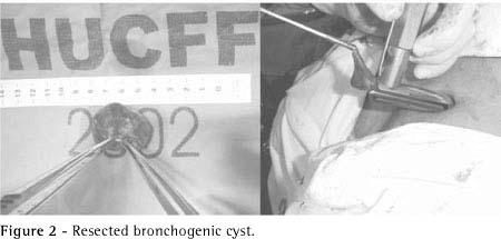
The patient was then submitted to a cervical mediastinoscopy, revealing a well-delineated cystic lesion, which released a viscous, mucoid whitish liquid upon puncture. A rhomboid flap was used to perform a complete resection of the lesion (Figure 2), sometimes manually, with constant traction.
The postoperative course was uneventful, and the patient was discharged on postoperative day two. The anatomopathological exam of the lesion confirmed the diagnosis of bronchogenic cyst, with respiratory epithelium in the cyst wall. The histopathological examination of the cystic lesion revealed it was internally lined by pseudostratified ciliated epithelium (of the respiratory type), supported by loose conjunctive stroma, permeated by bundles of smooth muscular cells and deep mucous glands, similar to the normal components of the bronchial walls, except for the absence of cartilage (Figure 3).
DISCUSSION
A BC is a congenital lesion derived from an abnormal budding of the foregut.(2,5) In general, when such an abnormality occurs precociously, mediastinal cysts are formed, usually presenting no communication with the tracheobronchial tree. When formed later, they result in intraparenchymal cysts, frequently in communication with the airways, and are more symptomatic and more frequently complicated by infection.(1,5,9-10)
A BC is composed of respiratory epithelium and is filled with whitish mucinous, or even hematic, material, almost always unilocular.(1-2) The presence of cartilage distinguishes the BCs from enteric cysts.(10) Depending on their location, BCs are divided into five groups: paratracheal, carinal, hilar, paraesophageal and miscellaneous, the last comprising the intrapulmonary and infradiaphragmatic forms.(9) In the three largest studies in the literature, the paratracheal location was the most commonly found.(1,3,10)
In the literature, the BC clinical manifestations reported include chest pain, cough, dyspnea, fever, hemoptysis and dysphagia.(1,3,5) Complications include infection, compression of adjacent structures (as in superior vena cava syndrome), fistulization and even malignant degeneration.(3, 11)
The diagnosis often results from the incidental finding of a mediastinal mass on a routine X-ray.(2) A computed tomography scan of the chest usually reveals a cystic unilocular lesion.(5) Other than allowing the identification of mucous secretion, fine needle aspiration has little diagnostic value.(3)
The treatment proposed for mediastinal BC is total resection of the lesion. This is classically achieved through thoracotomy. However, with the popularization of video-assisted thoracoscopy in recent years, video-assisted thoracic resection has been increasingly employed.(1-3,5,8,11)
Resection by mediastinoscopy was described by Ginsberg in 1972, but did not become popular.(6) It is believed that this was due to concern that the resection of the lesion would be incomplete, thereby increasing the possibility of relapse, and that it would be technically impossible to safely resect subcarinal lesions. However, the efficacy and safety of the method has been proven, and it has been shown that it is possible to perform subtotal resection of the lesion and to treat the lesion with a sclerosing agent, both with very good results.(4,7) Obviously, this approach should be reserved for BCs located in regions easily accessed by conventional cervical mediastinoscopy and, as previously mentioned, a significant number of BCs are located in these regions.(1,3)
It is known that mediastinoscopy is a procedure that offers a minimum risk of complications when performed by experienced individuals.(7,12) The reduced surgical time, shorter hospital stay, minimal postoperative pain and lower incidence of the complications associated with thoracotomy and video-assisted thoracic surgery, have made the mediastinoscopic approach very attractive.
Therefore, in view of the possibility of diagnostic confirmation and adequate treatment of BCs through mediastinoscopy, we believe that this technique should always be considered for the treatment of the cysts located in regions easily accessed through cervical mediastinoscopy.
ACKNOWLEDGMENTS
We wish to thank Prof. Paulo Marcos Valiante for his valuable collaboration in the preparation of the slides and photographs.
REFERENCES
Submitted: 20 July 2004. Accepted, after review: 04 February 2005.
- 1. Ribet ME, Copin MC, Gosselin BH. Bronchogenic cysts of the mediastinum. J Thorac Cardiovasc Surg. 1995;109(5):1003-10.
- 2. Bolton JW, Shahian DM. Asymptomatic bronchogenic cysts: what is the best management? Ann Thorac Surg. 1992;53(6):1134-37.
- 3. Suen HC, Mathisen DJ, Grillo HC, LeBlanc J, McLoud TC, Moncure AC, et al. Surgical management and radiological characteristics of bronchogenic cysts. Ann Thorac Surg. 1993;55(2):476-81.
- 4. Urschel JD, Horan TA. Mediastinoscopic treatment of mediastinal cysts. Ann Thorac Surg. 1994;58(6):1698-700.
- 5. Duranceau ACH, Deslauriers J. Foregut cysts of the mediastinum in adults. In: Shields TW, Locicero J III, Ponn RB, editors. General thoracic surgery. 5th ed. Philadelphia, USA: Lippincott Williams; 2000.
- 6. Ginsberg RJ, Atkins RW, Paulson DL. A bronchogenic cyst successfully treated by mediastinoscopy. Ann Thorac Surg. 1972;13(3):266-8.
- 7. Smythe WR, Bavaria JE, Kaiser LR. Mediastinoscopic subtotal removal of mediastinal cysts. Chest. 1998;114(2):614-7.
- 8. Brichon PY, Brambilla E, Frassinetti E, Brambilla C, Latreille R. Excision by video-thoracoscopy of a mediastinal bronchogenic cyst. A case report. Rev Mal Resp. 1992;9:44-5. French.
- 9. Maier HC. Bronchogenic cysts of the mediastinum. Ann Thorac Surg. 1948;127: 476-502.
- 10. Takeda SI, Miyoshi S, Minami M, Ohta M, Masaoka A, Matsuda H. Clinical spectrum of mediastinal cysts. Chest. 2003;124(1):125-32.
- 11. Rammohan G, Berger HW, Lajam F, Buhain WJ. Superior vena cava syndrome by bronchogenic cyst. Chest. 1975;68(4):599-601.
- 12. Puhakka H. Complications of mediastinoscopy. J Laryngol Otol. 1989;103(3):312-5.
Publication Dates
-
Publication in this collection
31 Oct 2005 -
Date of issue
Aug 2005
History
-
Received
20 July 2004 -
Accepted
04 Feb 2005