Abstracts
OBJECTIVE: To present the computed tomography aspects of Rhodococcus equi pneumonia in seven patients with acquired immunodeficiency syndrome. METHODS: A retrospective study of the computed tomography scans of seven patients with acquired immunodeficiency syndrome and Rhodococcus equi infection. RESULTS: The most common findings were consolidation (n = 7), consolidation with cavitation (n = 6), ground glass opacities (n = 6), peribronchial nodules (n = 4) and centrilobular nodules presenting a "tree-in-bud" pattern (n = 3). CONCLUSION: The most common finding in patients with Rhodococcus equi pulmonary infection and acquired immunodeficiency syndrome was consolidation with cavitation.
Acquired immunodeficiency syndrome; Rhodococcus equi; Actimomycetales infections; Lung diseases, fungal; Tomography, emission-computed
OBJETIVO: Apresentar os aspectos na tomografia computadorizada da pneumonia pelo Rhodococcus equi em sete pacientes com síndrome da imunodeficiência adquirida. MÉTODOS: Estudo retrospectivo das tomografias de sete pacientes com síndrome da imunodeficiência adquirida e infecção pelo Rhodococcus equi. RESULTADOS: Os achados mais freqüentes foram: consolidação (n = 7) com escavação (n = 6), opacidades em vidro fosco (n = 6), nódulos do espaço aéreo (n = 4) e nódulos centrolobulares com árvore em brotamento (n = 3). CONCLUSÃO: Os achados mais comuns na infecção pulmonar pelo Rhodococcus equi em pacientes com síndrome da imunodeficiência adquirida foram as consolidações escavadas.
Síndrome da imunodeficiência adquirida; Rhodococcus equi; Infecções por actinomycetales; Pneumopatias fúngicas; Tomografia computadorizada de emissão
ORIGINAL ARTICLE
Rhodococcus equi infection in acquired immunodeficiency syndrome. Computed tomography aspects* * Study carried out in the Radiological Diagnosis Department at the Hospital Universitário Clementino Fraga Filho (HUCFF, Clementino Fraga Filho University Hospital), of the Universidade Federal do Rio de Janeiro (UFRJ, Federal University of Rio de Janeiro), Rio de Janeiro, Brazil.
Edson MarchioriI; Renato Gonçalves de MendonçaII; Domenico CaponeIII; Elza Maria de CerqueiraIV; Arthur Soares Souza JúniorV; Gláucia ZanettiVI; Dante EscuissatoVII; Emerson GasparettoVIII
IFull Professor and Head of the Department of Radiology at the Universidade Federal Fluminense (UFF, Federal Fluminense University). Adjunct Coordinator of the Postgraduate Department of Radiology at the Universidade Federal do Rio de Janeiro (UFRJ, Federal University of Rio de Janeiro), Rio de Janeiro, Brazil
IIAttending Radiologist at the Copa D'Or and Barra D'Or Hospitals, Rio de Janeiro, Brazil
IIIProfessor of Pulmunology at the Universidade do Estado do Rio de Janeiro(UERJ, Rio de Janeiro State University). Attending Radiologist at the Clementino Fraga Filho University Hospital of the Universidade Federal do Rio de Janeiro (UFRJ, Federal University of Rio de Janeiro), Rio de Janeiro, Brazil
IVSpecialist in the Department of Radiology at the Universidade Estadual de Campinas(UNICAMP, State University of Campinas), Campinas, Brazil
VAdjunct Professor of Radiology at the Faculdade de Medicina de São José do Rio Preto (FAMERP, Sao Jose do Rio Preto School of Medicine), Sao Jose do Rio Preto, Brazil
VIDoctoral student in Radiology at the Universidade Federal do Rio de Janeiro (UFRJ, Federal University of Rio de Janeiro). Professor of Pulmunology at the Petropolis School of Medicine, Rio de Janeiro, Brazil
VIIAssistant Professor of Radiology at the Universidade Federal do Paraná(UFPR, Federal University of Parana), Londrina, Brazil
VIIIMedical Researcher at the Institute of Radiology of the Universidade de São Paulo(USP, University of Sao Paulo), Sao Paulo, Brazil
Correspondence to Correspondence to: Edson Marchiori Rua Thomaz Cameron, 438, Valparaíso CEP: 25685-120, Petrópolis, RJ, Brasil Tel: 55 24 2249-2777 Email: edmarchiori@bl.com.br
ABSTRACT
OBJECTIVE: To present the computed tomography aspects of Rhodococcus equi pneumonia in seven patients with acquired immunodeficiency syndrome.
METHODS: A retrospective study of the computed tomography scans of seven patients with acquired immunodeficiency syndrome and Rhodococcus equi infection.
RESULTS: The most common findings were consolidation (n = 7), consolidation with cavitation (n = 6), ground glass opacities (n = 6), peribronchial nodules (n = 4) and centrilobular nodules presenting a "tree-in-bud" pattern (n = 3).
CONCLUSION: The most common finding in patients with Rhodococcus equi pulmonary infection and acquired immunodeficiency syndrome was consolidation with cavitation.
Keywords: Acquired immunodeficiency syndrome; Rhodococcus equi; Actimomycetales infections; Lung diseases, fungal; Tomography, emission-computed
INTRODUCTION
Rhodococcus equi is a common cause of infection in horses and other animals. The infection is rare in humans,(1) primarily affecting individuals with a high degree of immunodeficiency. Approximately 80% of cases occur among patients with acquired immunodeficiency syndrome, notably those who present CD4 lymphocyte counts lower than 200 cells/mm3.(2-3)
Among humans, the principal site of infection is the lung.(1) The most frequent form of clinical presentation is that of a slowly evolving profile of pneumonia, with cough, fever and constitutional symptoms. R. equi is a frequent cause of bacterial infection, and extrapulmonary symptoms can occur. The etiologic agent is easily isolated from the sites of infection.(2) The most common radiological presentation in cases of pulmonary infection is consolidation with cavitation, with or without air-fluid levels, most commonly affecting the upper lobes.(2,4-5)
Despite the great number of studies in the literature that focus on the clinical aspects, treatment and conventional chest X-ray findings, there is little data concerning the aspects of infection caused by R. equi in computed tomography of the chest which, like X-rays, reveal consolidation with cavitation and air-fluid levels. Computed tomography is capable of better characterizing the findings and of identifying alterations that are not visible on X-rays: centrilobular nodules, ground glass opacities, areas of bronchiolar obstruction, minor pleural effusion and mediastinal lymph node enlargement.(5-6)
The objective of this study is to present the computed tomography aspects observed in seven patients with acquired immunodeficiency syndrome and infected with R. equi.
METHODS
A retrospective study was carried out, in which the computed tomography scans of seven patients with pulmonary infection caused by R. equi were evaluated. The scans, performed between 1996 and 2004, were obtained through a search of the radiological archives of three university hospitals in Rio de Janeiro (the Clementino Fraga Filho University Hospital, of the Federal University of Rio de Janeiro; the Pedro Ernesto University Hospital, of the Rio de Janeiro State University; and the Antonio Pedro University Hospital, of the Fluminense Federal University), as well as that of the State University of Campinas University Hospital in Campinas.
Six patients were males, and one was female. The mean age was 38 years (range, 22-49 years). All the patients had acquired immunodeficiency syndrome and presented CD4 lymphocyte counts lower than 100 cells/mm3. The diagnosis of R. equi infection was made through the culture of material obtained from bronchoalveolar lavage or transthoracic puncture. Two patients also presented positive blood culture. All of the patients presented only pulmonary manifestations of the infection, with a slowly evolving profile of pneumonia for more than one month.
Six of the seven scans were performed using Siemens Somatom AR computed tomography scanners (Siemens Medical Solutions, Munich, Germany). The remaining scan was performed using a GE 9800 S computed tomography scanner (General Electric Medical Systems, Milwaukee, WI, USA). Computed tomography scans of the chest were carried out using a conventional acquisition technique, with slice thicknesses varying from 5 to 10 mm. Five patients were also submitted to a high-resolution acquisition technique, with slice thicknesses varying between 1 and 2 mm. The exams were documented using the appropriate lung windows ranging from 1000 to 1300 HU, with the center varying from -500 to -700 HU. The mediastinum windows varied between 200 and 600 HU, with the center ranging from 60 to 120 HU. No intravenous contrast medium was administered to any of the patients.
The computed tomography scans were independently assessed by two observers. The final decisions were made by consensus.
RESULTS
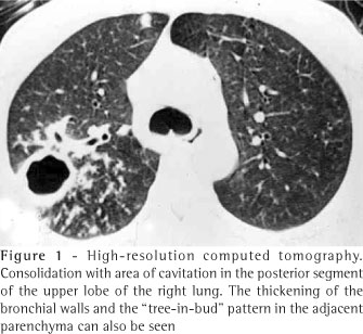
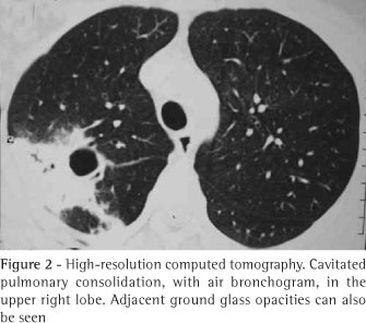
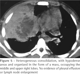
The most frequent finding in the imaging, present in the seven patients, was ill-defined consolidation (Figures 1, 2, 3 and 4). Five of patients presented air bronchogram, principally at the periphery of the lesion. In three cases, the consolidation was organized in the form of a mass. In six of the seven patients, irregular areas of cavitation were observed within the consolidations. Multiple cavitations were seen in five patients, and a singular cavitation was observed in one patient. In the patient presenting no cavitation, there were hypodense areas suggestive of necrosis (Figure 5). Only one of the patients presented air-fluid levels within the cavities.
Cavitated lesions predominated in the upper lobes, four in the right lobe and one in the left lobe. In one patient, the middle lobe was also affected. In two cases, such lesions occurred in the lower lobes, one in the right lobe and one in the left lobe. In four cases, the lesions affected only the lobes mentioned above and were therefore unilateral. In the remaining three cases, the contralateral lung was also affected, albeit to a lesser degree. In one case, there was also relatively thin-walled, multiloculated cavitation, without an air-fluid level, in the middle lobe.
Ground glass opacities, primarily surrounding the cavitated lesions, were reported in six patients, although this was not the predominant finding in any of the cases.
Small centrilobular nodular opacities presenting a 'tree-in-bud' pattern, predominantly encircling the area of consolidation, were observed in three patients. In one of those patients, the lesions were also found in the hanging portions of the lower lobes. Peribronchial nodules, tending toward confluence and located in the periphery of the pulmonary consolidations, were observed in four cases.
None of the other findings described in the literature (pulmonary coin lesion, pleural effusion, pneumothorax, lymph node enlargement and soft tissue involvement) were observed in any of the patients.
DISCUSSION
In patients with acquired immunodeficiency syndrome, the differential diagnosis of R. equi pulmonary infection includes cavitated infections (tuberculosis, nocardiosis, fungal diseases and pulmonary abscess), pulmonary neoplasms and, more rarely, Pneumocystis carinii pneumonia.(1,2,7) Infection caused by Mycobacterium tuberculosis was the principal differential diagnosis proposed for the patients evaluated in the present study.
In a study(5) using X-rays and computed tomography scans of the chest, nine patients with acquired immunodeficiency syndrome were evaluated. The computed tomography scans of the chest, in which an intravenous contrast medium was used, revealed masses with heterogeneous impregnation, together with dense pulmonary consolidations (with or without cavitations), in eight patients. The tomography confirmed the cavitations and air-fluid levels identified in the X-rays and belied the results of three other exams in which such findings had not been previously suspected. Mediastinal lymph node enlargements were present in eight patients. In one case, small centrilobular nodules and areas of bronchiolar obstruction, indicating bronchogenic dissemination, were observed. In two other cases, multiple nodules, approximately 5 to 20 mm in size, most of which were cavitated, were observed.
In our study, the principal pattern of pulmonary involvement was air bronchogram and cavitation, which was present in six patients. There were multiple cavitations in five cases and a singular cavitation in one. The consolidations were large and had irregular contours. The air bronchograms were located in the peripheral portions of the lesions. The pulmonary lobe most frequently involved was the upper right lobe, which was affected in four patients. Most of the case review studies confirm these findings.(2,5) Some studies have reported that, although cavitation might not be present at the time of diagnosis, it develops over the course of the disease.(4) In the sample evaluated in the present study, air-fluid levels within the cavitations were seen in only one patient.
Other findings were ground glass opacities, peribronchial nodules, small nodules of predominantly centrilobular distribution, and the "tree-in-bud" pattern. These alterations were predominantly seen surrounding the cavitated consolidations. In one case, they were also present in both of the lower lobes. It is believed that this finding indicates bronchogenic dissemination of the infection.
The typical histopathological finding in infection caused by R. equi is a necrotic cavitation, composed of a dense histiocytic infiltrate with abundant granular eosinophilic cytoplasm (von Hansemann cells) and affecting consolidations or soft tissue masses. Polymorphonuclear leukocytes are abundant in disseminated micro-abscesses. The periodic acid-Schiff staining procedure reveals highly positive histiocytes, similar to those encountered in Whipple's disease. Gram-positive coccobacilli are easily seen in the tissue staining performed in the Gram stain method. Another finding reported in infection caused by R. equi is pulmonary malacoplakia.(8)
In general, computed tomography scans are is more efficient than chest X-rays in characterizing pulmonary lesions, since air bronchograms and cavitated areas in the consolidations, as well as air-fluid levels, are more easily seen in the former. Computed tomography scans are also capable of detecting small centrilobular nodular opacities, peribronchial nodules, areas of bronchiolar obstruction and ground glass opacities, notably in the high-resolution slices. Lymph node enlargements, small pleural effusions and pneumothorax, as well as soft tissue involvement, could be safely ruled out through the use of computed tomography scans.
In conclusion, infection caused by R. equi should be considered in the differential diagnosis of cavitated consolidations among patients with acquired immunodeficiency syndrome, since it can be easily mistaken for lesions caused by tuberculosis.
REFERENCES
Submitted: 25 September 2005. Accepted, after review: 6 December 2005
- 1. Alves JA, Cerqueira EMFP, Nanni L, Toniani MP. Rhodococcus equi: infecção pulmonar em patientes imunocomprometidos - relato de um caso e revisão da literatura. Radiol Bras. 1997;30(6):347-9.
- 2. Capdevila JA, Buján S, Gavalda J, Ferrer A, Pahissa A. Rhodococcus equi pneumonia in pacients infected with human immudeficiency virus. Report of 2 cases and review of the literature. Scand J Infect Dis. 1997;29(6):535-41.
- 3. Haramati LB, Jenny-Avital ER. Approach to the diagnosis of pulmonary disease in patients infected with the human immunodeficiency virus. J Thorac Imaging. 1998;13(4): 247-60.
- 4. Arlotti M, Zoboli G, Moscatelli GL, Magnani G, Maserati R, Borghi V, et al. Rhodococcus equi infection in HIV-positive subjects: A retrospective analysis of 24 cases. Scand J Infect Dis. 1996;28(5):463-7.
- 5. Wicky S, Cartei F, Mayor B, Frija J, Genevois PA, Giron J, et al. Radiological findings in nine AIDS patients with Rhodococcus equi pneumonia. Eur Radiol. 1996;6(6): 826-30.
- 6. Marchiori E, Muller NL, de Mendonca RG, Capone D, Souza AS Jr, et al. Rhodococcus equi pneumonia in AIDS: high-resolution CT findings in five patients. Br J Radiol. 2005;78(933):783-6.
- 7. Marchiori E, Muller NL, Soares Souza A Jr, Escuissato DL, Gasparetto EL, Franquet T. Pulmonary disease in patients with AIDS: high-resolution CT and pathologic findings. AJR Am J Roentgenol. 2005;184(3):757-64.
- 8. Scott MA, Graham BS, Verral R, Dixon R, Schaffner, Tham KT. Rhodococcus equi - An increasingly recognized opportunistic pathogen. Report of 12 cases and review of 65 cases in the literature. Am J Clin Pathol. 1995;103(5):649-55.
Publication Dates
-
Publication in this collection
15 Mar 2007 -
Date of issue
Oct 2006
History
-
Accepted
06 Dec 2005 -
Received
25 Sept 2005