Abstracts
Community-acquired pneumonia continues to be the acute infectious disease that has the greatest medical and social impact regarding morbidity and treatment costs. Children and the elderly are more susceptible to severe complications, thereby justifying the fact that the prevention measures adopted have focused on these age brackets. Despite the advances in the knowledge of etiology and physiopathology, as well as the improvement in preliminary clinical and therapeutic methods, various questions merit further investigation. This is due to the clinical, social, demographical and structural diversity, which cannot be fully predicted. Consequently, guidelines are published in order to compile the most recent knowledge in a systematic way and to promote the rational use of that knowledge in medical practice. Therefore, guidelines are not a rigid set of rules that must be followed, but first and foremost a tool to be used in a critical way, bearing in mind the variability of biological and human responses within their individual and social contexts. This document represents the conclusion of a detailed discussion among the members of the Scientific Board and Respiratory Infection Committee of the Brazilian Thoracic Association. The objective of the work group was to present relevant topics in order to update the previous guidelines. We attempted to avoid the repetition of consensual concepts. The principal objective of creating this document was to present a compilation of the recent advances published in the literature and, consequently, to contribute to improving the quality of the medical care provided to immunocompetent adult patients with community-acquired pneumonia.
Pneumonia; Diagnosis; Epidemiology; Practice guideline; Primary prevention
A pneumonia adquirida na comunidade mantém-se como a doença infecciosa aguda de maior impacto médico-social quanto à morbidade e a custos relacionados ao tratamento. Os grupos etários mais suscetíveis de complicações graves situam-se entre os extremos de idade, fato que tem justificado a adoção de medidas de prevenção dirigidas a esses estratos populacionais. Apesar do avanço no conhecimento no campo da etiologia e da fisiopatologia, assim como no aperfeiçoamento dos métodos propedêuticos e terapêuticos, inúmeros pontos merecem ainda investigação adicional. Isto se deve à diversidade clínica, social, demográfica e estrutural, que são tópicos que não podem ser previstos em sua totalidade. Dessa forma, a publicação de diretrizes visa agrupar de maneira sistematizada o conhecimento atualizado e propor sua aplicação racional na prática médica. Não se trata, portanto, de uma regra rígida a ser seguida, mas, antes, de uma ferramenta para ser utilizada de forma crítica, tendo em vista a variabilidade da resposta biológica e do ser humano, no seu contexto individual e social. Esta diretriz constitui o resultado de uma discussão ampla entre os membros do Conselho Científico e da Comissão de Infecções Respiratórias da Sociedade Brasileira de Pneumologia e Tisiologia. O grupo de trabalho propôs-se a apresentar tópicos considerados relevantes, visando a uma atualização da diretriz anterior. Evitou-se, tanto quanto possível, uma repetição dos conceitos considerados consensuais. O objetivo principal do documento é a apresentação organizada dos avanços proporcionados pela literatura recente e, desta forma, contribuir para a melhora da assistência ao paciente adulto imunocompetente portador de pneumonia adquirida na comunidade.
Pneumonia; Diagnóstico; Epidemiologia; Guia de prática clínica; Prevenção primária
BTA GUIDELINES
Brazilian guidelines for community-acquired pneumonia in immunocompetent adults - 2009*
Ricardo de Amorim CorrêaI; Fernando Luiz Cavalcanti LundgrenII,V; Jorge Luiz Pereira-SilvaIII,V; Rodney Luiz Frare e SilvaIV; Alexandre Pinto CardosoV; Antônio Carlos Moreira LemosV; Flávia RossiV; Gustavo MichelV; Liany RibeiroV; Manuela Araújo de Nóbrega CavalcantiV; Mara Rúbia Fernandes de FigueiredoV; Marcelo Alcântara HolandaV; Maria Inês Bueno de André ValeryV; Miguel Abidon AidêV; Moema Nudilemon ChatkinV; Octávio MessederV; Paulo José Zimermann TeixeiraV; Ricardo Luiz de Melo MartinsV; Rosali Teixeira da RochaV
IAdjunct Professor. Universidade Federal de Minas Gerais - UFMG, Federal University of Minas Gerais - School of Medicine, Belo Horizonte, Brazil
IIPhysician. Otávio de Freitas General Hospital, Recife, Brazil
IIIHead of the Pulmonology Department. Jorge Valente Hospital, Salvador, Brazil
IVAdjunct Professor. Federal University of Curitiba Hospital de Clínicas, Curitiba, Brazil
VGrupo de Trabalho da Diretriz em nome da Comissão de Infecções Respiratórias e Micoses - Sociedade Brasileira de Pneumologia e Tisiologia
Correspondence to
ABSTRACT
Community-acquired pneumonia continues to be the acute infectious disease that has the greatest medical and social impact regarding morbidity and treatment costs. Children and the elderly are more susceptible to severe complications, thereby justifying the fact that the prevention measures adopted have focused on these age brackets. Despite the advances in the knowledge of etiology and physiopathology, as well as the improvement in preliminary clinical and therapeutic methods, various questions merit further investigation. This is due to the clinical, social, demographical and structural diversity, which cannot be fully predicted. Consequently, guidelines are published in order to compile the most recent knowledge in a systematic way and to promote the rational use of that knowledge in medical practice. Therefore, guidelines are not a rigid set of rules that must be followed, but first and foremost a tool to be used in a critical way, bearing in mind the variability of biological and human responses within their individual and social contexts. This document represents the conclusion of a detailed discussion among the members of the Scientific Board and Respiratory Infection Committee of the Brazilian Thoracic Association. The objective of the work group was to present relevant topics in order to update the previous guidelines. We attempted to avoid the repetition of consensual concepts. The principal objective of creating this document was to present a compilation of the recent advances published in the literature and, consequently, to contribute to improving the quality of the medical care provided to immunocompetent adult patients with community-acquired pneumonia.
Keywords: Pneumonia; Diagnosis; Epidemiology; Practice guideline; Primary prevention.
Methodology of the guidelines
This is a review and update of the previous guidelines published in 2004 by the Brazilian Thoracic Association. The present document presents certain topics that had not been discussed previously, as well as recently published data, and focuses exclusively on community-acquired pneumonia (CAP) in immunocompetent patients.
At the end of each section of this update are the principal recommendations and respective levels of evidence, according to the current guidelines of the Brazilian Medical Association.
The participants of this edition of 2008 were divided into four work groups. In each group, an editor was responsible for distributing the topics among the members of the group.
Group I: Definition, incidence, mortality, etiology, diagnostic criteria and radiological diagnosis
Group II: Diagnostic and complementary tests, etiologic investigation, severity and locale of treatment
Group III: Treatment, treatment failure and prevention
Group IV: Severe CAP: adjuvant treatment
Levels of evidence
The present guidelines were based on up-to-date evidence in the literature, classified according to the recommendations of the Brazilian Medical Association (Chart 1). The final work of each group was extensively discussed among the editors and participants of the work groups.
Definition and clinical manifestations
Pneumonia is an acute infectious disease that is inflammatory, affects the air spaces and is caused by viruses, bacteria or fungi. The term CAP refers to pneumonia acquired outside the hospital environment or special health care units, or to pneumonia that manifests within the first 48 h after admission to a health care facility.(1) However, there is a special group of pneumonia patients (those who were hospitalized for 48 h or more in the 90 days preceding the disease; those residing in nursing homes or other health care facilities; those who received intravenous antibiotics, chemotherapy or scar treatment in the 30 days preceding the disease; and those undergoing treatment in dialysis clinics) that are more appropriately classified as having hospital-acquired pneumonia.(2,3)
The diagnosis is based on the following: symptoms of acute lower respiratory tract infection (cough and one or more of the following symptoms: expectoration, shortness of breath and chest pain); focal findings on physical examination of the chest; and systemic manifestations (confusion, headache, sweating, chills, myalgia and temperatures higher than 37.8ºC). These indications can be corroborated by a chest X-ray finding of new pulmonary opacity. Other clinical conditions can manifest in a similar manner, which can pose difficulties for primary and emergency care physicians in making a diagnosis of CAP. The clinical findings are only moderately accurate and do not allow a diagnosis of CAP to be confirmed or excluded with any degree of certainty. In addition, the heterogeneity of the physical examination conducted by primary and emergency care physicians, as well as the relative lack of experience of such health professionals in comparison with those who specialize in detecting radiological alterations, contributes to the difficulty in diagnosing CAP.(4,5)
Incidence and mortality
Most of the studies of CAP conducted in Brazil have focused on the etiology and treatment of the disease, and official statistics are a valuable source of information regarding the occurrence of CAP.
According to the Hospital Information Service of the Unified Health Care System, pneumonia was the leading non-obstetric cause of disease-related hospitalization in Brazil in 2007, accounting for 733,209 hospitalizations. Among such hospitalizations, there was a predominance of males, as well as greater occurrence between the months of March and July.(6)
The rate of hospitalization for pneumonia has decreased over the last decade,(7) whereas the rate of in-hospital pneumonia-related mortality has increased, which might be due to various factors, such as hospitalization of patients with pneumonia that was more severe and the aging of the population. The highest rates of hospitalization for pneumonia are observed among individuals less than 5 years of age and among those over 80 years of age, showing opposite temporal trends: a downward trend among the former and an upward trend among the latter.
Respiratory diseases constitute the fifth leading cause of death in Brazil. Pneumonia is the second most common respiratory disease, causing 35,903 deaths in 2005, 8.4% of which occurred among patients less than 5 years of age and 61% of which occurred among patients over 70 years of age. The pneumonia-related mortality rate increased in the period between 2001 and 2004. In 2005, however, it fell to levels below 20/100,000 population, according to the latest mortality statistics provided by the National Ministry of Health.
The pneumonia-related mortality rate differs among age groups. Over the last 5 years, the rate has increased significantly among patients over 70 years of age, reaching levels above 500/100,000 population among those over 80 years of age. The lowest rates are found in the 5-49 year age bracket (less than 10/100,000 population); among those less than 5 years of age, the mortality rate remains at approximately 17/100,000 population, showing a slight downward trend (Figure 1). These data are similar to those obtained in other Latin American countries such as Chile.(8)
Relevant points
Hospitalizations for pneumonia showed a predominance of males and greater occurrence between the months of March and July (level II evidence).
The rate of hospitalizations for pneumonia has been decreasing since the last decade (level II evidence).
The rate of in-hospital mortality has increased, which might be due to various factors, such as hospitalization of patients with pneumonia that was more severe and the aging of the population (level IV evidence).
The pneumonia-related mortality rate differs among age groups and, as in other Latin American countries, has increased over the last decade among patients over 70 years of age (level II evidence).
Diagnostic and complementary tests
Radiological diagnosis
The present guidelines reiterate the previous recommendation that anteroposterior and lateral chest X-rays be taken, because, in addition to being central to the diagnosis of CAP, this aids in assessing severity, identifies multilobar involvement and might suggest alternative etiologies, such as abscess and TB. Chest X-rays can reveal concomitant conditions, e.g., bronchial obstruction or pleural effusion, as well as being useful in monitoring treatment response. However, the classification of CAP according to radiological patterns (lobar pneumonia, bronchopneumonia and interstitial pneumonia) is of limited use in predicting the causal agent: it cannot distinguish between the groups of agents (bacterial and nonbacterial).(9-15) Specific agents can cause different manifestations that can change or become more intense throughout the course of the disease, being frequently influenced by the immunological status.(13)
Chest X-ray is the imaging method of choice for the initial approach to CAP because its cost-effectiveness is excellent, the doses of radiation employed are low and the method is widely available.
Half of the cases diagnosed as CAP in Brazil in reality are not.(16) The greatest diagnostic difficulty lies in the interpretation of the chest X-rays by non-specialists.
Cavitation is suggestive of an anaerobic etiologic agent, Staphylococcus aureus and, occasionally, gram-negative bacilli. In such cases, screening for TB should always be performed. Swollen lobes causing bulging fissures is a nonspecific finding that reflects intense inflammatory reaction.(10)
A CT scan of the chest is useful in a number of situations: when a chest X-ray alone does not clearly show whether or not there is infiltrate; when the clinical profile is unfavorable but chest X-ray is normal; when the objective is to detect complications such as loculated pleural effusion and encapsulated abscess in the airways; and when it is necessary to differentiate inflammatory infiltrate from lung masses.(17,18)
In cases of pleural effusion greater than 5 cm in the posterior mediastinum revealed by lateral chest X-ray in the orthostatic position, or in cases of loculated pleural effusion, thoracentesis should be performed in order to exclude the diagnoses of empyema and complicated parapneumonic effusion. Thoracentesis is strongly recommended in cases of pleural effusion that occupy more than 20% of the hemithorax.(19) Ultrasound is useful in cases of small pleural effusion or in suspected cases of loculated pleural effusion because it can determine the exact location of the effusion for subsequent drainage.(9,20)
Radiological progression after hospital admission might occur regardless of the etiology, and the therapeutic regimen should not be changed unless the clinical profile has shown no improvement.(10) Radiological resolution is relatively slow and occurs after clinical recovery. Complete resolution of radiological alterations occurs two weeks after the initial presentation in half of all cases and six weeks after the initial presentation in two thirds.(11) Advanced age, COPD, immunosuppression, alcoholism, diabetes and multilobar pneumonia are independently associated with slower resolution. Pneumonia caused by Mycoplasma sp. resolves more rapidly. Pneumonia caused by Legionella sp. resolves in a particularly slow manner. Residual lesions are found in 25% of the cases of Legionella sp. and bacteremic pneumococcal pneumonia.(10) Chest X-ray should be repeated six weeks after the onset of symptoms in smokers over 50 years of age (risk of bronchial carcinoma) and in cases in which the symptoms persist or physical examination reveals abnormal findings.(11,21)
Recommendations
Anteroposterior and lateral chest X-rays should be taken for the initial approach to patients suspected of having CAP (level III evidence).
Chest X-ray is the only complementary test to which low-risk CAP outpatients should be submitted (level I evidence).
The radiological patterns cannot be used to predict the causal agent or to distinguish between the groups of agents (level III evidence).
A CT scan of the chest should be performed when there is doubt regarding the presence of inflammatory infiltrate; in order to detect complications; and in suspected cases of neoplasia (level III evidence).
Significant pleural effusion (> 5 cm, seen on lateral chest X-ray in the orthostatic position from the posterior ridge) should be drained.
Ultrasound is useful in cases of small pleural effusion or in suspected cases of loculated pleural effusion (level III evidence).
Chest X-ray should be repeated six weeks after the onset of symptoms in smokers over 50 years of age and in cases in which the symptoms persist or physical examination reveals abnormal findings.
Persistence of radiological findings after six weeks requires additional investigation (level IV evidence).
Peripheral oxygen saturation and arterial blood gas analysis
Peripheral oxygen saturation (SpO2) should be monitored routinely before recommending oxygen therapy. Arterial blood gas analysis should be conducted in cases of SpO2< 90% on room air or in cases of severe pneumonia. Hypoxemia is an indication for supplemental oxygen and hospital admission.(22-24)
Recommendations
The SpO2 should be monitored routinely before the initiation of oxygen therapy (level I evidence).
Arterial blood gas analysis should be conducted in cases of SpO2< 90% on room air or in cases of severe pneumonia (level I evidence).
Hypoxemia is an indication for supplemental oxygen and hospital admission (level I evidence).
Complementary tests
Urea levels higher than 65 mg/dL (corresponding to a value > 11 mmol/L) constitute a strong marker of disease severity.(25-27) The blood workup shows low sensitivity and specificity, being useful as a criterion for severity and therapeutic response. Leukopenia (< 4,000 leukocytes/mm3) denotes poor prognosis.(28,29) Glycemia, electrolyte and transaminase levels have no diagnostic value; they can, however, influence the decision of hospitalizing a patient by allowing the identification of concomitant diseases.(30,31)
C-reactive protein
As a marker of inflammatory activity, C-reactive protein has prognostic value in follow-up treatment. High C-reactive protein levels after 3-4 days of treatment and a < 50% reduction in initial C-reactive protein levels suggest worse prognosis or complications. The impact of C-reactive protein levels on the diagnosis requires further investigation and definition of cut-off values before it can be used routinely in clinical practice. There is a lack of consistent data as to whether or not C-reactive protein can aid in the decision of using antibiotics.(32-35)
Procalcitonin
Procalcitonin is a marker of inflammatory activity that can be determined in various manners: using a commercially available monoclonal immunoluminometric assay, considered a less sensitive method; using a polyclonal immunoassay system (Kryptor; BRAHMS Aktiengesellschaft, Hennigsdorf, Germany), considered a more sensitive method but not widely available in practice; and more recently, using the VIDAS system of ELISA (bioMérieux, Marcy l'Étoile, France), which is almost as sensitive as the Kryptor system and more readily available, although the assay kit is expensive. Studies involving patients at different levels of risk of death have demonstrated that procalcitonin levels tend to be higher in bacterial pneumonia patients with pneumonia severity index (PSI) scores of I or II than in nonbacterial pneumonia patients with the same PSI scores.(36-38) No etiology-related differences were found among patients with pneumonia that was more severe; however, higher procalcitonin levels correlated with complications and death.(37) Procalcitonin is a better marker of severity than are C-reactive protein, IL-6 and lactate. Elevated serum procalcitonin levels are also found in other lung diseases such as chemical pneumonitis and smoke inhalation injury in burn patients.(34,36,39,40)
Etiologic investigation
The methods used for determining the etiology of CAP have low immediate yield and are unnecessary in outpatients, which is due to the high efficacy of empirical treatment and the low mortality among such patients (< 1%). The elucidation of the etiology of CAP does not result in lower mortality when compared with early, appropriate empirical antibiotic therapy.(41) When empirical treatment fails in severe CAP patients, etiologic diagnosis and specific treatment correlate with lower mortality. Treatment initiation should not be delayed in order to perform tests to determine the etiology of CAP.(41,42) The most common etiologic agents according to CAP severity and treatment locale are shown in Chart 2.
Sputum examination
Although sputum examination is commonly used for establishing the etiologic diagnosis of CAP, the beneficial role of this examination in the initial management of CAP is still controversial.(43,44) Some of the obstacles to the performance of sputum examination include the need for collecting the samples in an appropriate manner, the lack of standardization of the techniques used for sample preparation, the variable examiner ability to interpret the results and the lack of a gold standard for the microbiological diagnosis of CAP.(45) Valid sputum samples are defined as those that contain < 10 epithelial cells and > 25 polymorphonuclear cells per low-power microscopic field.
Due to the high prevalence of pulmonary TB and mycoses in Brazil, screening for acid-fast bacilli in the sputum using the Ziehl-Neelsen technique, as well as screening for fungi, should be performed in suspected cases of CAP, according to the Tuberculosis Control Guidelines.(46)
Blood culture
Because blood culture commonly shows a low yield, it should be reserved for severe CAP patients and for inpatients who do not respond to the therapeutic approach used. False-positive results are common, especially if antibiotics have previously been used, and rarely result in a change of approach. Sputum samples should be collected before initiation or change of treatment and should not delay the administration of the first dose of antibiotics.(28,29,47,48)
Other techniques for the collection of samples for microbiological examination
Other available techniques for obtaining samples from the lower airways include tracheal aspirate, mini-bronchoalveolar lavage and bronchoscopic protected specimen brush or bronchoalveolar lavage, as well as transthoracic needle aspiration biopsy.
These procedures should not be routinely recommended for most CAP patients. However, such procedures are useful in patients admitted to the intensive care unit (ICU) and in those who do not respond to empirical treatment. Percutaneous puncture lung biopsy is contraindicated in patients on invasive mechanical ventilation.(49-51)
When tracheal intubation and initiation of mechanical ventilation are indicated, material should be collected from the lower airways using tracheal aspirate or bronchoscopic techniques, in order to perform quantitative culture.
Secretion collection through bronchoscopy poses lower risks to patients than does transtracheal aspiration and lung puncture.(52-57)
Serologic tests
Serologic tests should not be routinely requested. Serologic tests allow the establishment of retrospective diagnosis of infection caused by certain microorganisms that are difficult to culture (Mycoplasma, Coxiella, Chlamydophila and Legionella, as well as viruses). Serologic tests are considered positive when the titer obtained in the convalescent phase, i.e., four to six weeks after defervescence, is four times greater than that obtained in the acute phase. Because of this technical characteristic, serologic tests are not useful in treating patients individually; they are, however, useful in establishing the epidemiological profile of a given region or an epidemic outbreak.(1,58)
Urinary antigen tests
Urinary antigen tests are simple, rapid and not influenced by the use of antibiotics. The test for Legionella pneumophila becomes positive from day 1 of disease and remains so for weeks. Its sensitivity ranges from 70 to 90%, and its specificity is approximately 100%. Since the test detects the L. pneumophila serogroup 1 antigen (the most prevalent serogroup), infections caused by other serogroups, although less common, might not be identified.(59-62)
The test for pneumococci has a sensitivity ranging from 50% to 80% (greater than that of sputum examination and blood culture) and a specificity of 90%.(63,64) Previous use of antibiotics does not affect the results. False-positive results can occur in cases of colonization of the oropharynx, especially among children with chronic lung diseases. The test is rapid and effective, with good sensitivity and specificity.(65-68)
PCR
The greatest potential of PCR lies in the identification of L. pneumophila, Mycoplasma pneumoniae and Chlamydophila pneumoniae, as well as of other habitually noncolonizing pathogens.
The use of PCR, for the detection of one causative agent, or multiplex PCR, for the detection of M. pneumoniae, C. pneumoniae and Legionella spp., shows good sensitivity and specificity; however, these tests are not available in most clinical laboratories.(68-71)
The etiologic tests recommended for use in specific situations are shown in Chart 3.
Recommendations
Although glycemia, electrolyte and transaminase levels have no diagnostic value, they can influence the decision to hospitalize a patient by allowing the identification of concomitant diseases (level II evidence).
Because blood culture commonly has a low yield, it should be reserved for use in severe CAP patients and for inpatients who do not respond to the therapeutic approach used (level III evidence).
Serologic tests are not useful in treating patients individually; they are, however, useful in establishing the epidemiological profile of a given region or epidemic outbreak (level III evidence).
Screening for the etiologic agent should be performed in severe CAP patients and inpatients who do not respond to the initial treatment (level III evidence).
In cases of severe CAP, microbiological investigation should be performed through blood culture, sputum culture, culture of tracheal aspirate or culture of samples obtained by bronchoscopy in patients on mechanical ventilation (level II evidence).
Screening for the S. pneumoniae urinary antigen should be performed in severe CAP patients, and screening for the L. pneumophila urinary antigen should be performed specifically in all patients who do not respond to the initial treatment (level II evidence).
Classification of severity and choice of treatment locale
Patients diagnosed with CAP should be evaluated for disease severity. This evaluation will aid in the choice of treatment locale, in the extent of etiologic investigation and in the choice of antibiotic therapy. Socioeconomic factors should also be taken into consideration when making this decision.
Disease severity scores(72) or prognostic models(73) evaluate the 30-day mortality risk and are used to identify low-risk patients, who are therefore candidates for outpatient treatment.
Pneumonia severity index
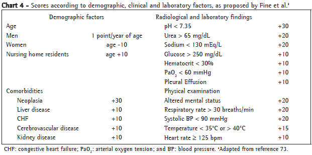
The PSI score comprises 20 variables that include demographic characteristics, concomitant diseases, abnormal laboratory test results, radiological alterations and physical examination findings. The points attributed to each variable allow the severity to be stratified into five classes, based on the risk of death (Charts 4 and 5). However, the primary objective of the original study was to identify low-risk patients.(30,73,74) The PSI score might underestimate CAP severity among young patients without concomitant diseases. In addition, because the PSI is complex and requires extensive laboratory evaluation, it is not considered ideal for routine use in clinical practice.
The British Thoracic Society disease severity score
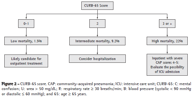
The disease severity score proposed by the British Thoracic Society is based on variables indicative of severe CAP, as follows: mental confusion (defined as an abbreviated mental test score < 8); urea > 50 mg/dL; respiratory rate > 30 breaths/min; systolic blood pressure < 90 mmHg or diastolic blood pressure < 60 mmHg; and age > 65 years. The name of this score (CURB-65) is an acronym based on the key term of each risk factor assessed (confusion, urea, respiratory and blood) and can be presented in its simplified form, CRB-65 (without urea levels). In this score, each variable represents 1 point, and the total possible score comprises 4 points (CRB-65) or 5 points (CURB-65), as shown in Figures 2 and 3.(25,72,75-77)
The greatest limitation of the CURB-65 and CRB-65 is the fact that they do not include concomitant diseases that can increase the risk of CAP, e.g., alcoholism, heart failure, liver failure and neoplasia.
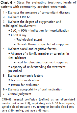
The present guidelines reiterate the recommendation of the previous guidelines regarding the need to evaluate concomitant diseases, the extent of radiological involvement, the degree of oxygenation, psychosocial factors, socioeconomic factors and the viability of oral medication use in terms of their influence on the choice of treatment locale. Due to the simplicity of the CURB-65 and CRB-65 scores, as well as the fact that they are immediately applicable and easy to use, we recommend their use as appropriate criteria for the stratification of CAP severity at the primary and emergency care levels (Chart 6).
If not contraindicated by socioeconomic complications, decompensated concomitant diseases, hypoxemia or inability to take oral medication, hospital admission is indicated for patients with a score of at least two points on the CURB-65 or at least one point on the CRB-65. When such contraindications exist, the attending physician should prescribe outpatient treatment.
Severe community-acquired pneumonia
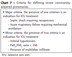
From a practical standpoint, severe CAP is defined as CAP in which the clinical profile is more likely to worsen or the risk of death is higher. Admission to the ICU is mandatory for the appropriate management of severe CAP patients. Septic shock and the need for mechanical ventilation are absolute criteria for ICU admission.(78)
The currently used criteria were defined by Ewig et al. and have a sensitivity of 78%, a specificity of 94%, a negative predictive value of 95% and a positive predictive value of 75% in selecting patients who require ICU admission.(78-80)
The present guidelines corroborate the adoption of such criteria in order to define severe CAP and to recommend ICU admission (Chart 7). If a patient meets two of the minor criteria or one of the major criteria for CAP severity, ICU admission is indicated.(81,82)
Recommendations
The decision of whether to hospitalize a CAP patient is the prerogative of the attending physician, and the currently available evaluation scores are tools that inform that decision (level III evidence).
The use of the CURB-65 or CRB-65 scores is recommended to aid in the choice of treatment locale (level III evidence).
Psychosocial and socioeconomic factors should be taken into consideration when selecting the treatment locale (level III evidence).
CAP patients presenting with septic shock, requiring vasopressors or with acute respiratory failure (requiring mechanical ventilation) should be admitted to the ICU, as should patients who meet at least two of the minor criteria for CAP severity (level III evidence).
The possibility of treatment reevaluation should be guaranteed for CAP patients receiving home treatment (level III evidence).
CAP patients who meet major or minor criteria for severe CAP should receive ICU treatment (level I evidence).
Treatment
Empirical treatment vs. specific treatment
In the vast majority of CAP cases, it is not possible to determine the etiologic agent prior to the therapeutic moment of decision. Empirical antibiotic therapy is habitually targeted at the most prevalent microorganisms. More often than not, more than one pathogen is present and atypical pathogens are included. This requires wider empirical coverage, particularly in cases that are more severe. Specific therapy has the potential to minimize adverse effects, decrease the induction of resistance to antimicrobial agents and reduce costs.(42,81,83,84) Specific therapy can replace empirical treatment of hospitalized patients when the specific pathogen is identified within the first 48-72 h after treatment initiation. In this context, the identification of the agent can narrow the empirical regimen or influence the choice of antimicrobial agent to be administered orally in the subsequent therapeutic approach.(81)
Recommendations
The selection of the initial therapeutic regimen for CAP patients takes into consideration the most prevalent microorganisms (level III evidence).
Specific treatment targeted at a previously identified pathogen or pathogens is to be preferred over empirical treatment. However, specific treatment is typically unfeasible at the therapeutic moment of decision (level III evidence).
The identification of the agent(s) allows the selection of specific therapy targeted at the pathogen(s) and the selection of antimicrobial agent to be used in the subsequent therapeutic approach, potentially reducing treatment cost, adverse effects and resistance induction (level II evidence).
Empirical antibiotic coverage of atypical pathogens
When CAP patients are submitted to specific diagnostic tests, atypical pathogens are frequently identified. Depending on the methodology employed, these agents can appear in isolation or as an integral part of a polymicrobial etiology.(85) In a large study based on the secondary analysis of international registries of hospitalized patients, the incidence of CAP caused by atypical pathogens showed that values were similar in North America, Europe, Latin America and Asia (22%, 28%, 21% and 20%, respectively).(86) However, in that study, the proportion of patients who received empirical antibiotic therapy with coverage of atypical pathogens was 91%, 74%, 53% and 10%, respectively. Comparing those patients with those not receiving such therapy, the authors observed the following: less time to achieve clinical stability (3.2 vs. 3.7 days; p < 0.001); shorter hospital stay (6.1 vs. 7.1 days; p < 0.01); lower overall mortality rate (7.0% vs. 11.1%; p < 0.01); and lower CAP-related mortality (3.8% vs. 6.4%; p < 0.05).
In general, the empirical antibiotic therapy for CAP covers atypical pathogens.(87) Although most guidelines recommend empirical antibiotic coverage of atypical pathogens, there is controversy regarding the level of scientific evidence on which this practice is based. A recently published observational study involving hospitalized patients (n = 201) revealed that previous outpatient treatment with a beta-lactam antibiotic correlated with an increased possibility (approximately three times greater) of an atypical pathogen being present, whereas the possibility of pneumococci being present was reduced to one third.(88) The need for and effectiveness of empirical antibiotic coverage of atypical pathogens in hospitalized patients with mild CAP have been recently reevaluated in three studies (systematic review or meta-analysis), the outcome measures of which were the efficacy of the practice and on the related mortality rates.(83,89,90) These reviews principally compared quinolone monotherapy with beta-lactam monotherapy. In the most recent of the three, which included 5,244 patients from 25 randomized studies, no significant difference in mortality was observed between patients treated with antibiotics that cover atypical pathogens (quinolones) and those treated with a beta-lactam (relative risk = 1.15; 95% CI: 0.85-1.56). No significant difference was observed regarding adverse effects or the need to discontinue treatment. In these reviews, the overall frequency of adverse effects was similar; however, patients treated with beta-lactam antibiotics presented more adverse effects in the gastrointestinal tract than did those treated with quinolones.(90) The two systematic reviews presented similar results.(83,89) In the three reviews previously cited, the use of empirical antibiotic coverage of atypical pathogens showed better results only in the subgroup of patients in which Legionella sp. was subsequently detected. Quinolone coverage did not result in worse disease progression in patients with CAP caused by pneumococci.(83,90) However, the validity of such observational studies is questionable.(87) Further prospective studies of such patients, comparing beta-lactam monotherapy with beta-lactam-macrolide combination therapy and electing the mortality rate as the primary outcome measure, are needed.
Recommendation
Although there is no conclusive evidence that empirical antibiotic coverage of atypical pathogens is superior, this therapeutic approach to hospitalized patients might result in lower mortality rates among patients with pneumonia caused by
Legionella
sp. It might also result in reduced hospital stay, overall mortality and mortality from pneumonia caused by atypical pathogens (level II evidence).
Combination therapy vs. monotherapy
Combination antibiotic therapy for CAP patients aims to broaden the coverage of atypical pathogens and of potentially resistant bacteria, as well as to reduce mortality in cases of bacteremia caused by Streptococcus pneumoniae. Few studies have compared the two most widely recommended antimicrobial regimens for CAP patients requiring hospitalization: combination therapy (a beta-lactam associated with a macrolide or quinolone); and monotherapy with coverage of atypical pathogens (quinolone or macrolide). In a retrospective study involving 515 patients (of which 261 received combined therapy) with severe CAP (cases of severe CAP were defined as those with a PSI score of V), lower 14-day and 30-day mortality rates were found among the patients who received combination antibiotic therapy, a result that was not observed among patients with CAP that was less severe.(91) In another observational study, survival rates were higher among patients with severe CAP and septic shock who received combination therapy than they were among patients who received monotherapy, even when the latter was considered appropriate for the etiologic agent. Combination therapy showed no advantage over monotherapy in patients without septic shock.(92) Combination therapy also seems to be more beneficial than monotherapy in cases of pneumococcal CAP with bacteremia.(93) Possible explanations include a possibly unapparent coinfection caused by atypical pathogens (found in 18-38% of the cases in some studies) or the immunomodulatory effects of macrolides, or a combination of the two.(94)
Recommendations
Combination therapy is not superior to monotherapy among low-risk patients (level II evidence).
Combination therapy should be recommended for patients with severe CAP, particularly for those with accompanying bacteremia, respiratory failure or septic shock (level II evidence).
In comparison with monotherapy, combination therapy with two effective antibiotics reduces mortality among bacteremic pneumococcal pneumonia (level II evidence).
Predictors of specific pathogens
The principal predictive factors of CAP caused by penicillin-resistant pneumococci are age < 4 years (OR = 5.3; 95% CI: 2.2-12.6), immunosuppression (OR = 3.0; 95% CI: 1.5-6.0) and recent use of a beta-lactam (OR = 2.1; 95% CI: 1.0-4.5).(95) The principal independent predictors of CAP caused by gram-negative bacilli include likely aspiration (OR = 2.3; 95% CI: 1.02-5.20; p = 0.04), hospitalization within the past 30 days (OR = 3.5; 95% CI: 1.7-7.1; p < 0.001), antibiotic use for more than 48 h within the past 30 days (OR = 1.9; 95 CI%: 1.01-3.70; p = 0.049) and parenchymal lung diseases (OR = 2.8; 95% CI: 1.5-5.5; p = 0.02).(96) A specific analysis of a subgroup of patients with CAP caused by Pseudomonas aeruginosa showed that a history of lung disease (OR = 5.8; 95% CI: 2.2-15.3; p < 0.001) and recent hospitalization (OR = 3.8; 95% CI: 1.8-8.3; p = 0.02) were independent predictors of CAP caused by P. aeruginosa. In that study, infection caused by gram-negative bacilli was an independent variable for risk of death (OR = 3.4; 95% CI: 1.6-7.4; p = 0.002).(96)
Recommendation
The predictors of risk for specific pathogens should be taken into consideration when selecting the empirical antibiotic therapy for CAP patients (level II evidence).
Potential benefits of early initiation of antibiotic therapy
At least two retrospective studies involving CAP patients aged > 65 years who were hospitalized in the USA demonstrated lower mortality among patients who received earlier antibiotic therapy.(97,98) One of the studies(98) proposed an interval of 8 h between patient admission and the administration of the first dose, whereas the other(97) established a 4-h limit for the administration of the first dose. Another group of authors reported that early antibiotic therapy can reduce the length of hospital stays.(99) Among patients with bacteremic pneumococcal pneumonia, initiation of antibiotic therapy within the first 4 h after hospital admission reduces mortality and length of hospital stay.(100) However, in a prospective observational study involving 409 patients with moderate to severe CAP, no direct correlation was found between early antibiotic therapy and the time required to achieve clinical stability, considering periods of less than 4 h, of 4-6 h and of more than 6 h.(101) In a recent review of the topic, it was suggested that early antibiotic therapy is potentially more beneficial for elderly patients who are antimicrobial treatment-naïve.(102) However, the pressure to administer the first dose in less than 4 h rather than in 8 h might lead to errors in the diagnosis of CAP.(103) Therefore, the ideal time frame between admission and the first dose of antibiotics remains controversial. It is recommended, however, that the first dose be administered to hospitalized patients "as early as possible", preferably at admission, particularly in the emergency room.(81)
Recommendation
Antibiotic therapy for CAP patients should be initiated as early as possible, since it can reduce mortality rates, shorten hospital stays and control costs (level III evidence).
Antibiotic resistance in S. pneumoniae: major changes in the Clinical Laboratory Standards Institute criteria
The selection of an antimicrobial agent to treat infections caused by S. pneumoniae is based on various factors, such as the site of infection, resistance to penicillin (and other agents), degree of severity, pharmacokinetics/pharmacodynamics of the drug and patient age. In community-acquired infections, empirical treatment can be guided by local epidemiological surveillance studies. In severe infections, it is important that culture and antibiogram be performed when treatment adjustment is needed. The interpretation of the minimum inhibitory concentration (MIC) of penicillin for S. pneumoniae should always consider the clinical sample in which the agent was isolated, as well as clinical suspicion of the type of infection. The disk method routinely used in laboratories is not definitive in detecting penicillin resistance, and the results obtained should always be confirmed by determining the MIC (mg/L).
The definition of penicillin resistance based on the MIC for S. pneumoniae strains was changed in 2008 by the Clinical Laboratory Standards Institute (CLSI) as a result of evidence of the pharmacokinetics and pharmacodynamics of penicillin and its derivatives (amoxicillin and ampicillin) in respiratory tract infections.(104) The original definition of penicillin resistance (MIC > 2 mg/L) was based on populations of patients with meningitis. This criterion was-and is-indistinctly applied to pneumonia isolates. In various regions, this has given rise to policies regarding the use of non-beta-lactam antibiotics that are based on questionable resistance rate data. These resistance rates need to be definitively reevaluated. For pneumonia-related S. pneumoniae strains (i.e., those unrelated to meningitis) isolated from the respiratory tract, the blood, or both, separate categories of i.v. penicillin sensitivity, based on the MIC, have been established: sensitive, < 2 mg/L; intermediate, > 4 mg/L < 8 mg/L; and resistant, > 8 mg/L. It should be borne in mind that these criteria do not apply to penicillin V (oral). According to the new CLSI guidelines, meningitis-related S. pneumoniae strains (isolated from the cerebrospinal fluid, blood, or both) are classified (based on the MIC) as penicillin-sensitive (< 0.06 mg/L) or penicillin-resistant (> 0.12 mg/L).
Local rules for the use of antibiotic therapy in CAP patients need to be revised, since they were based on earlier studies. Although routine laboratory tests can screen for penicillin resistance using an oxacillin disk, which indicates decreased sensitivity when it reveals halos < 20 mm, this interpretation should be confirmed by a quantitative method for the determination of penicillin (MIC), because sensitive strains might be present even if halos of 20 mm are shown. Only the MIC value can definitively classify a S. pneumoniae strain as being resistant.(105) Knowledge of the distinct local mechanisms of resistance and their clinical and laboratory interpretation are of utmost importance in order to select the appropriate treatment and contribute to the development of strategies for the use of the different classes of antimicrobial agents. The Brazilian Epidemiological Surveillance Program evaluated 6,470 invasive S. pneumoniae strains (isolated from respiratory samples, cerebrospinal fluid samples and blood samples) between 1993 and 2004.(106) The proportion of penicillin-resistant strains was found to range from 10.2% to 27.9%. In 1993 and 2004, respectively, 9.1% and 22.0% of the strains were classified as intermediate, compared with 1.1% and 5.9% that were classified as resistant, the rates for both categories presenting a temporal increase. These rates were cross-referenced with the CLSI criteria of the year of publication and will need to be recalculated according to the 2008 classification of resistance. It is of note, however, that none of the strains showed a MIC > 4 mg/L. Therefore, according to the new CLSI criteria, none of the invasive pneumonia-related strains evaluated in that study were resistant to penicillin (MIC > 8 mg/L). Most of those isolates were from patients less than 5 years of age and belonged to serotype 14, which is part of the heptavalent pneumococcal conjugate vaccine. Other resistance rates shown in that study were as follows: trimethoprim-sulfamethoxazole (65%), tetracycline (14.6%), erythromycin (6.2%), chloramphenicol (1.3%) and rifampicin (0.7%). None of the strains showed resistance to levofloxacin. Based on the new CLSI criteria, pneumonia-related strains isolated in Brazil show higher rates of resistance to macrolides than they do to penicillin.
Recommendations
Surveillance studies show that, according to the 2008 CLSI criteria, invasive strains of S. pneumoniae isolated in Brazil are uniformly sensitive to penicillin (level III evidence).
Penicillin resistance can only characterized by determination of the MIC, interpreted in view of the new cut-off values established in the 2008 CLSI criteria.
Monotherapy with intravenous azithromycin for hospitalized patients
The recommendation of empirical treatment with intravenous azithromycin as monotherapy for low-risk hospitalized patients is based on two studies.(107,108) One of the studies, an open randomized study, compared azithromycin with cefuroxime and erythromycin, as well as with the cefuroxime-erythromycin combination, in terms of efficacy and safety. In that study, 80% of the patients were low-risk patients (Fine score I-III). In the other study, which involved two groups of patients, azithromycin and the cefuroxime-erythromycin combination were both effective in 91% of the patients (61/67 and 71/78, respectively). Despite the similar effectiveness of the two treatments (p = 0.95), the incidence of adverse effects was higher in the latter group (p < 0.001). The degree of CAP severity was not reported in that study.
Recommendation
In hospitalized patients, empirical treatment with azithromycin as monotherapy should be limited to patients with mild CAP (level II evidence).
Ertapenem for hospitalized patients
Ertapenem is a beta-lactam that is structurally similar to meropenem (possessing a 1-beta-methyl group) and is indicated for patients with moderate to severe infection caused by gram-positive pathogens, as well as infection caused by aerobic or anaerobic gram-negative bacteria. It is administered i.m. or i.v. in a single daily dose of 1 g. It can be used to treat infections (soft tissue infections, abdominal infections, acute pelvic infections and complicated urinary tract infections), CAP and sepsis. It is particularly useful in cases of infection caused by bacteria producing extended spectrum beta-lactamases. It is not indicated in cases of infection caused by penicillin-resistant S. pneumoniae, methicillin-resistant S. aureus, Pseudomonas spp. and Acinetobacter spp.(109)
Two studies compared ertapenem with ceftriaxone in terms of their efficacy and safety when used in the treatment of CAP patients. One of the studies (n = 658), a double-blind, randomized, prospective multicenter study, showed no significant difference between the two regimens regarding their efficacy (91.9% vs. 92.0%, respectively; 95% CI: 4.5-4.4). The incidence of adverse effects was similar for the two treatments.(110)
The overall analysis of two studies involving a total of 857 severe CAP patients (defined as those with PSI scores of IV or V), 31% of whom had accompanying COPD and were treated with ertapenem or ceftriaxone, revealed that clinical responses were similar between patients with COPD and those without (90% vs. 93%; OR = 0.7; 95% CI: 0.4-1.2).(111)
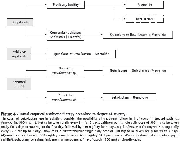
The initial antibiotic therapy for CAP outpatients recommended in the present guidelines takes into consideration three important aspects: the high proportion of CAP agents that are sensitive to beta-lactams in Brazil; the lack of definitive data with regard to empirical antibiotic coverage of atypical pathogens in cases of mild CAP; and the predominance of studies involving inpatients, rather than outpatients. However, it is of note that the coverage of macrolides has been broader than has that of amoxicillin (Figure 4). In all situations, patients undergoing treatment should be reevaluated at 48-72 h after treatment initiation. This reevaluation should be based almost exclusively on clinical data, and the repetition, in this period, of radiological examinations is not justified in stable patients showing satisfactory clinical evolution.
Recommendations
Ertapenem constitutes an acceptable alternative for CAP patients presenting risk factors for gram-negative pathogens, except Pseudomonas spp. and Acinetobacter spp. (level III evidence).
Ertapenem might be useful in CAP patients who have recently used antibiotics and in those with polymicrobial infection (level III evidence).
The initial antibiotic therapy for CAP outpatients recommended in the present guidelines takes into consideration the high proportion of CAP agents that are sensitive to beta-lactams in Brazil and the lack of definitive data with regard to empirical antibiotic coverage of atypical pathogens in cases of mild CAP; in addition, the coverage of macrolides is broader than is that of amoxicillin (level II evidence).
Treatment duration
Reduced treatment duration reduces CAP patient exposure to antibiotics, limits the risk of induction of resistance and reduces costs, as well as improving treatment adherence and tolerability. The bactericidal activity of fluoroquinolones is concentration-dependent. The pharmacodynamic properties of fluoroquinolones (the ratio between the area under the curve and the MIC required to inhibit 90% of the colonies [MIC90] and the ratio between maximum concentration [Cmax] and MIC90) allow a reduction in treatment duration without jeopardizing treatment efficacy. A double-blind, randomized, multicenter, phase III study demonstrated that gemifloxacin mesylate, when used at a dose of 320 mg/day for 7 days, shows the same clinical, bacteriological and radiological efficacy as the amoxicillin-clavulanate combination (1 g/125 mg three times a day for 10 days).(112) Another study compared the efficacy of gemifloxacin (320 mg/day) for 5 days is not inferior to 7 days with respect to clinical, bacteriological and radiological efficacy in the CAP population studied.(113) Yet another study demonstrated that the use of moxifloxacin (400 mg/day) and levofloxacin (500 mg/day), for 7-14 days, produced comparable results in a population of elderly CAP inpatients, except for the fact that treatment duration was shorter when moxifloxacin was used.(114) Because the Cmax/MIC90 of levofloxacin is inferior to that of moxifloxacin, the use of higher doses (750 mg/day) makes it possible to reduce treatment duration without jeopardizing treatment efficacy. A double-blind, randomized, multicenter study compared the efficacy of levofloxacin at 750 mg/day for 5 days with that of levofloxacin at 500 mg/day for 10 days in patients with moderate to severe CAP and obtained similar results (92.4% vs. 91.1%, respectively).(115) A meta-analysis performed to evaluate the efficacy of regimens of short duration (< 7 days) in adult patients with mild to moderate CAP, totaling 2,796 patients in 15 selected studies, showed that the efficacy of such regimens is similar to that of traditional regimens.(116)
Two recent randomized comparative studies demonstrated the efficacy and safety of a new microsphere formulation of azithromycin (not available in Brazil), used orally in a single dose, in patients with low-risk CAP.(117,118) A double-blind, randomized, multicenter, phase III study (n = 499) compared the efficacy (bacteriological and clinical cure) of the treatment of patients with low-risk CAP (Fine score I or II) using 2 g of azithromycin microspheres taken orally in a single dose (efficacy of 92.6%) with the efficacy of slow-release clarithromycin at 1 g/day for 7 days (efficacy of 94.7%). No significant difference was found between the treatments regarding the incidence of adverse effects.(117) Another study, which involved 427 CAP patients (Fine scores I-III), compared the efficacy of azithromycin microspheres taken orally in a single dose (n = 213, azithromycin group) with that of levofloxacin at 500 mg/day for 7 days (n = 214, levofloxacin group). The rate of clinical cure was similar in the two groups (89.7% and 93.7%, respectively). The two groups behaved similarly with regard to bacterial eradication (90.7% vs. 92.3%; 95% CI: 5.5-8.8). No significant difference was observed between the two groups regarding the incidence of adverse effects (19.9% vs. 12.3%).(118)
The Cmax and the area under the curve 24 h after a 2-g dose of azithromycin microspheres have been found to be twice and three times greater, respectively, than those observed when the traditional regimens of 1.5 g of azithromycin (500 mg/day for 3 days or 500 mg on day 1 and 250 mg/day for 4 more days) are used.(117) Therefore, higher serum and tissue concentration, reduced treatment duration and greater treatment adherence can be achieved, maintaining efficacy without jeopardizing tolerability.
Recommendations
Adult patients with mild to moderate CAP can be effectively treated with antibiotics administered for a period < 7 days (level I evidence).
This recommendation is consistent with the classes of antibiotics habitually recommended (level I evidence).
Although the results have been promising, there is still no solid clinical evidence for the empirical use of azithromycin microspheres, taken in a single dose, in the treatment of patients with low-to-moderate-risk CAP (level II evidence).
Treatment failure: definition, predictors of risk, markers and practice
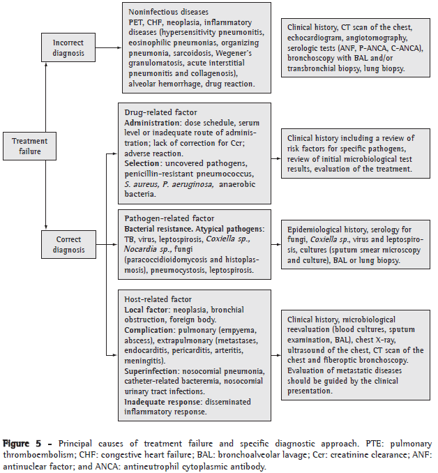
Most patients diagnosed with CAP respond adequately to treatment; however, it has been shown that 10-24% of inpatients(119-122) and 7% of outpatients(123,124) do not present an adequate clinical response. Treatment failure is an important prognostic factor for mortality among CAP patients, CAP-related mortality rates being approximately 40%.(119) The relevance of treatment failure can also be measured by the morbidity associated with it, which translates to longer hospital stays, need for ICU admission, complications and, indirectly, increased treatment costs.(121) There is no consensus regarding the definition of treatment failure. Among outpatients, treatment failure is defined as the need for hospitalization or for a change in the antibiotic therapy.(123,124) Among inpatients, treatment failure is classified as early or late.(120-122) Early treatment failure is characterized by respiratory failure (requiring mechanical ventilation) or septic shock, or both, within the first 72 h after admission. Late treatment failure occurs after 72 h of treatment and is characterized by persistent or recurrent fever associated with respiratory symptoms, by respiratory failure (requiring mechanical ventilation), by septic shock or by a combination of respiratory failure and septic shock. The principal causes of early and late treatment failure are summarized in Chart 8.
Few prospective studies have been conducted with the specific objective of evaluating the risk factors related to treatment failure by means of multivariate analysis.(120-122) The independent risk factors for early treatment failure include age > 65, PSI score > 90, inappropriate treatment, infection caused by Legionella sp. or by gram-negative bacteria, presence of multilobar infiltrate, pleural effusion and cavitation.(121,122) Late treatment failure has been linked to neoplasm, high PSI score, neurological disease, aspiration, liver cirrhosis, multilobar infiltrate and pleural effusion.(120,122) Influenza vaccination, initial treatment with fluoroquinolones and the presence of COPD have been considered protective factors against treatment failure.(120)
Biological markers have been shown to be useful in identifying patients at increased risk of treatment failure. In a recent study, C-reactive protein levels higher than 21.9 mg/dL on the first day of treatment were an independent predictor of treatment failure (OR = 2.6).(125) In the same study, C-reactive protein levels higher than 21.9 mg/dL and procalcitonin levels higher than 2.2 ng/mL on the first day of treatment showed high positive predictive value in diagnosing early treatment failure.
Treatment failure should be approached in an early, practical and systematized manner; potential causes and patterns of treatment failure should be investigated before changes in the therapeutic regimen are made. The principal causes of treatment failure and its diagnostic approach are summarized in Figure 5. A reevaluation of the therapeutic regimen and of the diagnosis before 72 h of treatment should only be considered in cases of progressive clinical deterioration, conflicting culture results or alternative microbiological diagnosis.
Recommendations
After the initiation of treatment for CAP, all patients (including outpatients) should be systematically evaluated for clinical evolution (level III evidence).
In suspected cases of treatment failure, clinical history of patients and initial results of microbiological tests should be initially reviewed.
Microbiological reevaluation can be performed using noninvasive or invasive techniques or a combination of the two (level III evidence).
Procalcitonin and C-reactive protein can be used as biological and inflammatory markers for the identification, since the initial evaluation, of patients at risk for treatment failure (level II evidence).
Prevention through vaccination
Influenza vaccine
The influenza vaccine can be administered to all those who wish to reduce the risk of becoming infected with the flu or of transmitting the virus to others. However, systematic annual vaccination should be aimed at groups of individuals at higher risk of becoming infected with influenza and of experiencing the complications thereof.(126)
The following are considered adult individuals at high risk of flu-related complications(126):
Adults aged > 50 years
Patients with chronic lung diseases (including asthma), cardiovascular diseases (except systemic arterial hypertension), kidney diseases, liver diseases, hematologic diseases and metabolic diseases (including diabetes mellitus)
Immunosuppressed adults, including those immunosuppressed by medication use and by HIV
Patients with neuromuscular disorders, because lung function is affected and because it is difficult to clear secretions from the respiratory tract
Pregnant (or breastfeeding) women and women who plan to become pregnant during the influenza season
Nursing home residents and individuals receiving home care
Potential transmitters of the virus to individuals at higher risk
Health professionals
Home caregivers of children (< 5 years of age) and of adults (> 50 years of age), particularly in the presence of other diseases with risk of complications
Professionals that provide health care services to patients receiving home care
The following individuals should not be vaccinated:
Individuals with known hypersensitivity to egg protein or to certain components of the vaccine
Individuals with acute febrile illness
Recommendations
All individuals aged > 50 years, individuals at increased risk of flu-related complications, people in household contact with others at high risk and health professionals should be given inactivated influenza vaccine (level I evidence).
The following individuals are particularly susceptible to flu-related complications: patients with chronic lung diseases (including asthma); patients with chronic cardiovascular diseases; patients with metabolic diseases (including diabetes mellitus); patients with renal dysfunction; patients with hemoglobinopathies; immunosuppressed patients (including patients with treatment-induced or HIV-related immunosuppression); pregnant women; and nursing home residents (level I evidence).
Individuals with hypersensitivity to egg protein should not be given influenza vaccine (level III evidence).
Patients with acute febrile illness should only be given influenza vaccine after the resolution of the symptoms (level III evidence).
Pneumococcal vaccine
Pneumonia, bacteremia and meningitis are the principal clinical syndromes associated with invasive pneumococcal disease. In approximately 50% of all adult cases of CAP, S. pneumonia is the causative agent. Together with influenza, pneumococcal disease constitutes one of the leading causes of death among elderly individuals. Bacteremia occurs in up to one third of the cases. Approximately 20% of individuals aged 80 years or older die from pneumococcal bacteremia.(127) The bacterial capsule, composed of polysaccharides, is the principal element responsible for the virulence of the pneumococcus. Vaccines composed of polysaccharides have been used to prevent invasive pneumococcal infections. The current pneumococcal polysaccharide vaccine provides protection against 23 of the 90 known pneumococcal serotypes (23-valent pneumococcal polysaccharide vaccine).
According to the current recommendation of the Advisory Committee of Immunization Practices of the Centers for Disease Control, target populations include the following(127):
Individuals aged > 65 years
Individuals with chronic disease, ranging in age from 2 to 64 years, particularly vulnerable to invasive infections, e.g., chronic cardiovascular diseases and chronic lung diseases (except asthma), and the complications thereof, as well as those with diabetes mellitus, alcoholism, chronic liver disease, cerebrospinal fluid fistula, cochlear implants or asplenia (anatomic or functional)
Immunocompromised individuals: patients with HIV/AIDS; patients with malignant disease or hematologic malignancies; patients with chronic renal failure or nephrotic syndrome; patients using corticosteroids or immunosuppressants; and transplant recipients
Nursing home residents
Most individuals require only one application of the vaccine. Revaccination is recommended after a minimum of 5 years for immunocompromised individuals and for those who received the first dose before 65 years of age. The vaccine is well-tolerated. Local reactions might occur (edema, pain and hyperemia); systemic manifestations (fever, myalgia and arthralgia) are rare and self-limited.
Recommendations
The pneumococcal vaccine is recommended for all individuals aged > 65 years (level II evidence).
The vaccine should also be given to individuals (aged 2-64 years) with high-risk comorbidities that make such individuals vulnerable to invasive pneumococcal infections and the complications thereof (level II evidence).
The pneumococcal vaccine should be given to immunocompromised patients and nursing home residents (level II evidence).
Severe community-acquired pneumonia: adjuvant treatment
Fluid resuscitation in severe pneumonia
Within the first hours after the onset of sepsis, venodilation, the transudation of fluid from the vascular space to the tissues and the reduction in oral ingestion, together with the increase in insensible water loss, lead to hypovolemia. Tissue perfusion is further compromised by arteriolar dilation, ventricular dysfunction and vascular obstruction.(128,129)
Fluid resuscitation in cases of severe sepsis or septic shock should be initiated as early as possible, preferably in the emergency room, before ICU admission. Hypoperfusion in patients without hypotension, as evidenced by serum lactate levels > 4 mmol/dL, indicates higher-risk patients. These patients should be submitted, as soon as possible, to arterial and central venous catheterization, aiming to achieve the following hemodynamic objectives: central venous pressure > 8 cmH2O; mean arterial pressure (MAP) > 65 mmHg; urinary output > 0.5 mL/kg/h and central venous saturation (ScvO2) > 70%. Such objectives should be achieved within the first 6 h after the diagnosis (in many cases, even before ICU admission). If an ScvO2> 70% is not achieved with aggressive fluid resuscitation within the first 6 h after the diagnosis, a packed red blood cell transfusion should be given until hematocrit is > 30%, and dobutamine should be administered if necessary (maximum dose of 20 mg/kg per min).
There is no definitive evidence that aggressive fluid resuscitation is effective in individuals with severe sepsis showing MAP > 65 mmHg or lactate < 4 mmol/L, or a combination of the two. However, ScvO2 and low urinary output might be early and independent markers of the low flow that precedes hypotension and the significant increase in serum lactate. Therefore, each variable and its respective target should be evaluated in conjunction, respecting the clinical context.(128-131)
Fluid resuscitation can be performed by i.v. injection of crystalloids or colloids, which should be administered early.(131)
Crystalloids are isotonic or hypertonic ionic solutions. The most commonly used are 0.9% saline solution and lactated Ringer's solution (both isotonic). Crystalloids are inexpensive and nontoxic; however, they produce less volume expansion than do colloids, which are more expensive and pose a higher risk of side effects.(132)
Colloids are solutions of high molecular weight that can exert oncotic pressure. The most commonly used are albumin, dextran, gelatins and hydroxyethyl starch. In addition to replacing blood volume, the ideal colloid should have a favorable impact on the modulation of the inflammatory process of sepsis, e.g., increased capillary permeability, tissue edema formation and impaired vasomotor control, as well as rheological alterations caused by abnormalities in the activation and adhesion of neutrophils. The physiological characteristics and clinical effects of colloids and crystalloids are well known. However, a detailed description of crystalloid solutions would be outside the scope of the present guidelines. It should be borne in mind that the effect of colloids, their expansion and their duration depend on molecular weight, molecule size, electric charge and metabolization. Some points should be highlighted: all colloids have anti-oncotic properties, not yet fully understood, which can influence vascular integrity and inflammation; all colloids affect the coagulation system, dextran and starch having a greater antithrombotic effect; colloids replace intravascular volume more rapidly than do crystalloids in all shock states, regardless of vascular permeability.
Initial fluid resuscitation in patients with sepsis can be performed with both types of volume expanders, i.e., crystalloids and colloids, since there is no definitive evidence in the literature that one is superior to the other regarding the outcomes of different types and etiologies of shock.
Recommendation
Volume replacement should be promptly initiated in patients with severe sepsis and hypotension (MAP
<
65 mmHg), and perfusion parameters should be monitored in order to achieve stable levels within the first 6 h (level III evidence).
Activated protein C
The use of drotrecogin alfa (activated), or activated protein C, albeit still controversial, has been recommended for patients in whom initial therapeutic measures do not result in sepsis control.(131,133) In such cases, the use of activated protein C correlates with reduced mortality and improved organ function.(133) In one study, patients with severe sepsis and at high risk of death, as indicated by an Acute Physiology and Chronic Health Evaluation score > 25, as well as patients presenting dysfunction of two or more organs, benefitted from activated protein C treatment. Apparently, the best results were observed in a subpopulation composed of patients with pneumonias. The activated protein C dose used was 24 µg/kg per min for 96 h. However, because it was an analysis of subgroups of patients, such results need to be confirmed by prospective randomized studies focusing specifically on pneumonia patients.(131)
Recommendations
The current controversy regarding the benefits of activated protein C reported in early studies weighs against its use, which is not recommended until further data regarding the cost-benefit ratio of the treatment is obtained (level II evidence).
Activated protein C is not recommended for patients with sepsis and at low risk of death (level II evidence).
Systemic corticosteroid in community-acquired pneumonia
A large multicenter clinical trial demonstrated that hydrocortisone at doses of 200-300 mg/day had a beneficial effect on vasopressor-dependent patients with septic shock who did not adequately respond to the cortisol test.(134) However, in a recent randomized controlled clinical trial, this therapeutic approach was found to offer no advantages, because although septic shock was more easily reversed in the group that received hydrocortisone than it was in the placebo group, episodes of superinfection (including sepsis and septic shock) were more common in the former.(135) Four clinical trials evaluated the use of systemic corticosteroids for the treatment of CAP patients. Two of those studies showed a significant improvement of markers of systemic inflammation(136,137); however, due to the small size of the sample, the findings were insufficient to demonstrate improvements in clinical outcomes. One clinical study demonstrated that a 7-day course of treatment with low-dose hydrocortisone injection correlated with significantly reduced duration of mechanical ventilation and of ICU stay, as well as with reduced in-hospital mortality.(138) Another study demonstrated that the use of systemic corticosteroids was independently associated with reduced mortality, and severe pneumonia was the only independent factor associated with increased mortality.(139) The limiting factors of that study included the retrospective design, the absence of determination of cortisol levels and the administration of corticosteroids at different time points throughout the treatment.
To date, there have been no studies employing adequate methodologies and samples that can warrant the recommendation of systematic use of systemic corticosteroid in all CAP patients. However, in patients with severe pneumonia and septic shock who have previously received adequate fluid replacement and vasopressors and have not responded satisfactorily, the possibility of suprarenal failure should be considered.
Recommendation
Despite having undergone adequate volume replacement and being dependent on vasoactive drugs, patients with severe CAP and arterial hypotension can receive i.v. injection of hydrocortisone (level II evidence).
Noninvasive ventilation
Patients with severe CAP are known to be at greater risk of early death due to circulatory collapse and of late death due to hypoxemic respiratory failure type.(140)
Therefore, noninvasive ventilation might be useful for severe CAP patients with hypoxemia or respiratory failure, unless immediate intubation is needed (PaO2/FiO2 < 150 and bilateral alveolar infiltrate).(81,141) Patients should be closely monitored in the first 2 h. If respiratory rate, oxygenation or hypercarbia do not improve, invasive ventilation is clearly indicated.
It has also been shown that the use of tidal volume at low levels (6 cm3/kg of ideal weight) can have beneficial effects on the survival of severe CAP patients requiring invasive ventilation.(81,141)
Recommendations
Noninvasive ventilation can be beneficial to severe CAP patients with hypoxemia or hypoxemic respiratory failure (level I evidence).
The use of low tidal volumes can be beneficial to severe CAP patients requiring invasive ventilation (level I evidence).
References
- 1. Sociedade Brasileira de Pneumologia e Tisiologia. Diretrizes para Pneumonias Adqüiridas na Comunidade (PAC) em Adultos Imunocompetentes. J Pneumol. 2004;30(Suppl 4):S1-S24.
- 2. American Thoracic Society; Infectious Diseases Society of America. Guidelines for the management of adults with hospital-acquired, ventilator-associated, and healthcare-associated pneumonia. Am J Respir Crit Care Med. 2005;171(4):388-416.
- 3. Sociedade Brasileira de Pneumologia e Tisiologia. Diretrizes brasileiras para tratamento das pneumonias adquiridas no hospital e das associadas à ventilação mecânica e Diretrizes brasileiras em pneumonia adquirida na comunidade em pediatria - 2007. J Bras Pneumol. 2007;33(Suppl 1S):S1-S50.
- 4. Wipf JE, Lipsky BA, Hirschmann JV, Boyko EJ, Takasugi J, Peugeot RL, et al. Diagnosing pneumonia by physical examination: relevant or relic? Arch Intern Med. 1999;159(10):1082-7.
- 5. Saldías F, Méndez JI, Ramírez D, Díaz O. Predictive value of history and physical examination for the diagnosis of community-acquired pneumonia in adults: a literature review [Article in Spanish]. Rev Med Chil. 2007;135(4):517-28.
- 6. Datasus [homepage on the Internet]. Brasília: Ministério da Saúde. [cited 2008 Mar 17]. Informações de Saúde. Available from: http://w3.datasus.gov.br/datasus/datasus.php?area=359A1B0C0D0E0F359G3H0I1Jd1L2M0N&VInclude=../site/texto.php
- 7. Toyoshima MT, Ito GM, Gouveia N. Trends in morbidity for respiratory diseases among hospitalized patients in the city of São Paulo [Article in Portuguese]. Rev Assoc Med Bras. 2005;51(4):209-13.
- 8. Valdivia CG. Epidemiología de la neumonía del adulto adquirida en la comunidad. Rev Chil Enf Respir. 2005;21:73-80.
- 9. Kearney SE, Davies CW, Davies RJ, Gleeson FV. Computed tomography and ultrasound in parapneumonic effusions and empyema. Clin Radiol. 2000;55(7):542-7.
- 10. Macfarlane JT, Miller AC, Roderick Smith WH, Morris AH, Rose DH. Comparative radiographic features of community acquired Legionnaires' disease, pneumococcal pneumonia, mycoplasma pneumonia, and psittacosis. Thorax. 1984;39(1):28-33.
- 11. Mittl RL Jr, Schwab RJ, Duchin JS, Goin JE, Albeida SM, Miller WT. Radiographic resolution of community-acquired pneumonia. Am J Respir Crit Care Med. 1994;149(3 Pt 1):630-5.
- 12. Rocha RT, Vital AC, Silva CO, Pereira CA, Nakatami J. Pneumonia adquirida na comunidade em pacientes tratados ambulatorialmente: aspectos epidemiológicos, clínicos e radiológicos das pneumonias atípicas e não atípicas. J Pneumol. 2000;26(1):5-14.
- 13. Tarver RD, Teague SD, Heitkamp DE, Conces DJ Jr. Radiology of community-acquired pneumonia. Radiol Clin North Am. 2005;43(3):497-512, viii.
- 14. Tew J, Calenoff L, Berlin BS. Bacterial or nonbacterial pneumonia: accuracy of radiographic diagnosis. Radiology. 1977;124(3):607-12.
- 15. Vilar J, Domingo ML, Soto C, Cogollos J. Radiology of bacterial pneumonia. Eur J Radiol. 2004;51(2):102-13.
- 16. Morrone N, Freire JA, Pereira CA, Saito M, Mendes ES, Dourado AD. Erros diagnósticos em pneumonia adquirida na comunidade: freqüência, causas e conseqüências. J Pneumol. 1987;13(1):1-6.
- 17. Beall DP, Scott WW Jr, Kuhlman JE, Hofmann LV, Moore RD, Mundy LM. Utilization of computed tomography in patients hospitalized with community-acquired pneumonia. Md Med J. 1998;47(4):182-7.
- 18. Syrjälä H, Broas M, Suramo I, Ojala A, Lähde S. High-resolution computed tomography for the diagnosis of community-acquired pneumonia. Clin Infect Dis. 1998;27(2):358-63.
- 19. Metersky ML. Is the lateral decubitus radiograph necessary for the management of a parapneumonic pleural effusion? Chest. 2003;124(3):1129-32.
- 20. Colice GL, Curtis A, Deslauriers J, Heffner J, Light R, Littenberg B, et al. Medical and surgical treatment of parapneumonic effusions : an evidence-based guideline. Chest. 2000;118(4):1158-71. Erratum in: Chest. 2001;119(1):319.
- 21. Woodhead MA, Macfarlane JT, McCracken JS, Rose DH, Finch RG. Prospective study of the aetiology and outcome of pneumonia in the community. Lancet. 1987;1(8534):671-4.
- 22. Blot SI, Rodriguez A, Solé-Violán J, Blanquer J, Almirall J, Rello J, et al. Effects of delayed oxygenation assessment on time to antibiotic delivery and mortality in patients with severe community-acquired pneumonia. Crit Care Med. 2007;35(11):2509-14.
- 23. Levin KP, Hanusa BH, Rotondi A, Singer DE, Coley CM, Marrie TJ, et al. Arterial blood gas and pulse oximetry in initial management of patients with community-acquired pneumonia. J Gen Intern Med. 2001;16(9):590-8.
- 24. Mower WR, Sachs C, Nicklin EL, Safa P, Baraff LJ. Effect of routine emergency department triage pulse oximetry screening on medical management. Chest. 1995;108(5):1297-302.
- 25. Capelastegui A, España PP, Quintana JM, Areitio I, Gorordo I, Egurrola M, et al. Validation of a predictive rule for the management of community-acquired pneumonia. Eur Respir J. 2006;27(1):151-7.
- 26. Fine MJ, Smith MA, Carson CA, Mutha SS, Sankey SS, Weissfeld LA, et al. Prognosis and outcomes of patients with community-acquired pneumonia. A meta-analysis. JAMA. 1996;275(2):134-41.
- 27. Suchyta MR, Dean NC, Narus S, Hadlock CJ. Effects of a practice guideline for community-acquired pneumonia in an outpatient setting. Am J Med. 2001;110(4):306-9.
- 28. Metersky ML, Ma A, Bratzler DW, Houck PM. Predicting bacteremia in patients with community-acquired pneumonia. Am J Respir Crit Care Med. 2004;169(3):342-7.
- 29. Paganin F, Lilienthal F, Bourdin A, Lugagne N, Tixier F, Génin R, et al. Severe community-acquired pneumonia: assessment of microbial aetiology as mortality factor. Eur Respir J. 2004;24(5):779-85.
- 30. Arnold FW, Ramirez JA, McDonald LC, Xia EL. Hospitalization for community-acquired pneumonia: the pneumonia severity index vs clinical judgment. Chest. 2003;124(1):121-4.
- 31. Marras TK, Gutierrez C, Chan CK. Applying a prediction rule to identify low-risk patients with community-acquired pneumonia. Chest. 2000;118(5):1339-43.
- 32. Almirall J, Bolíbar I, Toran P, Pera G, Boquet X, Balanzó X, et al. Contribution of C-reactive protein to the diagnosis and assessment of severity of community-acquired pneumonia. Chest. 2004;125(4):1335-42.
- 33. Chalmers JD, Singanayagam A, Hill AT. C-reactive protein is an independent predictor of severity in community-acquired pneumonia. Am J Med. 2008;121(3):219-25.
- 34. Niederman MS. Recent advances in community-acquired pneumonia: inpatient and outpatient. Chest. 2007;131(4):1205-15.
- 35. van der Meer V, Neven AK, van den Broek PJ, Assendelft WJ. Diagnostic value of C reactive protein in infections of the lower respiratory tract: systematic review. BMJ. 2005;331(7507):26.
- 36. Masiá M, Gutiérrez F, Shum C, Padilla S, Navarro JC, Flores E, et al. Usefulness of procalcitonin levels in community-acquired pneumonia according to the patients outcome research team pneumonia severity index. Chest. 2005;128(4):2223-9.
- 37. Müller B, Harbarth S, Stolz D, Bingisser R, Mueller C, Leuppi J, et al. Diagnostic and prognostic accuracy of clinical and laboratory parameters in community-acquired pneumonia. BMC Infect Dis. 2007;7:10.
- 38. Christ-Crain M, Müller B. Biomarkers in respiratory tract infections: diagnostic guides to antibiotic prescription, prognostic markers and mediators. Eur Respir J. 2007;30(3):556-73.
- 39. Christ-Crain M, Stolz D, Bingisser R, Müller C, Miedinger D, Huber PR, et al. Procalcitonin guidance of antibiotic therapy in community-acquired pneumonia: a randomized trial. Am J Respir Crit Care Med. 2006;174(1):84-93.
- 40. Ewig S, Welte T. Biomarkers in the diagnosis of pneumonia in the critically ill: don't shoot the piano player. Intensive Care Med. 2008;34(6):981-4.
- 41. Sanyal S, Smith PR, Saha AC, Gupta S, Berkowitz L, Homel P. Initial microbiologic studies did not affect outcome in adults hospitalized with community-acquired pneumonia. Am J Respir Crit Care Med. 1999;160(1):346-8.
- 42. van der Eerden MM, Vlaspolder F, de Graaff CS, Groot T, Bronsveld W, Jansen HM, et al. Comparison between pathogen directed antibiotic treatment and empirical broad spectrum antibiotic treatment in patients with community acquired pneumonia: a prospective randomised study. Thorax. 2005;60(8):672-8.
- 43. Ewig S, Schlochtermeier M, Göke N, Niederman MS. Applying sputum as a diagnostic tool in pneumonia: limited yield, minimal impact on treatment decisions. Chest. 2002;121(5):1486-92.
- 44. Rosón B, Carratalà J, Verdaguer R, Dorca J, Manresa F, Gudiol F. Prospective study of the usefulness of sputum Gram stain in the initial approach to community-acquired pneumonia requiring hospitalization. Clin Infect Dis. 2000;31(4):869-74.
- 45. Morris CG, Safranek S, Neher J. Clinical inquiries. Is sputum evaluation useful for patients with community-acquired pneumonia? J Fam Pract. 2005;54(3):279-81.
- 46. Sociedade Brasileira de Pneumologia e Tisiologia. II Consenso Brasileiro de Tuberculose - Diretrizes Brasileiras para Tuberculose 2004. J Pneumol. 2004;30(Suppl 1):S4-S56.
- 47. Bordón J, Peyrani P, Brock GN, Blasi F, Rello J, File T, et al. The presence of pneumococcal bacteremia does not influence clinical outcomes in patients with community-acquired pneumonia: results from the Community-Acquired Pneumonia Organization (CAPO) International Cohort study. Chest. 2008;133(3):618-24.
- 48. Theerthakarai R, El-Halees W, Ismail M, Solis RA, Khan MA. Nonvalue of the initial microbiological studies in the management of nonsevere community-acquired pneumonia. Chest. 2001;119(1):181-4.
- 49. Bartlett JG. Diagnostic test for etiologic agents of community-acquired pneumonia. Infect Dis Clin North Am. 2004;18(4):809-27.
- 50. Menendez R, Torres A. Treatment failure in community-acquired pneumonia. Chest. 2007;132(4):1348-55.
- 51. Weyers CM, Leeper KV. Nonresolving pneumonia. Clin Chest Med. 2005;26(1):143-58.
- 52. Torres A, Puig de la Bellacasa J, Xaubet A, Gonzalez J, Rodríguez-Roisin R, Jiménez de Anta MT, et al. Diagnostic value of quantitative cultures of bronchoalveolar lavage and telescoping plugged catheters in mechanically ventilated patients with bacterial pneumonia. Am Rev Respir Dis. 1989;140(2):306-10.
- 53. el-Ebiary M, Torres A, González J, de la Bellacasa JP, García C, Jiménez de Anta MT, et al. Quantitative cultures of endotracheal aspirates for the diagnosis of ventilator-associated pneumonia. Am Rev Respir Dis. 1993;148(6 Pt 1):1552-7.
- 54. Valencia Arango M, Torres Martí A, Insausti Ordeñana J, Alvarez Lerma F, Carrasco Joaquinet N, Herranz Casado M, et al. Diagnostic value of quantitative cultures of endotracheal aspirate in ventilator-associated pneumonia: a multicenter study [Article in Spanish]. Arch Bronconeumol. 2003;39(9):394-9.
- 55. Feinsilver SH, Fein AM, Niederman MS, Schultz DE, Faegenburg DH. Utility of fiberoptic bronchoscopy in nonresolving pneumonia. Chest. 1990;98(6):1322-6.
- 56. Ortqvist A, Kalin M, Lejdeborn L, Lundberg B. Diagnostic fiberoptic bronchoscopy and protected brush culture in patients with community-acquired pneumonia. Chest. 1990;97(3):576-82.
- 57. Jiménez P, Saldías F, Meneses M, Silva ME, Wilson MG, Otth L. Diagnostic fiberoptic bronchoscopy in patients with community-acquired pneumonia. Comparison between bronchoalveolar lavage and telescoping plugged catheter cultures. Chest. 1993;103(4):1023-7.
- 58. Littman AJ, Jackson LA, White E, Thornquist MD, Gaydos CA, Vaughan TL. Interlaboratory reliability of microimmunofluorescence test for measurement of Chlamydia pneumoniae-specific immunoglobulin A and G antibody titers. Clin Diagn Lab Immunol. 2004;11(3):615-7.
- 59. Murdoch DR. Diagnosis of Legionella infection. Clin Infect Dis. 2003;36(1):64-9.
- 60. Roig J, Sabria M, Pedro-Botet ML. Legionella spp.: community acquired and nosocomial infections. Curr Opin Infect Dis. 2003;16(2):145-51.
- 61. Rantakokko-Jalava K, Jalava J. Development of conventional and real-time PCR assays for detection of Legionella DNA in respiratory specimens. J Clin Microbiol. 2001;39(8):2904-10.
- 62. Diederen BM, Bruin JP, den Boer JW, Peeters MF, Yzerman EP. Sensitivity of Legionella pneumophila DNA detection in serum samples in relation to disease severity. J Med Microbiol. 2007;56(Pt 9):1255.
- 63. Domínguez J, Galí N, Blanco S, Pedroso P, Prat C, Matas L, et al. Detection of Streptococcus pneumoniae antigen by a rapid immunochromatographic assay in urine samples. Chest. 2001;119(1):243-9.
- 64. Porcel JM, Ruiz-González A, Falguera M, Nogués A, Galindo C, Carratalá J, et al. Contribution of a pleural antigen assay (Binax NOW) to the diagnosis of pneumococcal pneumonia. Chest. 2007;131(5):1442-7.
- 65. Briones ML, Blanquer J, Ferrando D, Blasco ML, Gimeno C, Marín J. Assessment of analysis of urinary pneumococcal antigen by immunochromatography for etiologic diagnosis of community-acquired pneumonia in adults. Clin Vaccine Immunol. 2006;13(10):1092-7.
- 66. Jacobs JA, Stobberingh EE, Cornelissen EI, Drent M. Detection of Streptococcus pneumoniae antigen in bronchoalveolar lavage fluid samples by a rapid immunochromatographic membrane assay. J Clin Microbiol. 2005;43(8):4037-40.
- 67. Marcos MA, Jiménez de Anta MT, de la Bellacasa JP, González J, Martínez E, García E, et al. Rapid urinary antigen test for diagnosis of pneumococcal community-acquired pneumonia in adults. Eur Respir J. 2003;21(2):209-14.
- 68. Strålin K, Törnqvist E, Kaltoft MS, Olcén P, Holmberg H. Etiologic diagnosis of adult bacterial pneumonia by culture and PCR applied to respiratory tract samples. J Clin Microbiol. 2006;44(2):643-5.
- 69. Ginevra C, Barranger C, Ros A, Mory O, Stephan JL, Freymuth F, et al. Development and evaluation of Chlamylege, a new commercial test allowing simultaneous detection and identification of Legionella, Chlamydophila pneumoniae, and Mycoplasma pneumoniae in clinical respiratory specimens by multiplex PCR. J Clin Microbiol. 2005;43(7):3247-54.
- 70. Loens K, Ursi D, Goossens H, Ieven M. Molecular diagnosis of Mycoplasma pneumoniae respiratory tract infections. J Clin Microbiol. 2003;41(11):4915-23.
- 71. Loens K, Beck T, Ursi D, Overdijk M, Sillekens P, Goossens H, et al. Evaluation of different nucleic acid amplification techniques for the detection of M. pneumoniae, C. pneumoniae and Legionella spp. in respiratory specimens from patients with community-acquired pneumonia. J Microbiol Methods. 2008;73(3):257-62.
- 72. Lim WS, van der Eerden MM, Laing R, Boersma WG, Karalus N, Town GI, et al. Defining community acquired pneumonia severity on presentation to hospital: an international derivation and validation study. Thorax. 2003;58(5):377-82.
- 73. Fine MJ, Auble TE, Yealy DM, Hanusa BH, Weissfeld LA, Singer DE, et al. A prediction rule to identify low-risk patients with community-acquired pneumonia. N Engl J Med. 1997;336(4):243-50.
- 74. Atlas SJ, Benzer TI, Borowsky LH, Chang Y, Burnham DC, Metlay JP, et al. Safely increasing the proportion of patients with community-acquired pneumonia treated as outpatients: an interventional trial. Arch Intern Med. 1998;158(12):1350-6.
- 75. Barlow G, Nathwani D, Davey P. The CURB65 pneumonia severity score outperforms generic sepsis and early warning scores in predicting mortality in community-acquired pneumonia. Thorax. 2007;62(3):253-9.
- 76. Myint PK, Kamath AV, Vowler SL, Harrison BD. Simple modification of CURB-65 better identifies patients including the elderly with severe CAP. Thorax. 2007;62(11):1015-6; author reply 1016.
- 77. Schaaf B, Kruse J, Rupp J, Reinert RR, Droemann D, Zabel P, et al. Sepsis severity predicts outcome in community-acquired pneumococcal pneumonia. Eur Respir J. 2007;30(3):517-24.
- 78. Ewig S, Schäfer H, Torres A. Severity assessment in community-acquired pneumonia. Eur Respir J. 2000;16(6):1193-201.
- 79. Buising KL, Thursky KA, Black JF, MacGregor L, Street AC, Kennedy MP, et al. A prospective comparison of severity scores for identifying patients with severe community acquired pneumonia: reconsidering what is meant by severe pneumonia. Thorax. 2006;61(5):419-24.
- 80. British Thoracic Society; Myint PK, Kamath AV, Vowler SL, Maisey DN, Harrison BD. Severity assessment criteria recommended by the British Thoracic Society (BTS) for community-acquired pneumonia (CAP) and older patients. Should SOAR (systolic blood pressure, oxygenation, age and respiratory rate) criteria be used in older people? A compilation study of two prospective cohorts. Age Ageing. 2006;35(3):286-91.
- 81. Mandell LA, Wunderink RG, Anzueto A, Bartlett JG, Campbell GD, Dean NC, et al. Infectious Diseases Society of America/American Thoracic Society consensus guidelines on the management of community-acquired pneumonia in adults. Clin Infect Dis. 2007;44 Suppl 2:S27-72.
- 82. Valencia M, Badia JR, Cavalcanti M, Ferrer M, Agustí C, Angrill J, et al. Pneumonia severity index class v patients with community-acquired pneumonia: characteristics, outcomes, and value of severity scores. Chest. 2007;132(2):515-22.
- 83. Shefet D, Robenshtok E, Paul M, Leibovici L. Empirical atypical coverage for inpatients with community-acquired pneumonia: systematic review of randomized controlled trials. Arch Intern Med. 2005;165(17):1992-2000.
- 84. Strålin K. Usefulness of aetiological tests for guiding antibiotic therapy in community-acquired pneumonia. Int J Antimicrob Agents. 2008;31(1):3-11.
- 85. Lim WS, Macfarlane JT, Boswell TC, Harrison TG, Rose D, Leinonen M, et al. Study of community acquired pneumonia aetiology (SCAPA) in adults admitted to hospital: implications for management guidelines. Thorax. 2001;56(4):296-301.
- 86. Arnold FW, Summersgill JT, Lajoie AS, Peyrani P, Marrie TJ, Rossi P, et al. A worldwide perspective of atypical pathogens in community-acquired pneumonia. Am J Respir Crit Care Med. 2007;175(10):1086-93.
- 87. Paul M, Nielsen AD, Gafter-Gvili A, Tacconelli E, Andreassen S, Almanasreh N, et al. The need for macrolides in hospitalised community-acquired pneumonia: propensity analysis. Eur Respir J. 2007;30(3):525-31.
- 88. van de Garde EM, Endeman H, van Hemert RN, Voorn GP, Deneer VH, Leufkens HG, et al. Prior outpatient antibiotic use as predictor for microbial aetiology of community-acquired pneumonia: hospital-based study. Eur J Clin Pharmacol. 2008;64(4):405-10.
- 89. Mills GD, Oehley MR, Arrol B. Effectiveness of beta lactam antibiotics compared with antibiotics active against atypical pathogens in non-severe community acquired pneumonia: meta-analysis. BMJ. 2005;330(7489):456.
- 90. Robenshtok E, Shefet D, Gafter-Gvili A, Paul M, Vidal L, Leibovici L. Empiric antibiotic coverage of atypical pathogens for community acquired pneumonia in hospitalized adults. Cochrane Database Syst Rev. 2008;(1):CD004418.
- 91. Lodise TP, Kwa A, Cosler L, Gupta R, Smith RP. Comparison of beta-lactam and macrolide combination therapy versus fluoroquinolone monotherapy in hospitalized Veterans Affairs patients with community-acquired pneumonia. Antimicrob Agents Chemother. 2007;51(11):3977-82.
- 92. Rodríguez A, Mendia A, Sirvent JM, Barcenilla F, de la Torre-Prados MV, Solé-Violán J, et al. Combination antibiotic therapy improves survival in patients with community-acquired pneumonia and shock. Crit Care Med. 2007;35(6):1493-8.
- 93. Baddour LM, Yu VL, Klugman KP, Feldman C, Ortqvist A, Rello J, et al. Combination antibiotic therapy lowers mortality among severely ill patients with pneumococcal bacteremia. Am J Respir Crit Care Med. 2004;170(4):440-4.
- 94. Amsden GW. Anti-inflammatory effects of macrolides--an underappreciated benefit in the treatment of community-acquired respiratory tract infections and chronic inflammatory pulmonary conditions? J Antimicrob Chemother. 2005;55(1):10-21.
- 95. Nava JM, Bella F, Garau J, Lite J, Morera MA, Martí C, et al. Predictive factors for invasive disease due to penicillin-resistant Streptococcus pneumoniae: a population-based study. Clin Infect Dis. 1994;19(5):884-90.
- 96. Arancibia F, Bauer TT, Ewig S, Mensa J, Gonzalez J, Niederman MS, et al. Community-acquired pneumonia due to gram-negative bacteria and pseudomonas aeruginosa: incidence, risk, and prognosis. Arch Intern Med. 2002;162(16):1849-58.
- 97. Houck PM, Bratzler DW, Nsa W, Ma A, Bartlett JG. Timing of antibiotic administration and outcomes for Medicare patients hospitalized with community-acquired pneumonia. Arch Intern Med. 2004;164(6):637-44.
- 98. Meehan TP, Fine MJ, Krumholz HM, Scinto JD, Galusha DH, Mockalis JT, et al. Quality of care, process, and outcomes in elderly patients with pneumonia. JAMA. 1997;278(23):2080-4.
- 99. Battleman DS, Callahan M, Thaler HT. Rapid antibiotic delivery and appropriate antibiotic selection reduce length of hospital stay of patients with community-acquired pneumonia: link between quality of care and resource utilization. Arch Intern Med. 2002;162(6):682-8.
- 100. Berjohn CM, Fishman NO, Joffe MM, Edelstein PH, Metlay JP. Treatment and outcomes for patients with bacteremic pneumococcal pneumonia. Medicine (Baltimore). 2008;87(3):160-6.
- 101. Silber SH, Garrett C, Singh R, Sweeney A, Rosenberg C, Parachiv D, et al. Early administration of antibiotics does not shorten time to clinical stability in patients with moderate-to-severe community-acquired pneumonia. Chest. 2003;124(5):1798-804.
- 102. Houck PM, Bratzler DW. Administration of first hospital antibiotics for community-acquired pneumonia: does timeliness affect outcomes? Curr Opin Infect Dis. 2005;18(2):151-6.
- 103. Welker JA, Huston M, McCue JD. Antibiotic timing and errors in diagnosing pneumonia. Arch Intern Med. 2008;168(4):351-6.
- 104. Wikler MA; Clinical and Laboratory Standards Institute. Performance Standards for Antimicrobial Susceptibility Testing: Eighteenth Informational Supplement. Wayne: Clinical and Laboratory Standards Institute, 2008.
- 105. Rossi F, Andreazzi DB. Resistência bacteriana interpretando o antibiograma. São Paulo: Atheneu; 2005.
- 106. Brandileone MC, Casagrande ST, Guerra ML, Zanella RC, Andrade AL, Di Fabio JL. Increase in numbers of beta-lactam-resistant invasive Streptococcus pneumoniae in Brazil and the impact of conjugate vaccine coverage. J Med Microbiol. 2006;55(Pt 5):567-74.
- 107. Vergis EN, Indorf A, File TM Jr, Phillips J, Bates J, Tan J, et al. Azithromycin vs cefuroxime plus erythromycin for empirical treatment of community-acquired pneumonia in hospitalized patients: a prospective, randomized, multicenter trial. Arch Intern Med. 2000;160(9):1294-300.
- 108. Plouffe J, Schwartz DB, Kolokathis A, Sherman BW, Arnow PM, Gezon JA, et al. Clinical efficacy of intravenous followed by oral azithromycin monotherapy in hospitalized patients with community-acquired pneumonia. The Azithromycin Intravenous Clinical Trials Group. Antimicrob Agents Chemother. 2000;44(7):1796-802.
- 109. Marchese A, Gualco L, Schito AM, Debbia EA, Schito GC. In vitro activity of ertapenem against selected respiratory pathogens. J Antimicrob Chemother. 2004;54(5):944-51.
- 110. Ortiz-Ruiz G, Vetter N, Isaacs R, Carides A, Woods GL, Friedland I. Ertapenem versus ceftriaxone for the treatment of community-acquired pneumonia in adults: combined analysis of two multicentre randomized, double-blind studies. J Antimicrob Chemother. 2004;53 Suppl 2:ii59-66.
- 111. Vetter N, Cambronero-Hernandez E, Rohlf J, Simon S, Carides A, Oliveria T, et al. A prospective, randomized, double-blind multicenter comparison of parenteral ertapenem and ceftriaxone for the treatment of hospitalized adults with community-acquired pneumonia. Clin Ther. 2002;24(11):1770-85.
- 112. Léophonte P, File T, Feldman C. Gemifloxacin once daily for 7 days compared to amoxicillin/clavulanic acid thrice daily for 10 days for the treatment of community-acquired pneumonia of suspected pneumococcal origin. Respir Med. 2004;98(8):708-20.
- 113. File TM Jr, Mandell LA, Tillotson G, Kostov K, Georgiev O. Gemifloxacin once daily for 5 days versus 7 days for the treatment of community-acquired pneumonia: a randomized, multicentre, double-blind study. J Antimicrob Chemother. 2007;60(1):112-20.
- 114. Anzueto A, Niederman MS, Pearle J, Restrepo MI, Heyder A, Choudhri SH, et al. Community-Acquired Pneumonia Recovery in the Elderly (CAPRIE): efficacy and safety of moxifloxacin therapy versus that of levofloxacin therapy. Clin Infect Dis. 2006;42(1):73-81. Erratum in: Clin Infect Dis. 2006;42(9):1350.
- 115. Dunbar LM, Wunderink RG, Habib MP, Smith LG, Tennenberg AM, Khashab MM, et al. High-dose, short-course levofloxacin for community-acquired pneumonia: a new treatment paradigm. Clin Infect Dis. 2003;37(6):752-60. Erratum in: Clin Infect Dis. 2003;37(8):1147.
- 116. Li JZ, Winston LG, Moore DH, Bent S. Efficacy of short-course antibiotic regimens for community-acquired pneumonia: a meta-analysis. Am J Med. 2007;120(9):783-90.
- 117. Drehobl MA, De Salvo MC, Lewis DE, Breen JD. Single-dose azithromycin microspheres vs clarithromycin extended release for the treatment of mild-to-moderate community-acquired pneumonia in adults. Chest. 2005;128(4):2230-7.
- 118. D'Ignazio J, Camere MA, Lewis DE, Jorgensen D, Breen JD. Novel, single-dose microsphere formulation of azithromycin versus 7-day levofloxacin therapy for treatment of mild to moderate community-acquired Pneumonia in adults. Antimicrob Agents Chemother. 2005;49(10):4035-41.
- 119. Arancibia F, Ewig S, Martinez JA, Ruiz M, Bauer T, Marcos MA, et al. Antimicrobial treatment failures in patients with community-acquired pneumonia: causes and prognostic implications. Am J Respir Crit Care Med. 2000;162(1):154-60.
- 120. Genné D, Sommer R, Kaiser L, Saaïdia A, Pasche A, Unger PF, et al. Analysis of factors that contribute to treatment failure in patients with community-acquired pneumonia. Eur J Clin Microbiol Infect Dis. 2006;25(3):159-66.
- 121. Menéndez R, Torres A, Zalacaín R, Aspa J, Martín Villasclaras JJ, Borderías L, et al. Risk factors of treatment failure in community acquired pneumonia: implications for disease outcome. Thorax. 2004;59(11):960-5.
- 122. Rosón B, Carratalà J, Fernández-Sabé N, Tubau F, Manresa F, Gudiol F. Causes and factors associated with early failure in hospitalized patients with community-acquired pneumonia. Arch Intern Med. 2004;164(5):502-8.
- 123. Fantin B, Aubert JP, Unger P, Lecoeur H, Carbon C. Clinical evaluation of the management of community-acquired pneumonia by general practitioners in France. Chest. 2001;120(1):185-92.
- 124. Minogue MF, Coley CM, Fine MJ, Marrie TJ, Kapoor WN, Singer DE. Patients hospitalized after initial outpatient treatment for community-acquired pneumonia. Ann Emerg Med. 1998;31(3):376-80.
- 125. Menéndez R, Cavalcanti M, Reyes S, Mensa J, Martinez R, Marcos MA, et al. Markers of treatment failure in hospitalised community acquired pneumonia. Thorax. 2008;63(5):447-52.
- 126. Fiore AE, Shay DK, Broder K, Iskander JK, Uyeki TM, Mootrey G, et al. Prevention and control of influenza: recommendations of the Advisory Committee on Immunization Practices (ACIP), 2008. MMWR Recomm Rep. 2008;57(RR-7):1-60.
- 127. Whitney CG. Preventing pneumococcal disease. ACIP recommends pneumococcal polysaccharide vaccine for all adults age > or = 65. Geriatrics. 2003;58(10):20-2, 25.
- 128. Durairaj L, Schmidt GA. Fluid therapy in resuscitated sepsis: less is more. Chest. 2008;133(1):252-63.
- 129. Russell JA. Management of sepsis. N Engl J Med. 2006;355(16):1699-713.
- 130. Rivers E, Nguyen B, Havstad S, Ressler J, Muzzin A, Knoblich B, et al. Early goal-directed therapy in the treatment of severe sepsis and septic shock. N Engl J Med. 2001;345(19):1368-77.
- 131. Dellinger RP, Levy MM, Carlet JM, Bion J, Parker MM, Jaeschke R, et al. Surviving Sepsis Campaign: international guidelines for management of severe sepsis and septic shock: 2008. Intensive Care Med. 2008;34(1):17-60.
- 132. American Thoracic Society. Evidence-based colloid use in the critically ill: American Thoracic Society Consensus Statement. Am J Respir Crit Care Med. 2004;170(11):1247-59.
- 133. Laterre PF, Garber G, Levy H, Wunderink R, Kinasewitz GT, Sollet JP, et al. Severe community-acquired pneumonia as a cause of severe sepsis: data from the PROWESS study. Crit Care Med. 2005;33(5):952-61.
- 134. Annane D, Sébille V, Charpentier C, Bollaert PE, François B, Korach JM, et al. Effect of treatment with low doses of hydrocortisone and fludrocortisone on mortality in patients with septic shock. JAMA. 2002;288(7):862-71. Erratum in: JAMA. 2008;300(14):1652.
- 135. Sprung CL, Annane D, Keh D, Moreno R, Singer M, Freivogel K, et al. Hydrocortisone therapy for patients with septic shock. N Engl J Med. 2008;358(2):111-24.
- 136. Meduri GU, Tolley EA, Chrousos GP, Stentz F. Prolonged methylprednisolone treatment suppresses systemic inflammation in patients with unresolving acute respiratory distress syndrome: evidence for inadequate endogenous glucocorticoid secretion and inflammation-induced immune cell resistance to glucocorticoids. Am J Respir Crit Care Med. 2002;165(7):983-91.
- 137. Marik P, Kraus P, Sribante J, Havlik I, Lipman J, Johnson DW. Hydrocortisone and tumor necrosis factor in severe community-acquired pneumonia. A randomized controlled study. Chest. 1993;104(2):389-92.
- 138. Confalonieri M, Urbino R, Potena A, Piattella M, Parigi P, Puccio G, et al. Hydrocortisone infusion for severe community-acquired pneumonia: a preliminary randomized study. Am J Respir Crit Care Med. 2005;171(3):242-8.
- 139. Garcia-Vidal C, Calbo E, Pascual V, Ferrer C, Quintana S, Garau J. Effects of systemic steroids in patients with severe community-acquired pneumonia. Eur Respir J. 2007;30(5):951-6.
- 140. Luján M, Gallego M, Rello J. Optimal therapy for severe pneumococcal community-acquired pneumonia. Intensive Care Med. 2006;32(7):971-80.
- 141. Sociedade Brasileira de Pneumologia e Tisiologia. III Consenso Brasileiro de Ventilação Mecânica. J Bras Pneumol. 2007;33(Suppl 2):S1-S150.
Publication Dates
-
Publication in this collection
13 July 2009 -
Date of issue
June 2009
History
-
Received
18 Apr 2009 -
Accepted
23 Apr 2009


