Abstracts
The aim of this work was to analyze the neuron morphology and morphometry of cervical, thoracic and lumbar areas of nonsymptomatic seropositive dogs’ spinal cord for toxoplasmosis. Twenty indefinite-breed adult dogs were used; ten dogs were healthy, with negative serology for toxoplasmosis, and were used as the control group (group 1), and ten dogs were nonsymptomatic but seropositive for toxoplasmosis (group 2). After the microtomy, with interval of 100 micrometers (µm), the histological 5-µm-thick cuts were dyed by hematoxylin-eosin and Masson's trichrome techniques. The glass slides were analyzed under light microscope to examine the neuron morphology. The parameters considered for the morphometric analysis were area, perimeter, maximum diameter, minimum diameter and shape factor of cytoplasm and nucleus of neuron. The results were statistically analyzed by Student’s t test at 5% probability level. The morphological characteristics between the two groups were similar and according to literature. The morphometric results showed that there were changes in neurons size and structure, and increase and loss of star shape were noticed in seropositive animals. The results suggest that the neurons of these dogs, yet nonsymptomatic, can have lost their conductor function.
canine; central nervous system; histology; Toxoplasma gondii
Este trabalho objetivou analisar a morfologia e a morfometria dos neurônios das regiões cervical, torácica e lombar da medula espinhal de cães assintomáticos soropositivos para toxoplasmose. Utilizaram-se 20 cães sem raça definida, adultos, sendo dez cães hígidos, com sorologia negativa, utilizados como controle (grupo 1) e dez cães assintomáticos mas soropositivos para toxoplasmose (grupo 2). Após microtomia semi-seriada, com intervalos de 100 micrômetros (µm), os cortes histológicos de medula espinhal, à espessura de 5 µm, foram corados pelas técnicas da hematoxilina-eosina e do tricrômico de Masson. As lâminas foram analisadas à microscopia de luz para verificar a morfologia dos neurônios. Para o estudo morfométrico, os parâmetros analisados foram: área, perímetro, diâmetro máximo, diâmetro mínimo e fator de forma do citoplasma e núcleo dos neurônios. Os resultados obtidos foram analisados estatisticamente, mediante o teste t de Student ao nível de 5% de probabilidade. As características morfológicas entre os dois grupos foram semelhantes e em conformidade com a literatura. Os resultados morfométricos demonstraram que há alteração no tamanho e estrutura dos neurônios, com aumento e perda do formato estrelado nos animais soropositivos. Os resultados sugerem que os neurônios destes cães, ainda que assintomáticos, possam ter perdido sua função condutora.
canino; histologia; sistema nervoso central; Toxoplasma gondii
Introduction
Inflammatory diseases of the central nervous system (CNS) are important causes of neurologic disorder in dogs( 11 Moretti LD, Ueno TE, Ribeiro MG. Toxoplasmose em cães co-infectados com o vírus da cinomose. Semina: Ciências Agrárias. 2002; 23:85-91. ). Although neurologic canine diseases caused by protozoa are not very common( 22 Thomas WB. Inflammatory diseases of the central nervous system in dogs. Clinical Techniques in Small Animal Practice. 1998; 13(3):167-178. ), Toxoplasma gondii (T. gondii) and Neospora caninum ( 33 Paixão TA, Santos RL. Encefalite por Neospora caninum e Toxoplasma gondii em cães. Clínica Veterinária. 2004; 9(48):44-52. ) are the agents normally responsible for the cases of encephalitis caused by protozoa.
Canine toxoplasmosis has captured researchers’ attention since it was described in 1910 by Mello in Italy( 44 Vidotto O. Toxoplasmose: epidemiologia e importância da doença na saúde animal. Semina: Ciências Agrárias. 1992; 13(1):69-75. ) and by Carini( 55 Carini A. Infection spontanée du pigeon et du chien due au Toxoplasma cuniculi. Bulletin de la Societé de Pathologie Exotique. 1911; 4:518-519. ) in Brazil, and the desease’s life cycle and different modes of transmission have been extensively discussed in veterinary literature( 66 Hass JA, Shell L, Saunders G. Neurological manifestations of toxoplasmosis: a literature review and case summary. Journal of America Animal Hospital Association. 1989; 25:253-260. ). Dogs are considered very receptive animals for this zoonosis, probably, due to their carnivorous eating habit, which facilitates the ingestion of tissues contaminated by cysts and the contact with sporulated oocysts in contaminated soil( 77 Germano PML. Estudo sorológico da toxoplasmose canina, pela prova de imunofluorêscencia indireta, na cidade de Campinas em 1981. Revista da Faculdade de Medicina Veterinária e Zootecnia. 1985; 22:53-58. ).
The infection has been noticed in cats and dogs in many countries, demonstrating its
cosmopolitan feature. Severe and fatal cases have been reported in spite of clinical
manifestations being uncommon(
33 Paixão TA, Santos RL. Encefalite por Neospora caninum e
Toxoplasma gondii em cães. Clínica Veterinária. 2004;
9(48):44-52.
,
66 Hass JA, Shell L, Saunders G. Neurological manifestations of
toxoplasmosis: a literature review and case summary. Journal of America Animal
Hospital Association. 1989; 25:253-260.
,
88 Lindsay SD. Serological prevalence of Neospora caninum
and Toxoplasma gondii in dogs from Kansas. Journal of the
Helminthological Society of Washington. 1990; 57(1):86-88.
,
99 Guimarães AM, Ribeiro MFB, Lima JD, Cury MC, Spiewak G. Freqüência de
anticorpos anti-Toxoplasma gondii em cães de Belo Horizonte, Minas
Gerais. Arquivo Brasileiro de Medicina Veterinária e Zootecnia. 1992;
44(1):67-68.
), and although many animals are serologically positive for
toxoplasmosis, few develop clinical signs of the disease(
1010 Dubey JP, Lappin MR. Toxoplasmosis and Neosporosis. In: Greene CE.
Infectious Diseases of the Dog and Cat. 4th ed. Saint Louis: Elsevier; 2012. p.
806-827.
). Ataxia, seizures, tremors, cranial
nerve disorders, progressive paresis and paralysis are the most common clinical
manifestations of CNS toxoplasmosis(
1111 Averill DR, Lahunta A. Toxoplasmosis of the canine nervous system:
clinicopathological findings in four cases. Journal of the American Veterinary
Medical Association. 1971; 159:1134-1141.
12 Nesbit JW, Lourens DC, Williams MC. Spastic paresis in two littermate
pups caused by Toxoplasma gondii. Journal of the South African
Veterinary Association. 1981; 52:243-246.
13 Suter MM, Hauser B, Palmer DG, Oettli P. Polymyositis - polyradiculitis
due to toxoplasmosis in the dog: Serology and tissue biopsy as diagnostic aids.
Zentralblatt fuer Veterinaermedizin. 1984; 31(10):792-798.
14 Braund KG, Blagburn BL, Toivio-Kinnucan M, Amling KA, Pidgeon GL.
Toxoplasma polymyositis/polyneuropathy – a new clinical variant
in two mature dogs. Journal of the American Animal Hospital Association. 1988;
24(1):93-97.
-
1515 Davidson MG. Toxoplasmosis. Veterinary Clinics of North America Small
Animal Practice. 2000; 30(5):1051-1062.
). It is
presumed that the disease manifestation occurs by local or systemic immune deficit of
the host organism, so that immunosuppressed patients may have primary or recurrent
infection(
1616 Swinger RL, Schimdt KA, Dubielzig RR. Keratoconjunctivitis associated
with Toxoplasma gondii in a dog. Veterinary Ophthalmology. 2009;
12(1):56-60.
).
Areas of discoloration, necrosis and cerebellar atrophy have been observed macroscopically in CNS of infected dogs. Lesions seen early in blood vessels consist of endothelial cell proliferation, necrosis, and perivascular cuffing. Neuronal necrosis, mild malacia, and some astrocytosis may be seen. Multifocal leptomeningeal infiltrates of macrophages, plasma cells, and some lymphocytes and neutrophils are found( 1010 Dubey JP, Lappin MR. Toxoplasmosis and Neosporosis. In: Greene CE. Infectious Diseases of the Dog and Cat. 4th ed. Saint Louis: Elsevier; 2012. p. 806-827. ). The most common histopathological alterations in the CNS are characterized by nonsuppurative meningoencephalomyelitis associated with vasculitis, necrosis, malacia and gliosis with possible involvement of peripheral nerves( 1010 Dubey JP, Lappin MR. Toxoplasmosis and Neosporosis. In: Greene CE. Infectious Diseases of the Dog and Cat. 4th ed. Saint Louis: Elsevier; 2012. p. 806-827. , 1717 Giraldi JH, Bracarense APFR, Vidotto O, Tudury EA, Navarro IT, Batista TN. Serology and histopathology of Toxoplasma gondii and Neospora caninum in dogs with neurologic disorders. Semina: Ciências Agrárias. 2002; 23:9-14. ). Histopathological evaluation of spinal cords of T. gondii-infected mice revealed high counts of infiltrated inflammatory cells, presence of cysts mostly in the gray matter, neuronal degeneration and hemorrhage, with no obvious changes in the myelin staining intensity or axonal density( 1818 Möhle L, Parlog A, Pahnke J, Dunay IR. Spinal cord pathology in chronic experimental Toxoplasma gondii infection. European Journal of Microbiology and Immunology. 2014; 4(1):65-75. ).
The neurons possess a big nucleus with a proeminent nucleolus and distended chromosomes. Cytoplasm of nuclear body, or perikaryon, is rich in ribosome and they might be concentrated in small cytoplasmatic areas, and under optical microscope, we can see granular basophilic bodies, known as Nissl bodies( 1919 Junqueira J, Carneiro F. Tecido nervoso. In: Junqueira J, Carneiro F. Histologia Básica: Texto & Atlas. 12th ed. Rio de Janeiro: Guanabara Koogan; 2013. p. 149-175. Portuguese. , 2020 Ross MH, Pawlina W. Nervous tissue In: Ross MH, Pawlina W. Histology: a text and atlas with correlated cell and molecular biology. 6rd ed. Baltimore: Lippincott Williams & Wilkins; 2011. p. 352-399. ). The shape of the cellular body varies greatly( 1919 Junqueira J, Carneiro F. Tecido nervoso. In: Junqueira J, Carneiro F. Histologia Básica: Texto & Atlas. 12th ed. Rio de Janeiro: Guanabara Koogan; 2013. p. 149-175. Portuguese. ), but also the size is extremely variable, and according to the type of neuron, it may vary from 3 to 150 µm( 1919 Junqueira J, Carneiro F. Tecido nervoso. In: Junqueira J, Carneiro F. Histologia Básica: Texto & Atlas. 12th ed. Rio de Janeiro: Guanabara Koogan; 2013. p. 149-175. Portuguese. , 2121 Machado CRS. Tecido nervoso. In: Machado A. Neuroanatomia Funcional. 2nd ed. São Paulo: Atheneu; 2002. p. 17-32. Portuguese. ). According to Carvalho et al.( 2222 Carvalho ACF, Pacheco MR, Baraldi-Artoni SM, Mateus O. Canine spinal cord neuron and axon myelin sheath morphometry. Anatomia, Histologia, Embryologia: Journal of Veterinary Medicine. 2006; 35(5):284-286. ), the size of cytoplasm and nucleus of the neurons from canine spinal cord is also very variable, depending on the location of neurons. Neurons placed in lumbar area are, statistically, bigger than those found in cervical and thoracic areas. They also mention that these lumbar neurons, statistically, are more rounded shaped than the neurons from other areas, this fact is proved by morphometric parameter analysis cellular shape factor.
It is known that important histopathological changes are observed in the CNS with clinical toxoplasmosis, so the objective of this research was to analyze the occurrence of early changes in the morphometry and morphology of neurons from spinal cord of apparently healthy dogs, but with high serological reactivity to toxoplasmosis, in order to better investigate and document the possible histological changes before clinical signs.
Material and Methods
For the proposed objectives we used twenty indefinite-breed adult dogs, weighing from seven to 15 kg, from the Zoonosis Control Center of Araraquara city, state of São Paulo, Brazil. The samples were collected in 2000 and the studied was concluded by 2007. The serology for T. gondii was performed by Enzyme-Linked Immunosorbent Assay (ELISA), with the technique described by Domingues et al.( 2323 Domingues LM, Machado RZ, Tinucci Costa M, Carvalho CS, Costa AJ, Malheiros EB. Canine toxoplasmosis: a comparative evaluation of the detection of anti-toxoplasma gondii antibodies by the indirect immunoenzymatic assay (ELISA) and the indirect immunofluorescence reaction (IFF). Revista Brasileira de Parasitologia Veterinária. 1998; 7(2):79-85. ), at the Department of Veterinary Pathology of the School of Agrarian and Veterinary Sciences, São Paulo State University, Jaboticabal, Brazil. The reactivity of sera was analyzed in terms of ELISA levels (from 0 to 9) and the animals used in this study were those that presented reactivity 8 or 9. Ten dogs, with negative serology for T. gondii, were used as control group (group 1) and ten nonsymptomatic dogs, with levels of reactivity for T. gondii over 8, detected by ELISA, were used as reagent group (group 2).
After the formation of groups 1 and 2, fragments of spinal cord corresponding to cervical, thoracic and lumbar area were collected post-mortem, being removed from the medullary canal using a blunt-pointed forceps, and fixed in Bouin’s solution during 24 hours and processed, as usual, to be included in paraffin.
After the microtomy, with interval of 100 micrometers (µm), the histological 5-µm-thick cuts were dyed by hematoxylin-eosin (HE) and Masson's trichrome (MT) techniques. The glass slides were analyzed under light microscope using a Olympus BX50 photo microscope.
The morphometric analysis was performed in cytoplasm and nucleus of neurons. The following parameters were analyzed, in micrometers (µm): area (µm²), perimeter (µm), maximum diameter (µm), minimum diameter (µm), and shape factor. The measurement, in µm, of cellular parameters was done using the image analysis software, Image Pro-plus, joined to a binocular microscope, both by Carl Zeiss.
The shape factor was expressed by the mathematic formula π.R, and also from the circle area, whose
equation is π.R
2. Substituting the
perimeter2 and the area we obtained: , which was programmed into the image
analysis software. This factor was calculated, indirectly, from the circle perimeter,
whose equation is 2
, which was programmed into the image
analysis software. This factor was calculated, indirectly, from the circle perimeter,
whose equation is 2
Shape factor = 
The smallest value of this factor was equal one, meaning that the shape of the cytoplasm and/or nucleus was similar to the shape of a circle. When this factor is bigger than one, we understood that the shape of the structure was irregular.
For each one of the parameters analyzed, 30 neurons in each area per animal were measured, totalizing 300 neurons per region of spinal cord in each experimental group. The results were statistically analyzed by Student’s t test at 5% probability level (p<0.05), using Statistic Analysis Software SAS.
Results and Discussion
The neuron morphological characteristics of cervical, thoracic and lumbar areas from the spinal cord of dogs showed similarities between groups 1 and 2 were compared. The average value of area, maximum diameter, minimum diameter, perimeter, and shape factor of cytoplasm and nucleus of neurons in cervical, thoracic and lumbar areas from spinal cord of dogs are respectively listed in Tables 1 to 5.
Comparison of the average values (μm2) of the variable cytoplasm area and nucleus of neurons, in cervical, thoracic and lumbar areas from spinal cord of negative (group 1) and seropositive (group 2) animals
Comparison of the average values (μm) of the variable maximum diameter of cytoplasm and nucleus of neurons, in cervical, thoracic and lumbar areas from spinal cord of negative (group 1) and seropositive (group 2) animals
Comparison of the average values (μm) of the variable minimum diameter of cytoplasm and nucleus of neurons, in cervical, thoracic and lumbar areas from spinal cord of negative (Group 1) and seropositive (Group 2) animals
Comparison of the average values (μm) of the variable perimeter of cytoplasm and nucleus of neurons, in cervical, thoracic and lumbar areas from spinal cord of negative (Group 1) and seropositive (Group 2) anitnals
Comparison of the average values of tlie variable shape factor of cytoplasm and nucleus of neurons, in cervical, thoracic and lumbar areas from spinal cord of negative (Group 1) and seropositive (Group 2) animals
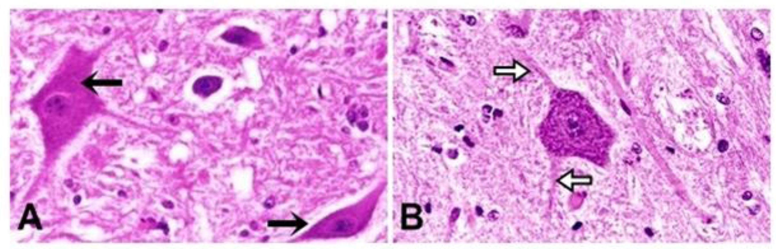
The nucleus was basophilic and the cytoplasm was acidophilic when stained with HE (Figures 1A, 2A and 2B), whereas the nucleus was acidophilic and the cytoplasm was basophilic when stained with MT (Figure 3). Photomicrography taken from these cells showed, in the different areas, cytoplasm with accumulation of deeply basophilic granules distributed in perikaryon, known as Nissl corpuscles, which correspond to rough endoplasmic reticulum, an organelle rich in polyribosomes (Figure 1B). The perikaryon also presented irregular projections of cytoplasm, providing great variation in shape and size of these cells was observed in both groups (Figure 2).
Photomicrographies of neurons from cervical area of spinal cord of control dogs (A, HE, 100x) with acidophilic cytoplasm (薔): and toxoplasmosis seropositive dogs (B, MT, 100x) showing Nissl corpuscles (蚀) in neuron's cytoplasm.
Photomicrographies from thoracic area of spinal cord of control dogs (A; HE, 40x) showing neurons with different sizes and shapes (薔); and toxoplasmosis seropositive dogs (B, HE: 40x) with neuron projections (蚀)
Photomicrographies from lumbar area of spinal cord of control dogs (A, MT, 100x) and toxoplasmosis seropositive dogs (B, MT, 100x) showing neurons with basophilic cytoplasm (薔) and nucleus with visible nucleolus (蚀).
We observed the nucleus of neurons was generally big, spherical or egg shaped, little stained, with loose chromatin and one or more apparent nucleolus, placed in the center of the cellular body (Figure 3A).
The neuron morphological characteristics from cervical, thoracic and lumbar areas from the spinal cord of dogs belonging to group 2 were preserved, and were similar to the neuron characteristics of dogs in group 1. The results of this study concerning the shape of the cytoplasm, quantity of projections of cytoplasm, number of visible nucleolus, and the chromatin pattern of these cells, were included, altogether, in the reports of Junqueira and Carneiro( 1919 Junqueira J, Carneiro F. Tecido nervoso. In: Junqueira J, Carneiro F. Histologia Básica: Texto & Atlas. 12th ed. Rio de Janeiro: Guanabara Koogan; 2013. p. 149-175. Portuguese. ) and Machado( 2121 Machado CRS. Tecido nervoso. In: Machado A. Neuroanatomia Funcional. 2nd ed. São Paulo: Atheneu; 2002. p. 17-32. Portuguese. ).
As stated by Junqueira and Carneiro( 1919 Junqueira J, Carneiro F. Tecido nervoso. In: Junqueira J, Carneiro F. Histologia Básica: Texto & Atlas. 12th ed. Rio de Janeiro: Guanabara Koogan; 2013. p. 149-175. Portuguese. ), Ross and Pawlina( 2020 Ross MH, Pawlina W. Nervous tissue In: Ross MH, Pawlina W. Histology: a text and atlas with correlated cell and molecular biology. 6rd ed. Baltimore: Lippincott Williams & Wilkins; 2011. p. 352-399. ), Machado( 2121 Machado CRS. Tecido nervoso. In: Machado A. Neuroanatomia Funcional. 2nd ed. São Paulo: Atheneu; 2002. p. 17-32. Portuguese. ), and Carvalho et al.( 2424 Carvalho ACF, Pacheco MR, Baraldi-Artoni SM, Mateus O. Morfologia de células neurológicas e imunológicas da medula espinhal de cães (Canis familiaris, Linnaeus, 1758). Acta Scientiarum Animal Science. 2005; 27(3):383-390. ), cytoplasm with accumulation of intensively basophilic granules were noticed, distributed in perikaryon and dendrites, but absent in axon and in its axon hillock.
As for the cytoplasm and neuron nucleus shape, the discoveries from this research agree with Junqueira and Carneiro( 1919 Junqueira J, Carneiro F. Tecido nervoso. In: Junqueira J, Carneiro F. Histologia Básica: Texto & Atlas. 12th ed. Rio de Janeiro: Guanabara Koogan; 2013. p. 149-175. Portuguese. ), Machado( 2121 Machado CRS. Tecido nervoso. In: Machado A. Neuroanatomia Funcional. 2nd ed. São Paulo: Atheneu; 2002. p. 17-32. Portuguese. ), and Carvalho et al.( 2424 Carvalho ACF, Pacheco MR, Baraldi-Artoni SM, Mateus O. Morfologia de células neurológicas e imunológicas da medula espinhal de cães (Canis familiaris, Linnaeus, 1758). Acta Scientiarum Animal Science. 2005; 27(3):383-390. ), when they reveal a cellular body with variable shape and a nucleus that is big, spherical, little stained and with loose chromatin and one or more apparent nucleolus.
The cytoplasmic projections, dendrites and axons, visible in this study, occurred in bigger neurons, confirming the findings by Machado( 2121 Machado CRS. Tecido nervoso. In: Machado A. Neuroanatomia Funcional. 2nd ed. São Paulo: Atheneu; 2002. p. 17-32. Portuguese. ), who reported that routine histological techniques show only the neuronal body and, in bigger neurons, the initial portions of their projections.
The morphological results indicate that there was no visible morphological change, under light microscope, in neurons of animals belonging to group 2 when they were compared to the animals in group 1, in that occasion, what allow us to conclude that the morphological characteristics observed agree with those described in classic literature.
When the cervical area was analyzed no significant difference was observed (p>0.05) for cytoplasm and nucleus of neuron, in both groups; however, the average values of thoracic and lumbar areas showed significant difference (p<0.05) in both groups, and group 2 presented the highest values for this parameter in these areas (Table 1).
Significant difference (p<0.05) was observed in maximum diameter of the nucleus of cervical area and cytoplasm in thoracic area, and the highest average values were found in group 2. There were no significant differences (p>0.05) between groups for the average values of maximum diameter of cytoplasm of cervical area, of nucleus of thoracic area, and of both cytoplasm and nucleus of lumbar area (Table 2).
Significant difference (p<0.05) was observed for minimum diameter between thoracic and lumbar areas, for both cytoplasm nucleus of neurons. The highest values were found in group 2. There were no significant differences (p>0.05) between the groups for minimum diameter of cytoplasm and nucleus of cervical area (Table 3).
No significant difference was observed (p>0.05) for perimeter of cytoplasm of cervical area and of nucleus of neuron from thoracic area forboth groups; however, significant difference (p<0.05) occurred between the groups for the perimeter of nucleus of cervical area, of cytoplasm of thoracic area, and for both cytoplasm and nucleus of lumbar area (Table 4).
Significant difference (p<0.05) was noticed in shape factor in the three areas for both cytoplasm and nucleus of neurons, in most of comparisons, except shape factor of nucleus of thoracic area, where there was no statistic difference (p>0.05) between the groups. The lowest average values were found in group 2, indicating that the neurons of these animals are more rounded shaped than the neurons of animals in group 1 (Table 5).
Concerning the morphometric analysis, we could observe that the neurons of animals belonging to group 2 showed statistically significant differences when compared to the neurons of dogs belonging to group 1. They had higher average values, suggesting that these neurons might be suffering process of edema, making the cell bigger, due to the presence of the parasite inside the neuron, as reported by Summers et al.( 2525 Summers BA, Cummings JF, Lahunta AD. Inflammatory diseases of the central nervous system. In: Summers BA, Cummings JF, Lahunta AD. Veterinary Neuropathology. Saint Louis: Mosby; 1995. p. 162-165. ) and Giraldi et al.( 1717 Giraldi JH, Bracarense APFR, Vidotto O, Tudury EA, Navarro IT, Batista TN. Serology and histopathology of Toxoplasma gondii and Neospora caninum in dogs with neurologic disorders. Semina: Ciências Agrárias. 2002; 23:9-14. ) about dogs with chronic toxoplasmosis with neurological signs, although the most common histopathological changes in CNS are characterized by perivascular cuffing associated to vasculitis, necrosis, malacia, and focal gliosis( 1111 Averill DR, Lahunta A. Toxoplasmosis of the canine nervous system: clinicopathological findings in four cases. Journal of the American Veterinary Medical Association. 1971; 159:1134-1141. , 1717 Giraldi JH, Bracarense APFR, Vidotto O, Tudury EA, Navarro IT, Batista TN. Serology and histopathology of Toxoplasma gondii and Neospora caninum in dogs with neurologic disorders. Semina: Ciências Agrárias. 2002; 23:9-14. ). The characteristics described were different from those of Möhle et al.( 1818 Möhle L, Parlog A, Pahnke J, Dunay IR. Spinal cord pathology in chronic experimental Toxoplasma gondii infection. European Journal of Microbiology and Immunology. 2014; 4(1):65-75. ), who observed high counts of inflammatory cells, cysts mostly in the gray matter, neuronal degeneration and hemorrhage in different segments of spinal cord of T. gondii-infected mice.
Regarding the average values of shape factor of cytoplasm and nucleus of neurons, we observed more rounded-shape neurons in animals of group 2, as they presented the lowest average values, which can be attributed to the possible cellular edema that gave these cells a rounder shape. Therefore, we could conjecture that these neurons lost their characteristic star shape, because, with support by Summers et al.( 2525 Summers BA, Cummings JF, Lahunta AD. Inflammatory diseases of the central nervous system. In: Summers BA, Cummings JF, Lahunta AD. Veterinary Neuropathology. Saint Louis: Mosby; 1995. p. 162-165. ), chronic toxoplasmosis caues areas with cellular edema, as well as loss of division between gray and white matter.
Conclusions
Although the morphological results have not showed differences between the groups and agree with those findings already described in classic literature, the morphometry demonstrated that the neurons of seropositive nonsymptomatic animals presented higher average values than those belonging to control group for all parameters studied, except for shape factor, which were lower. These observations indicate that there were changes in size and structure of these cells, occurring enlargement and loss of star shape, probably due to a cellular edema process. These results suggest that the neurons of these dogs, although nonsymptomatic, can have lost their conductor function.
References
-
1Moretti LD, Ueno TE, Ribeiro MG. Toxoplasmose em cães co-infectados com o vírus da cinomose. Semina: Ciências Agrárias. 2002; 23:85-91.
-
2Thomas WB. Inflammatory diseases of the central nervous system in dogs. Clinical Techniques in Small Animal Practice. 1998; 13(3):167-178.
-
3Paixão TA, Santos RL. Encefalite por Neospora caninum e Toxoplasma gondii em cães. Clínica Veterinária. 2004; 9(48):44-52.
-
4Vidotto O. Toxoplasmose: epidemiologia e importância da doença na saúde animal. Semina: Ciências Agrárias. 1992; 13(1):69-75.
-
5Carini A. Infection spontanée du pigeon et du chien due au Toxoplasma cuniculi Bulletin de la Societé de Pathologie Exotique. 1911; 4:518-519.
-
6Hass JA, Shell L, Saunders G. Neurological manifestations of toxoplasmosis: a literature review and case summary. Journal of America Animal Hospital Association. 1989; 25:253-260.
-
7Germano PML. Estudo sorológico da toxoplasmose canina, pela prova de imunofluorêscencia indireta, na cidade de Campinas em 1981. Revista da Faculdade de Medicina Veterinária e Zootecnia. 1985; 22:53-58.
-
8Lindsay SD. Serological prevalence of Neospora caninum and Toxoplasma gondii in dogs from Kansas. Journal of the Helminthological Society of Washington. 1990; 57(1):86-88.
-
9Guimarães AM, Ribeiro MFB, Lima JD, Cury MC, Spiewak G. Freqüência de anticorpos anti-Toxoplasma gondii em cães de Belo Horizonte, Minas Gerais. Arquivo Brasileiro de Medicina Veterinária e Zootecnia. 1992; 44(1):67-68.
-
10Dubey JP, Lappin MR. Toxoplasmosis and Neosporosis. In: Greene CE. Infectious Diseases of the Dog and Cat. 4th ed. Saint Louis: Elsevier; 2012. p. 806-827.
-
11Averill DR, Lahunta A. Toxoplasmosis of the canine nervous system: clinicopathological findings in four cases. Journal of the American Veterinary Medical Association. 1971; 159:1134-1141.
-
12Nesbit JW, Lourens DC, Williams MC. Spastic paresis in two littermate pups caused by Toxoplasma gondii Journal of the South African Veterinary Association. 1981; 52:243-246.
-
13Suter MM, Hauser B, Palmer DG, Oettli P. Polymyositis - polyradiculitis due to toxoplasmosis in the dog: Serology and tissue biopsy as diagnostic aids. Zentralblatt fuer Veterinaermedizin. 1984; 31(10):792-798.
-
14Braund KG, Blagburn BL, Toivio-Kinnucan M, Amling KA, Pidgeon GL. Toxoplasma polymyositis/polyneuropathy – a new clinical variant in two mature dogs. Journal of the American Animal Hospital Association. 1988; 24(1):93-97.
-
15Davidson MG. Toxoplasmosis. Veterinary Clinics of North America Small Animal Practice. 2000; 30(5):1051-1062.
-
16Swinger RL, Schimdt KA, Dubielzig RR. Keratoconjunctivitis associated with Toxoplasma gondii in a dog. Veterinary Ophthalmology. 2009; 12(1):56-60.
-
17Giraldi JH, Bracarense APFR, Vidotto O, Tudury EA, Navarro IT, Batista TN. Serology and histopathology of Toxoplasma gondii and Neospora caninum in dogs with neurologic disorders. Semina: Ciências Agrárias. 2002; 23:9-14.
-
18Möhle L, Parlog A, Pahnke J, Dunay IR. Spinal cord pathology in chronic experimental Toxoplasma gondii infection. European Journal of Microbiology and Immunology. 2014; 4(1):65-75.
-
19Junqueira J, Carneiro F. Tecido nervoso. In: Junqueira J, Carneiro F. Histologia Básica: Texto & Atlas. 12th ed. Rio de Janeiro: Guanabara Koogan; 2013. p. 149-175. Portuguese.
-
20Ross MH, Pawlina W. Nervous tissue In: Ross MH, Pawlina W. Histology: a text and atlas with correlated cell and molecular biology. 6rd ed. Baltimore: Lippincott Williams & Wilkins; 2011. p. 352-399.
-
21Machado CRS. Tecido nervoso. In: Machado A. Neuroanatomia Funcional. 2nd ed. São Paulo: Atheneu; 2002. p. 17-32. Portuguese.
-
22Carvalho ACF, Pacheco MR, Baraldi-Artoni SM, Mateus O. Canine spinal cord neuron and axon myelin sheath morphometry. Anatomia, Histologia, Embryologia: Journal of Veterinary Medicine. 2006; 35(5):284-286.
-
23Domingues LM, Machado RZ, Tinucci Costa M, Carvalho CS, Costa AJ, Malheiros EB. Canine toxoplasmosis: a comparative evaluation of the detection of anti-toxoplasma gondii antibodies by the indirect immunoenzymatic assay (ELISA) and the indirect immunofluorescence reaction (IFF). Revista Brasileira de Parasitologia Veterinária. 1998; 7(2):79-85.
-
24Carvalho ACF, Pacheco MR, Baraldi-Artoni SM, Mateus O. Morfologia de células neurológicas e imunológicas da medula espinhal de cães (Canis familiaris, Linnaeus, 1758). Acta Scientiarum Animal Science. 2005; 27(3):383-390.
-
25Summers BA, Cummings JF, Lahunta AD. Inflammatory diseases of the central nervous system. In: Summers BA, Cummings JF, Lahunta AD. Veterinary Neuropathology. Saint Louis: Mosby; 1995. p. 162-165.
Publication Dates
-
Publication in this collection
Apr-Jun 2015
History
-
Received
18 June 2012 -
Accepted
22 Jan 2015