Abstract
The aim of this study was to compare the muscle thickness (MT), muscle quality (MQ), and functionality of two different age groups of active elderly women. Twenty seven older women (between 60 and 80 years old), physically active was dividing in two age groups: old 60-66 years (Old) and old 73-80 years (Older) volunteered for the present study. Knee extension isometric peak torque (PT) was obtained during a muscle isometric voluntary contraction (MIVC), and the value was normalized by MT. The Quadriceps Echo Intensity (QEI) was defined as the mean Echo Intensity (EI) value founded in all quadriceps muscle portions in ultrasound imaging. Was found MIVC increased (p=0.024) in the O1 (120.26±24.77 N/m) group when compared with the O2 (99.25±20.95 N/m) group, but when the MIVC was normalized the difference did not exist between groups (p≥0.05). The QEI was found increased in the Older group (97.10±17.35 a.u.) when compared with the Old group (80.18±14.95 a.u.) (p<0.05). Contrary to that, the QMT did not differ significantly between the groups (p≥0.05). The Old group presented a significant higher number of repetitions in the 30SS (14.13±2.29 repetitions) when compared to Older group (11.25±1.95 repetitions) (p<0.05). We observed that even without significantly greater muscle size, the Old group presented the results of 30SS significantly greater when compared with the older group. Besides that, the QEI was found increased in the older group and suggests that EI may be the best predictor of functional capacity than muscle quantity in active older woman.
Key words:
Aged; Aging; Musculoskeletal system; Ultrasonography
Resumo
Comparar a espessura muscular (EM), qualidade muscular (QM), e a funcionalidade entre dois grupos de mulheres idosas. Vinte e sete mulheres idosas (entre 60 e 80 anos) fisicamente ativas foram divididas em dois grupos: Grupo Old (n=15, 60-66 anos) e grupo Older (n=12, 73-80 anos). O pico de torque (PT) isométrico dos extensores de joelho foi obtido por contração isométrica voluntária máxima (CIVM), e seu valor foi normalizado pela EM. A espessura muscular do quadríceps femoral (EMQ) foi definida como o somatório da espessura muscular de todas as porções do quadríceps, e a qualidade muscular foi estimada pela média da Eco Intensidade do quadríceps femoral (EIQ). O grupo Old apresentou maiores valores absolutos de PT (120,3±24,8 N.m) comparado ao grupo Older (99,2±20,9 N.m) (p<0,05), entretanto os valores relativizados por espessura muscular não apresentaram diferenças entre grupos (p≥0,05). A EIQ foi maior no grupo Old (97,1±17,3 u.a.) quando comparado ao grupo Older (80,2±14,9 u.a.) (p<0,05). Não houve diferença significante na EMQ entre os grupos (p≥0,05). O grupo Old realizou um número maior de repetições no teste 30-sit-to-stand (30SS) (14,1±2,3 repetições) comparado ao grupo Older (11,2±1,9 repetições) (p<0,05). O grupo Old realizou um maior número de repetições no teste 30SS quando comparado ao grupo Older, embora ambos os grupos tenham apresentado valores semelhantes de EMQ. A EIQ foi maior no grupo Older, o que sugere que a eco intensidade parece ser uma variável melhor preditora da capacidade funcional do que a massa muscular em mulheres idosas ativa.
Palavras-chave:
Envelhecimento; Idoso; Sistema musculoesquelético; Ultrassonografia
INTRODUCTION
Aging has been associated with loss of muscle quantity accompanied by reduced ability to generate maximal muscle strength, resulting in functional capacity impairments11 Metter EJ, Conwit R, Tobin J, Fozard JL. Age-associated loss of power and strength in the upper extremities in women and men. J Gerontol A Biol Sci Med Sci 1997;52(5):B267-76.,22 Doherty TJ. Invited review: Aging and sarcopenia. J Appl Physiol 2003;95(4):1717-27.. A reduction of almost 50% on skeletal muscle mass is expected to happen from 20 to 90 years of life33 Tzankoff SP, Norris AH. Longitudinal changes in basal metabolism in man. J Appl Physiol Respir Environ Exerc Physiol 1978;45(4):536-9.. Some subjects present an accelerated loss of skeletal muscle mass (that goes beyond the normal age effect), condition known as sarcopenia, which has long been regarded as largely responsible for the decline on muscle function and physical performance22 Doherty TJ. Invited review: Aging and sarcopenia. J Appl Physiol 2003;95(4):1717-27.,44 Evans JG, Bond J. The challenges of age research. Age Ageing 1997;26(Suppl 4):43-6.. The prevalence of sarcopenia is around 25% under the seventh decade of life and may reach 50% from the eighth onwards55 Candow DG, Chilibeck PD. Effect of creatine supplementation during resistance training on muscle accretion in the elderly. J Nutr Health Aging 2007;11(2):185-8..
The quantity of muscle mass may be estimated by skeletal muscle thickness (MT), which can be evaluated by ultrasonography66 Fukumoto Y, Ikezoe T, Yamada Y, Tsukagoshi R, Nakamura M, Mori N, et al. Skeletal muscle quality assessed from echo intensity is associated with muscle strength of middle-aged and elderly persons. Eur J Appl Physiol 2012;112(4):1519-25.. The measure of MT has been associated with the capacity to develop isometric strength in older men and women66 Fukumoto Y, Ikezoe T, Yamada Y, Tsukagoshi R, Nakamura M, Mori N, et al. Skeletal muscle quality assessed from echo intensity is associated with muscle strength of middle-aged and elderly persons. Eur J Appl Physiol 2012;112(4):1519-25.
7 Cadore EL, Izquierdo M, Conceicao M, Radaelli R, Pinto RS, Baroni BM, et al. Echo intensity is associated with skeletal muscle power and cardiovascular performance in elderly men. Exp Gerontol 2012;47(6):473-8.-88 Watanabe Y, Yamada Y, Fukumoto Y, Ishihara T, Yokoyama K, Yoshida T, et al. Echo intensity obtained from ultrasonography images reflecting muscle strength in elderly men. Clin Interv Aging 2013;8(1):993-8.. Furthermore, quadriceps MT is linked to functional performance in the same population66 Fukumoto Y, Ikezoe T, Yamada Y, Tsukagoshi R, Nakamura M, Mori N, et al. Skeletal muscle quality assessed from echo intensity is associated with muscle strength of middle-aged and elderly persons. Eur J Appl Physiol 2012;112(4):1519-25.,99 Rech A, Radaelli R, Goltz FR, da Rosa LH, Schneider CD, Pinto RS. Echo intensity is negatively associated with functional capacity in older women. Age (Dordr) 2014;36(5):9708.,1010 Wilhelm EN, Rech A, Minozzo F, Radaelli R, Botton CE, Pinto RS. Relationship between quadriceps femoris echo intensity, muscle power, and functional capacity of older men. Age (Dordr) 2014;36(3):9625.. However, the measurement of skeletal muscle quantity may overestimate the amount of contractile material available in elderly66 Fukumoto Y, Ikezoe T, Yamada Y, Tsukagoshi R, Nakamura M, Mori N, et al. Skeletal muscle quality assessed from echo intensity is associated with muscle strength of middle-aged and elderly persons. Eur J Appl Physiol 2012;112(4):1519-25.,1111 Goodpaster BH, Carlson CL, Visser M, Kelley DE, Scherzinger A, Harris TB, et al. Attenuation of skeletal muscle and strength in the elderly: The Health ABC Study. J Appl Physiol 2001;90(6):2157-65. and previous studies have reported that the loss of muscle tissue explains a modest part of the decreases in strength and power capacity in older subjects1212 Delmonico MJ, Harris TB, Visser M, Park SW, Conroy MB, Velasquez-Mieyer P, et al. Longitudinal study of muscle strength, quality, and adipose tissue infiltration. Am J Clin Nutr 2009;90(6):1579-85.
13 Clark BC, Fernhall B, Ploutz-Snyder LL. Adaptations in human neuromuscular function following prolonged unweighting: I. Skeletal muscle contractile properties and applied ischemia efficacy. J Appl Physiol 2006;101(1):256-63.-1414 Clark BC, Manini TM, Bolanowski SJ, Ploutz-Snyder LL. Adaptations in human neuromuscular function following prolonged unweighting: II. Neurological properties and motor imagery efficacy. J Appl Physiol 2006;101(1):264-72.. Hence, additional mechanisms may be involved in the age-related loss of skeletal muscle function. In this sense, the muscle quality (MQ) may play an important role in such aging-related changes.
A method widely used to assess the MQ is the evaluation of echo intensity (EI) by ultrasonography1515 Pillen S, Tak RO, Zwarts MJ, Lammens MM, Verrijp KN, Arts IM, et al. Skeletal muscle ultrasound: correlation between fibrous tissue and echo intensity. Ultrasound Med Biol 2009;35(3):443-6.
16 Arts IM, Pillen S, Schelhaas HJ, Overeem S, Zwarts MJ. Normal values for quantitative muscle ultrasonography in adults. Muscle Nerve 2010;41(1):32-41.
17 Nishihara K, Kawai H, Hayashi H, Naruse H, Kimura A, Gomi T, Hoshi F. Frequency analysis of ultrasonic echo intensities of the skeletal muscle in elderly and young individuals. Clin Interv Aging 2014;9(1):1471-8.-1818 Conte M, Vasuri F, Trisolino G, Bellavista E, Santoro A, Degiovanni A, et al. Increased Plin2 expression in human skeletal muscle is associated with sarcopenia and muscle weakness. PLoS One 2013;8(8):e73709.. The skeletal muscle EI has been suggested to reflect a morphological measurement of non-contractile tissue deposition, and has been reported to increase during aging1515 Pillen S, Tak RO, Zwarts MJ, Lammens MM, Verrijp KN, Arts IM, et al. Skeletal muscle ultrasound: correlation between fibrous tissue and echo intensity. Ultrasound Med Biol 2009;35(3):443-6.
16 Arts IM, Pillen S, Schelhaas HJ, Overeem S, Zwarts MJ. Normal values for quantitative muscle ultrasonography in adults. Muscle Nerve 2010;41(1):32-41.-1717 Nishihara K, Kawai H, Hayashi H, Naruse H, Kimura A, Gomi T, Hoshi F. Frequency analysis of ultrasonic echo intensities of the skeletal muscle in elderly and young individuals. Clin Interv Aging 2014;9(1):1471-8.. Accordingly, lipid droplets has also been shown to increase with aging, giving some support to the theory that EI alterations reflect intramuscular changes in adipose tissue composition1818 Conte M, Vasuri F, Trisolino G, Bellavista E, Santoro A, Degiovanni A, et al. Increased Plin2 expression in human skeletal muscle is associated with sarcopenia and muscle weakness. PLoS One 2013;8(8):e73709.. Previous studies have shown significant relationship between quadriceps EI (QEI) and knee extension strength in the older population66 Fukumoto Y, Ikezoe T, Yamada Y, Tsukagoshi R, Nakamura M, Mori N, et al. Skeletal muscle quality assessed from echo intensity is associated with muscle strength of middle-aged and elderly persons. Eur J Appl Physiol 2012;112(4):1519-25.,99 Rech A, Radaelli R, Goltz FR, da Rosa LH, Schneider CD, Pinto RS. Echo intensity is negatively associated with functional capacity in older women. Age (Dordr) 2014;36(5):9708.,1010 Wilhelm EN, Rech A, Minozzo F, Radaelli R, Botton CE, Pinto RS. Relationship between quadriceps femoris echo intensity, muscle power, and functional capacity of older men. Age (Dordr) 2014;36(3):9625.. Likewise, recent investigations suggested that EI may be an alternative parameter to predict the MIVC, dynamic peak torque, power performance and functionality in lower limbs when compared to quadriceps femoris muscle thickness (QMT)66 Fukumoto Y, Ikezoe T, Yamada Y, Tsukagoshi R, Nakamura M, Mori N, et al. Skeletal muscle quality assessed from echo intensity is associated with muscle strength of middle-aged and elderly persons. Eur J Appl Physiol 2012;112(4):1519-25.,77 Cadore EL, Izquierdo M, Conceicao M, Radaelli R, Pinto RS, Baroni BM, et al. Echo intensity is associated with skeletal muscle power and cardiovascular performance in elderly men. Exp Gerontol 2012;47(6):473-8.,1010 Wilhelm EN, Rech A, Minozzo F, Radaelli R, Botton CE, Pinto RS. Relationship between quadriceps femoris echo intensity, muscle power, and functional capacity of older men. Age (Dordr) 2014;36(3):9625..
Despite the large number of studies describing an association of EI with aging, the time-progression of EI while aging, and its ability in discriminate muscular performance has still to be determined. Although a previous study described the behavior of the MQ evaluated by EI in different age groups1616 Arts IM, Pillen S, Schelhaas HJ, Overeem S, Zwarts MJ. Normal values for quantitative muscle ultrasonography in adults. Muscle Nerve 2010;41(1):32-41., only the rectus femoris (RF) and vastus intermedius
(VI) were measured by these authors. Given the aforementioned, the aim of this study was to compare the maximal strength, MT, QEI measured by the sum of all portions of quadriceps femoris, and functional capacity of the lower limbs from active older women of two different age groups.
METHODOLOGICAL PROCEDURES
Participants
Twenty seven healthy elderly women from 59 to 83 years volunteered to participate in this study. All women were physically active and free from neurological, cardiovascular and orthopedics conditions that would restrict them from performing physical exercise. Participants were divided into two groups according to their age range: 59-66 years (Old) and 73-83 years (Older). All participants were carefully informed of the possible risks and discomforts associated with the study and written consent was obtained prior to participation. The study was conducted according the declaration of Helsinki and all procedures were approved by the local Institutional Research Ethics committee.
Experimental procedures
Each subject visited the exercise research laboratory on two non-consecutive days. On the first visit subjects were interviewed about their habits, classified regarding physical active levels according to the International Physical Activity Questionnaire (IPAQ)1919 Craig CL, Marshall AL, Sjostrom M, Bauman AE, Booth ML, Ainsworth BE, et al. International physical activity questionnaire: 12-country reliability and validity. Med Sci Sports Exerc 2003;35(8):1381-95., and familiarized with the study procedures. Participants characterized as physically active were included in the study. The familiarization session, involving all physical tests [i.e. isometric strength, and 30 seconds sit to stand up test (30SS)] was conducted to reduce possible learning effects associated with the experimental protocol. The second visit took part 48h after the familiarization, and anthropometric measurements, as well as B-mode ultrasound images of the quadriceps femoris were obtained to evaluate the MT and EI. Subsequently the maximal isometric strength of the knee extensors and 30SS were measured.
Anthropometric measurements and bioelectrical impedance
Total body mass, height, body mass index (BMI) and body composition were evaluated for participant characterization. Total body mass (kg) was measured using a standard scale (Filizola, Brazil), and a linked stadiometer was used to verify stature (m). The BMI was calculated as body mass (kg) divided by the squared height (m2). The percentage of body fat (%BF) was obtained by bioelectrical impedance with a Maltron BF-906 Body Fat Analyzer (Maltron International, UK). All the measurements were performed in the morning, between 7.30-9.00 a.m. Subjects arrived at the laboratory in a fasted state (8-10 hours), and were instructed to avoid physical exercise for at least 8 hours prior the arrival at the laboratory to avoid confounding factors such as hydration status and fatigue associated with the post exercise period.
Maximal Strength Measurement
Knee extension isometric peak torque were obtained during a maximal isometric voluntary contraction (MIVC) which was performed on a isokinetic dynamometer (Cybex Norm, USA), connected to a 2000 Hz analog digital (A/D) converter (Miotec Equipamentos Biomédicos, Brazil). Subjects were seated with hips flexed at 85° (0° = anatomic position) and the lateral femoral condyle of the right leg was aligned with the axis of rotation of the dynamometer. A warm-up (10 submaximal isokinetic knee extension/flexion repetitions at 120°/s) was performed and after 30 seconds subjects performed one submaximal isometric knee extension to remember with the testing commands and procedures. Thereafter, three 5-second knee extension MIVC were performed at knee joint angle of 60° (0° = knee fully extended), with 90 seconds of rest intervals between trials. The peak torque was determined by the dynamometer software (Humac version 9.6.2, Cybex Norm, USA) and the highest value was used for further analysis. Furthermore, the relative maximal strength was obtained by specific tension (ST), from quotient between the peak torque and subject's quadriceps muscle thickness. In this case, the ST was determined by the following formula: unilateral knee extension MIVC (N.m) / quadriceps muscle thickness (mm).
Muscle thickness and echo intensity measurements
Ultrasound images were obtained using real-time B-mode ultrasonography with a 38-mm, 9.0 MHz linear-array probe (Nemio XG ultrasound, Toshiba, Japan). Before any assessment, subjects rested in supine position with their lower limbs extended and relaxed for 15 minutes to allow fluid shifts to stabilize2020 Berg HE, Tedner B, Tesch PA. Changes in lower limb muscle cross-sectional area and tissue fluid volume after transition from standing to supine. Acta Physiol Scand 1993;148(4):379-85.. Transversal images of the right vastus lateralis (VL), RF, VI and vastus medialis (VM) muscles were acquired. The probe was coated with a water-soluble transmission gel to provide acoustic contact and care was taken to avoid the excessive compress of the dermal surface. The measurement sites were the same as those adopted in previous studies2121 Korhonen MT, Mero AA, Alen M, Sipila S, Hakkinen K, Liikavainio T, et al. Biomechanical and skeletal muscle determinants of maximum running speed with aging. Med Sci Sports Exerc 2009;41(4):844-56.. All images were acquired and analyzed by the same investigator.
Single images of each muscle were digitized and analyzed using ImageJ 1.42q software (National Institute of Health, USA). Regions of interest of each quadriceps muscle portion (VL, RF, VI and VM) were selected, including as much muscle as possible but avoiding other tissues (such as bone and surrounding fascia) for EI calculation of each component of quadriceps femoris. The mean EI was determined using a standard grayscale histogram function and expressed as a value between 0 (black) and 255 (white). Thereafter, the QEI were determined from the average EI value from all quadriceps muscle portions (QEI = [VL Echo Intensity + RF Echo Intensity + VI Echo Intensity + VM Echo Intensity] / 4). The MT was determined as the distance between adipose tissue-muscle interface for VL, RF, and VM, and as bone-muscle interface for VI. Whole QMT was obtained as the sum of the four individual quadriceps portions (QMT = [VL Muscle Thickness + RF Muscle Thickness + VI Muscle Thickness + VM Muscle Thickness])99 Rech A, Radaelli R, Goltz FR, da Rosa LH, Schneider CD, Pinto RS. Echo intensity is negatively associated with functional capacity in older women. Age (Dordr) 2014;36(5):9708.,1010 Wilhelm EN, Rech A, Minozzo F, Radaelli R, Botton CE, Pinto RS. Relationship between quadriceps femoris echo intensity, muscle power, and functional capacity of older men. Age (Dordr) 2014;36(3):9625.,2222 Pinto RS, Correa CS, Radaelli R, Cadore EL, Brown LE, Bottaro M. Short-term strength training improves muscle quality and functional capacity of elderly women. Age (Dordr) 2014;36(1):365-72..
30-s sit-to-stand test
To assess functional capacity, the 30SS test was performed with a standard chair (42 cm)2323 Jones CJ, Rikli RE, Beam WC. A 30-s chair-stand test as a measure of lower body strength in community-residing older adults. Res Q Exerc Sport 1999;70(2):113-9.. Subjects were seated on the chair with both hands crossed over their chest, and feet shoulder-width apart. Five sub-maximal repetitions were performed as a warm-up and, after a 30 second interval subjects completed the following functional test; after a verbal signal, subjects should rose to an upright position before returning to the initial position. The test was timed with a digital stopwatch by the researcher and subjects were instructed to perform as many repetitions as possible during the 30 second time-period.
Data Analysis
All values are reported as mean ± standard deviation (SD). Normal distribution and homogeneity parameters were checked with Shapiro-wilk and Levene's test respectively. After the data showed normality and homogeneity (p≥0.05), an independent sample t test was performed for comparison between Old and Older group and identify significant differences. The level of significance was set at p<0.05. Statistical analysis was performed using SPSS version 20.0 (IBM, Somers, NY, USA).
RESULTS
The physical characteristics are shown in Table 1.
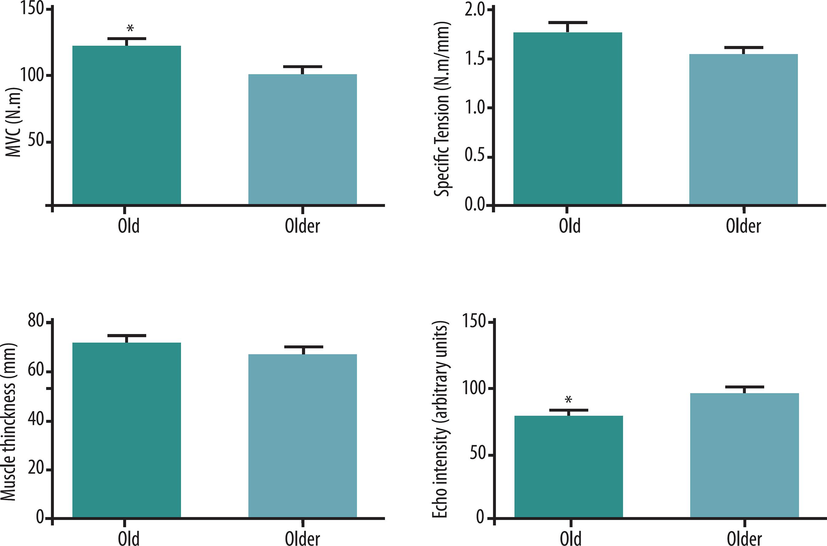
The MIVC was significantly greater (p<0.05) in the Old group (120.3±24.8 N/m) when compared to the Older group (99.2±20.9 N/m). However, when the value was normalized by ST, no significant difference (p≥0.05) was observed between the Old and Older group. A greater QEI value was found (p<0.05) in the Older (97.1±17.3 a.u.) compared to the Old group (80.2±14.9 a.u.). In contrast, the QMT did not differ significantly between groups (p≥0.05) (Figure 1).
Between group comparison of maximal isometric voluntary contraction (MIVC), specific tension (ST), quadriceps muscle thickness, and quadriceps echo intensity; Old group: aged between 60 and 66 years; Older group: aged between 73 and 83 years; *: Significant different from Older group (p<0.05).
The results of 30SS are presented in Figure 2. The Old group performed a significantly more (p<0.05) repetitions (14.1±2.3 repetitions) in the 30SS test than the older group (11.2±1.9 repetitions).
Total number of repetitions performed in the 30-sit-to-stand test; Old group: aged between 60 and 66 years; Older group: aged between 73 and 83 years; *:Significant different from Older group (p<0.05).
DISCUSSION
The aim of this study was to compare the maximal strength, muscle mass, muscle quality, and functional performance obtained in two different age groups of active elderly women. We observed that although muscle mass has been similar in both groups, the Old group showed better performance in the 30SS compared with the Older group. Besides that, the QEI was higher in the Older, suggesting that active women in the eighth decade of life may present impairments in QEI and functional performance when compared to active women at seventh decade of life, even though they presented similar MT. Furthermore, our results suggest that muscle composition may be more important to muscle functional performance than muscle size in active elderly women.
Muscle strength is recognized to be a powerful indicator of functional performance. Our results showed higher MIVC values in the Old group compared with the Older group. Differently from our results, Power et al.2424 Power GA, Allen MD, Booth WJ, Thompson RT, Marsh GD, Rice CL. The influence on sarcopenia of muscle quality and quantity derived from magnetic resonance imaging and neuromuscular properties. Age (Dordr) 2014; 36(3):9642. did not found significantly difference in MIVC of dorsiflexor muscles between, men aged from 60 to 73 years (39.8±6.5 N.m), and older men aged between 76 and 85 years (40.9±6.4 N.m)2424 Power GA, Allen MD, Booth WJ, Thompson RT, Marsh GD, Rice CL. The influence on sarcopenia of muscle quality and quantity derived from magnetic resonance imaging and neuromuscular properties. Age (Dordr) 2014; 36(3):9642.. The different gender and muscle group assessed may have contributed to these discrepant results. However, in our study no significant differences were observed when the strength was normalized by muscle thickness. That was an unexpected result since ST has been shown be higher affected by aging process2525 Tracy BL, Ivey FM, Hurlbut D, Martel GF, Lemmer JT, Siegel EL, et al. Muscle quality. II. Effects of strength training in 65-to 75-yr-old men and women. J Appl Physiol 2009; 86(1):195-201.. For possible explanation, this age difference may not present changes enough for significant alterations in force integrity per muscle thickness between groups.
Regarding muscle quantity, our results did not show significant group differences in QMT. Likewise, Arts et al.1616 Arts IM, Pillen S, Schelhaas HJ, Overeem S, Zwarts MJ. Normal values for quantitative muscle ultrasonography in adults. Muscle Nerve 2010;41(1):32-41. showed in regression analysis a small age-related decrease in RF MT and VI MT of women compared to men1616 Arts IM, Pillen S, Schelhaas HJ, Overeem S, Zwarts MJ. Normal values for quantitative muscle ultrasonography in adults. Muscle Nerve 2010;41(1):32-41.. The later had thicker muscles but presented a larger age-related decline in muscle mass, and strength. An interesting result was the slower decline in age-related muscle thickness in elderly women, suggesting gender differences in muscle quantity1616 Arts IM, Pillen S, Schelhaas HJ, Overeem S, Zwarts MJ. Normal values for quantitative muscle ultrasonography in adults. Muscle Nerve 2010;41(1):32-41.. These results indicate that the QMT may suffer little deleterious effects from sixth to ninth decade of life on women1616 Arts IM, Pillen S, Schelhaas HJ, Overeem S, Zwarts MJ. Normal values for quantitative muscle ultrasonography in adults. Muscle Nerve 2010;41(1):32-41.. However, the MT may not be representative by exclusively contractile tissue66 Fukumoto Y, Ikezoe T, Yamada Y, Tsukagoshi R, Nakamura M, Mori N, et al. Skeletal muscle quality assessed from echo intensity is associated with muscle strength of middle-aged and elderly persons. Eur J Appl Physiol 2012;112(4):1519-25., and hence careful should be taken when the muscle size is used to compare different populations.
Previous investigations reported significant associations between MT of knee extensors and maximal knee extension strength66 Fukumoto Y, Ikezoe T, Yamada Y, Tsukagoshi R, Nakamura M, Mori N, et al. Skeletal muscle quality assessed from echo intensity is associated with muscle strength of middle-aged and elderly persons. Eur J Appl Physiol 2012;112(4):1519-25.,88 Watanabe Y, Yamada Y, Fukumoto Y, Ishihara T, Yokoyama K, Yoshida T, et al. Echo intensity obtained from ultrasonography images reflecting muscle strength in elderly men. Clin Interv Aging 2013;8(1):993-8.
9 Rech A, Radaelli R, Goltz FR, da Rosa LH, Schneider CD, Pinto RS. Echo intensity is negatively associated with functional capacity in older women. Age (Dordr) 2014;36(5):9708.-1010 Wilhelm EN, Rech A, Minozzo F, Radaelli R, Botton CE, Pinto RS. Relationship between quadriceps femoris echo intensity, muscle power, and functional capacity of older men. Age (Dordr) 2014;36(3):9625.. Significant differences between groups were observed regarding muscle strength, it was expected in the current study that the older group would exhibit a lower QMT when compared to the old group. However, both groups showed similar muscle thickness. These differences between Arts et al.1616 Arts IM, Pillen S, Schelhaas HJ, Overeem S, Zwarts MJ. Normal values for quantitative muscle ultrasonography in adults. Muscle Nerve 2010;41(1):32-41. results and ours may be due to two factors: (1) different to Arts et al.1616 Arts IM, Pillen S, Schelhaas HJ, Overeem S, Zwarts MJ. Normal values for quantitative muscle ultrasonography in adults. Muscle Nerve 2010;41(1):32-41., in which QMT was measured by the sum of RF and VI1616 Arts IM, Pillen S, Schelhaas HJ, Overeem S, Zwarts MJ. Normal values for quantitative muscle ultrasonography in adults. Muscle Nerve 2010;41(1):32-41., whereas in the current study the sum of four quadriceps femoris portions was utilized to define QMT, what is believed to better represent the knee extensor muscles; and (2) Arts et al.1616 Arts IM, Pillen S, Schelhaas HJ, Overeem S, Zwarts MJ. Normal values for quantitative muscle ultrasonography in adults. Muscle Nerve 2010;41(1):32-41. study had 6 participants in each age groups, whereas at the present investigation counted with 12 and 15 participants in age groups. These differences and results may suggest that some neural component or changes in intramuscular composition may be more important for determination of the strength capacity than muscle size in active elderly women.
Some previous studies have reported that EI is a predictor of strength capacity66 Fukumoto Y, Ikezoe T, Yamada Y, Tsukagoshi R, Nakamura M, Mori N, et al. Skeletal muscle quality assessed from echo intensity is associated with muscle strength of middle-aged and elderly persons. Eur J Appl Physiol 2012;112(4):1519-25.
7 Cadore EL, Izquierdo M, Conceicao M, Radaelli R, Pinto RS, Baroni BM, et al. Echo intensity is associated with skeletal muscle power and cardiovascular performance in elderly men. Exp Gerontol 2012;47(6):473-8.-88 Watanabe Y, Yamada Y, Fukumoto Y, Ishihara T, Yokoyama K, Yoshida T, et al. Echo intensity obtained from ultrasonography images reflecting muscle strength in elderly men. Clin Interv Aging 2013;8(1):993-8.,2626 Strasser EM, Draskovits T, Praschak M, Quittan M, Graf A. Association between ultrasound measurements of muscle thickness, pennation angle, echogenicity and skeletal muscle strength in the elderly. Age (Dordr) 2013; 35(5):2025-6. and functionality99 Rech A, Radaelli R, Goltz FR, da Rosa LH, Schneider CD, Pinto RS. Echo intensity is negatively associated with functional capacity in older women. Age (Dordr) 2014;36(5):9708.,1010 Wilhelm EN, Rech A, Minozzo F, Radaelli R, Botton CE, Pinto RS. Relationship between quadriceps femoris echo intensity, muscle power, and functional capacity of older men. Age (Dordr) 2014;36(3):9625. in elderly population. The inverse relationship between QEI and functional capacity observed in previous studies99 Rech A, Radaelli R, Goltz FR, da Rosa LH, Schneider CD, Pinto RS. Echo intensity is negatively associated with functional capacity in older women. Age (Dordr) 2014;36(5):9708.,1010 Wilhelm EN, Rech A, Minozzo F, Radaelli R, Botton CE, Pinto RS. Relationship between quadriceps femoris echo intensity, muscle power, and functional capacity of older men. Age (Dordr) 2014;36(3):9625.,1717 Nishihara K, Kawai H, Hayashi H, Naruse H, Kimura A, Gomi T, Hoshi F. Frequency analysis of ultrasonic echo intensities of the skeletal muscle in elderly and young individuals. Clin Interv Aging 2014;9(1):1471-8. suggest that smaller EI values may indicate reduced deposition of non-contractile material on skeletal muscles and, thus, increase the strength capacity for the same muscle volume. In agreement with these findings, we found lower QEI and higher number of repetitions performed in the 30SS test in the Old group. Likewise, Wilhelm et al.1010 Wilhelm EN, Rech A, Minozzo F, Radaelli R, Botton CE, Pinto RS. Relationship between quadriceps femoris echo intensity, muscle power, and functional capacity of older men. Age (Dordr) 2014;36(3):9625. found a significant negative correlation (r=-0.502) between QEI and the number of repetitions in 30SS test, and the authors suggested a relevant influence of QEI on elderly population functionality. These findings are in accordance with the suggestion that non-contractile muscle composition (in this study estimated by QEI) may be more representative of the muscle functionality than muscle size99 Rech A, Radaelli R, Goltz FR, da Rosa LH, Schneider CD, Pinto RS. Echo intensity is negatively associated with functional capacity in older women. Age (Dordr) 2014;36(5):9708.,1010 Wilhelm EN, Rech A, Minozzo F, Radaelli R, Botton CE, Pinto RS. Relationship between quadriceps femoris echo intensity, muscle power, and functional capacity of older men. Age (Dordr) 2014;36(3):9625..
It has been reported that muscle size and muscle quality are determinant for muscle strength66 Fukumoto Y, Ikezoe T, Yamada Y, Tsukagoshi R, Nakamura M, Mori N, et al. Skeletal muscle quality assessed from echo intensity is associated with muscle strength of middle-aged and elderly persons. Eur J Appl Physiol 2012;112(4):1519-25.. Fukumoto et al.66 Fukumoto Y, Ikezoe T, Yamada Y, Tsukagoshi R, Nakamura M, Mori N, et al. Skeletal muscle quality assessed from echo intensity is associated with muscle strength of middle-aged and elderly persons. Eur J Appl Physiol 2012;112(4):1519-25. reported that MT and EI act independently on isometric knee extension in women of different ages (51-87 years). These results may point out that both muscle quantity and muscle quality contribute to muscle strength. Differently, our results did not show significant differences in QMT between groups, however a significantly higher QEI and lower MIVC were found in the Older group. As a possible explanation, Fukumoto et al.66 Fukumoto Y, Ikezoe T, Yamada Y, Tsukagoshi R, Nakamura M, Mori N, et al. Skeletal muscle quality assessed from echo intensity is associated with muscle strength of middle-aged and elderly persons. Eur J Appl Physiol 2012;112(4):1519-25. suggested that substitutions of contractile tissue per adipose and fibrous tissue are larger than muscle size decrease and this could influence QMT values. Moreover, the current findings suggest that for elderly women in sixth and seventh decade of life the quality of muscles may be more relevant to strength capacity than muscle size. Similarities may be found when the ST is carried out by muscle thickness.
In agreement with previous studies66 Fukumoto Y, Ikezoe T, Yamada Y, Tsukagoshi R, Nakamura M, Mori N, et al. Skeletal muscle quality assessed from echo intensity is associated with muscle strength of middle-aged and elderly persons. Eur J Appl Physiol 2012;112(4):1519-25.,1616 Arts IM, Pillen S, Schelhaas HJ, Overeem S, Zwarts MJ. Normal values for quantitative muscle ultrasonography in adults. Muscle Nerve 2010;41(1):32-41., we found that the QEI was greater in the Older group when compared with Old group, following a significant lower number of repetitions in the 30SS test. In recent studies from our research group99 Rech A, Radaelli R, Goltz FR, da Rosa LH, Schneider CD, Pinto RS. Echo intensity is negatively associated with functional capacity in older women. Age (Dordr) 2014;36(5):9708.,1010 Wilhelm EN, Rech A, Minozzo F, Radaelli R, Botton CE, Pinto RS. Relationship between quadriceps femoris echo intensity, muscle power, and functional capacity of older men. Age (Dordr) 2014;36(3):9625. it was observed that the muscle quality (estimated by QEI) may be more representative of the muscle functionality than muscle size99 Rech A, Radaelli R, Goltz FR, da Rosa LH, Schneider CD, Pinto RS. Echo intensity is negatively associated with functional capacity in older women. Age (Dordr) 2014;36(5):9708.,1010 Wilhelm EN, Rech A, Minozzo F, Radaelli R, Botton CE, Pinto RS. Relationship between quadriceps femoris echo intensity, muscle power, and functional capacity of older men. Age (Dordr) 2014;36(3):9625.. In current study, Older group performed about ~20% less repetitions on the 30SS compared to Old group. This finding corroborates with Rech et al.99 Rech A, Radaelli R, Goltz FR, da Rosa LH, Schneider CD, Pinto RS. Echo intensity is negatively associated with functional capacity in older women. Age (Dordr) 2014;36(5):9708. that found a significant negative correlation (r=-0.493; p<0.01) between QEI and the number of repetitions performed on 30SS test. In additional, they did not found association between maximal isometric knee extension and functional tests (30SS and usual gait speed) (r=0.247, p>0.05), suggesting that EI may be a relevant predictor of functional capacity more than maximal strength99 Rech A, Radaelli R, Goltz FR, da Rosa LH, Schneider CD, Pinto RS. Echo intensity is negatively associated with functional capacity in older women. Age (Dordr) 2014;36(5):9708.,1010 Wilhelm EN, Rech A, Minozzo F, Radaelli R, Botton CE, Pinto RS. Relationship between quadriceps femoris echo intensity, muscle power, and functional capacity of older men. Age (Dordr) 2014;36(3):9625.. However, more studies are necessary to investigate if QEI is more representative of functionality than muscle strength in both elderly men and women.
CONCLUSION
In summary, the current results demonstrate significant differences between Old and Older groups regarding EI and 30SS test, although no differences were observed in QMT. These findings suggest that QMT may be less sensitive than muscle quality (evaluated by EI) for track changes in functional performance. A putative explanation is that aging-related infiltration of non-contractile tissue in the skeletal muscles may hinder the decrease in muscle size, and therefore mask the actual lost in contractile tissue availability, although this remains speculative. Therefore, these results suggest that physical interventions for older women in the seventh decade of life should focus not only on increasing muscle size, but also on improving muscle quality.
Acknowledgments
We would like to thank the CNPq (Conselho Nacional de Desenvolvimento Científico e Tecnológico) and CAPES (Coordenação de Aperfeiçoamento de Pessoal de Nível Superior) for their financial support.
REFERENCES
-
1Metter EJ, Conwit R, Tobin J, Fozard JL. Age-associated loss of power and strength in the upper extremities in women and men. J Gerontol A Biol Sci Med Sci 1997;52(5):B267-76.
-
2Doherty TJ. Invited review: Aging and sarcopenia. J Appl Physiol 2003;95(4):1717-27.
-
3Tzankoff SP, Norris AH. Longitudinal changes in basal metabolism in man. J Appl Physiol Respir Environ Exerc Physiol 1978;45(4):536-9.
-
4Evans JG, Bond J. The challenges of age research. Age Ageing 1997;26(Suppl 4):43-6.
-
5Candow DG, Chilibeck PD. Effect of creatine supplementation during resistance training on muscle accretion in the elderly. J Nutr Health Aging 2007;11(2):185-8.
-
6Fukumoto Y, Ikezoe T, Yamada Y, Tsukagoshi R, Nakamura M, Mori N, et al. Skeletal muscle quality assessed from echo intensity is associated with muscle strength of middle-aged and elderly persons. Eur J Appl Physiol 2012;112(4):1519-25.
-
7Cadore EL, Izquierdo M, Conceicao M, Radaelli R, Pinto RS, Baroni BM, et al. Echo intensity is associated with skeletal muscle power and cardiovascular performance in elderly men. Exp Gerontol 2012;47(6):473-8.
-
8Watanabe Y, Yamada Y, Fukumoto Y, Ishihara T, Yokoyama K, Yoshida T, et al. Echo intensity obtained from ultrasonography images reflecting muscle strength in elderly men. Clin Interv Aging 2013;8(1):993-8.
-
9Rech A, Radaelli R, Goltz FR, da Rosa LH, Schneider CD, Pinto RS. Echo intensity is negatively associated with functional capacity in older women. Age (Dordr) 2014;36(5):9708.
-
10Wilhelm EN, Rech A, Minozzo F, Radaelli R, Botton CE, Pinto RS. Relationship between quadriceps femoris echo intensity, muscle power, and functional capacity of older men. Age (Dordr) 2014;36(3):9625.
-
11Goodpaster BH, Carlson CL, Visser M, Kelley DE, Scherzinger A, Harris TB, et al. Attenuation of skeletal muscle and strength in the elderly: The Health ABC Study. J Appl Physiol 2001;90(6):2157-65.
-
12Delmonico MJ, Harris TB, Visser M, Park SW, Conroy MB, Velasquez-Mieyer P, et al. Longitudinal study of muscle strength, quality, and adipose tissue infiltration. Am J Clin Nutr 2009;90(6):1579-85.
-
13Clark BC, Fernhall B, Ploutz-Snyder LL. Adaptations in human neuromuscular function following prolonged unweighting: I. Skeletal muscle contractile properties and applied ischemia efficacy. J Appl Physiol 2006;101(1):256-63.
-
14Clark BC, Manini TM, Bolanowski SJ, Ploutz-Snyder LL. Adaptations in human neuromuscular function following prolonged unweighting: II. Neurological properties and motor imagery efficacy. J Appl Physiol 2006;101(1):264-72.
-
15Pillen S, Tak RO, Zwarts MJ, Lammens MM, Verrijp KN, Arts IM, et al. Skeletal muscle ultrasound: correlation between fibrous tissue and echo intensity. Ultrasound Med Biol 2009;35(3):443-6.
-
16Arts IM, Pillen S, Schelhaas HJ, Overeem S, Zwarts MJ. Normal values for quantitative muscle ultrasonography in adults. Muscle Nerve 2010;41(1):32-41.
-
17Nishihara K, Kawai H, Hayashi H, Naruse H, Kimura A, Gomi T, Hoshi F. Frequency analysis of ultrasonic echo intensities of the skeletal muscle in elderly and young individuals. Clin Interv Aging 2014;9(1):1471-8.
-
18Conte M, Vasuri F, Trisolino G, Bellavista E, Santoro A, Degiovanni A, et al. Increased Plin2 expression in human skeletal muscle is associated with sarcopenia and muscle weakness. PLoS One 2013;8(8):e73709.
-
19Craig CL, Marshall AL, Sjostrom M, Bauman AE, Booth ML, Ainsworth BE, et al. International physical activity questionnaire: 12-country reliability and validity. Med Sci Sports Exerc 2003;35(8):1381-95.
-
20Berg HE, Tedner B, Tesch PA. Changes in lower limb muscle cross-sectional area and tissue fluid volume after transition from standing to supine. Acta Physiol Scand 1993;148(4):379-85.
-
21Korhonen MT, Mero AA, Alen M, Sipila S, Hakkinen K, Liikavainio T, et al. Biomechanical and skeletal muscle determinants of maximum running speed with aging. Med Sci Sports Exerc 2009;41(4):844-56.
-
22Pinto RS, Correa CS, Radaelli R, Cadore EL, Brown LE, Bottaro M. Short-term strength training improves muscle quality and functional capacity of elderly women. Age (Dordr) 2014;36(1):365-72.
-
23Jones CJ, Rikli RE, Beam WC. A 30-s chair-stand test as a measure of lower body strength in community-residing older adults. Res Q Exerc Sport 1999;70(2):113-9.
-
24Power GA, Allen MD, Booth WJ, Thompson RT, Marsh GD, Rice CL. The influence on sarcopenia of muscle quality and quantity derived from magnetic resonance imaging and neuromuscular properties. Age (Dordr) 2014; 36(3):9642.
-
25Tracy BL, Ivey FM, Hurlbut D, Martel GF, Lemmer JT, Siegel EL, et al. Muscle quality. II. Effects of strength training in 65-to 75-yr-old men and women. J Appl Physiol 2009; 86(1):195-201.
-
26Strasser EM, Draskovits T, Praschak M, Quittan M, Graf A. Association between ultrasound measurements of muscle thickness, pennation angle, echogenicity and skeletal muscle strength in the elderly. Age (Dordr) 2013; 35(5):2025-6.
Publication Dates
-
Publication in this collection
May-Jun 2015
History
-
Received
01 Feb 2015 -
Accepted
21 Apr 2015