Abstracts
A histological analysis was conducted on the gills of 15 Piaractus mesopotamicus and 19 Prochilodus lineatus specimens collected between April and November 2004 from the Aquidauana River, State of Mato Grosso do Sul, Central-West Brazil, to describe the anatomopathological characteristics of the gills of these freshwater fish. Gill samples were fixated in 10% buffered formalin and processed following histological routine procedures. The histological examination of the gills of P. mesopotamicus revealed intralamellar monogenean and mixosporean cysts of Henneguya piaractus at several developmental stages over the entire (basal, median and distal) lamella. Intraepithelial cysts caused lamella dilation and deformity of adjacent lamellae. In P. lineatus gills, monogenean cysts were detected. In both host species, hyperplasia of the gill epithelium and structural disorganization of secondary lamellae was seen diffusely in the gills, leading to fused lamellae in the gills. In few cases, there was found mononuclear inflammatory cells and hemorrhagic focal points distally in the lamellae.
Tissues alterations; parasites; gills; fish
Realizou-se análise histológica de brânquias de 15 espécimes de Piaractus mesopotamicus e 19 Prochilodus lineatus coletados de abril a novembro de 2004, no Rio Aquidauana, MS, com intuito de contribuir com achados anatomopatológicos em brânquias dessas espécies de peixes de água doce. Amostras de brânquias foram fixadas em formalina 10%, tamponadas e processadas conforme rotina histológica. Em P. mesopotamicus observou-se presença de monogênea e cistos de mixosporídio da espécie Henneguya piaractus, com localização intralamelar em vários estágios de desenvolvimento, localizados em todas as regiões (basal, mediana ou distal) das lamelas. Cistos intraepiteliais causaram dilatação e deformação das lamelas vizinhas. Em brânquias de P. lineatus, observou-se presença de monogênea. Nas duas espécies de hospedeiro foram registradas hiperplasia do epitélio branquial e desorganização estrutural das lamelas em extensas regiões, alterações que causaram a fusão lamelar. Em poucos casos registrou-se presença de células inflamatórias mononucleares e focos hemorrágicos na região distal das lamelas.
Alterações teciduais; parasitos; brânquias; peixes
RESEARCH NOTE
Histopathology of gills of Piaractus mesopotamicus (Holmberg, 1887) and Prochilodus lineatus (Valenciennes, 1836) infested by monogenean and myxosporea, caugth in Aquidauana River, State of Mato Grosso do Sul, Brazil
Histopatologia de brânquias de Piaractus mesopotamicus (Holmberg, 1887) e Prochilodus lineatus (Valenciennes, 1836) parasitados por monogêneas e mixosporídios, capturados no Rio Aquidauana, Mato Grosso do Sul, Brasil
Cristiane Meldau de CamposI; Julieta Rondini Engrácia de MoraesII; Flávio Ruas de MoraesII
IUnidade Universitária de Aquidauana, Universidade Estadual de Mato Grosso do Sul UEMS
IIDepartamento de Patologia Veterinária, Centro de Aqüicultura, Universidade Estadual Paulista UNESP
Corresponding author Corresponding author: Cristiane Meldau de Campos Unidade Universitária de Aquidauana Universidade Estadual de Mato Grosso do Sul UEMS Rod. Aquidauana-UEMS, Km 12, Zona Rural CEP 79200-000, Aquidauana - MS, Brazil e-mail: cmeldau@uems.br
ABSTRACT
A histological analysis was conducted on the gills of 15 Piaractus mesopotamicus and 19 Prochilodus lineatus specimens collected between April and November 2004 from the Aquidauana River, State of Mato Grosso do Sul, Central-West Brazil, to describe the anatomopathological characteristics of the gills of these freshwater fish. Gill samples were fixated in 10% buffered formalin and processed following histological routine procedures. The histological examination of the gills of P. mesopotamicus revealed intralamellar monogenean and mixosporean cysts of Henneguya piaractus at several developmental stages over the entire (basal, median and distal) lamella. Intraepithelial cysts caused lamella dilation and deformity of adjacent lamellae. In P. lineatus gills, monogenean cysts were detected. In both host species, hyperplasia of the gill epithelium and structural disorganization of secondary lamellae was seen diffusely in the gills, leading to fused lamellae in the gills. In few cases, there was found mononuclear inflammatory cells and hemorrhagic focal points distally in the lamellae.
Keywords: Tissues alterations, parasites, gills, fish.
RESUMO
Realizou-se análise histológica de brânquias de 15 espécimes de Piaractus mesopotamicus e 19 Prochilodus lineatus coletados de abril a novembro de 2004, no Rio Aquidauana, MS, com intuito de contribuir com achados anatomopatológicos em brânquias dessas espécies de peixes de água doce. Amostras de brânquias foram fixadas em formalina 10%, tamponadas e processadas conforme rotina histológica. Em P. mesopotamicus observou-se presença de monogênea e cistos de mixosporídio da espécie Henneguya piaractus, com localização intralamelar em vários estágios de desenvolvimento, localizados em todas as regiões (basal, mediana ou distal) das lamelas. Cistos intraepiteliais causaram dilatação e deformação das lamelas vizinhas. Em brânquias de P. lineatus, observou-se presença de monogênea. Nas duas espécies de hospedeiro foram registradas hiperplasia do epitélio branquial e desorganização estrutural das lamelas em extensas regiões, alterações que causaram a fusão lamelar. Em poucos casos registrou-se presença de células inflamatórias mononucleares e focos hemorrágicos na região distal das lamelas.
Palavras-chave: Alterações teciduais, parasitos, brânquias, peixes.
Introduction
Parasites may be found in every fish organ, among them, the gills, causing either little or no damage or serious structural changes in their hosts. Gill lesions caused by parasites generally include structural disorganization, hyperplasia of epithelial and mucous cells increasing mucous production, cellular hypertrophy, necrosis and inflammation (TAKASHIMA; HIBIYA, 1995). These abnormalities are affected by a number of factors, such as disease duration, infection intensity and degree of regeneration.
Among the causative parasites of fish diseases, mixosporeans (Myxozoa: Myxobolidae) and monogeneans (Platyhelminthes: Monogenea) infect mainly the gills (MARTINS et al., 1997, 2000, 2001). According to Kent et al. (2001 apud BARASSA et al., 2003), there are approximately 1,350 species of mixosporeans distributed in 52 genus, the majority of which parasite freshwater fish. Mixosporeans may be present in natural and fish farming environments and may have clinical signs only when there is a host, parasite and environment imbalance (LOM; NOBLE 1984). As for monogeneans, up to this day, about 250 species are known according to Pavanelli et al. (2002).
Among freshwater fish, Piaractus mesopotamicus, pacu, is a species of great economic importance in the Pantanal wetland, in the State of Mato Grosso do Sul, Central-West Brazil, for its commercial value, food source and potential for aquaculture. Prochilodus lineatus, curimbatá, besides being a food source, it is also very important for the fishing industry, either professional or sport, for use as bait.
The gills of Piaractus mesopotamicus and Prochilodus lineatus from the Aquidauana River, infected by mixosporean and monogenean parasites, were carefully examined and their anatomopathological characteristics described.
Material and Methods
An histological analysis was conducted in 15 pacu, Piaractus mesopotamicus Holmberg, 1887 and 19 curimbatá, Prochilodus lineatus Valenciennes, 1836 specimens that were collected between April and November 2004 from the Aquidauana River, Camisão District (20º 29' 08.7" S and 55º 38' 42.0" W), in the municipality of Aquidauana, State of Mato Grosso do Sul. The fish were killed by cerebral commotion, weighed (g) and measured (cm) to determine total and standard length.
After necropsy, the external examination consisted of a careful observation of the fish external surface with the naked eye and with stereoscopic microscope. Gill fragments were collected and compressed between microscope blades for fresh examination to search for spores, cysts or adult parasites. For the histological study, gill samples were collected and fixated in 10% buffered formalin, followed by dehydration, diaphanization and inclusion in paraffin. The 5 µm thick sections were dyed with hematoxylin- eosin (HE) and examined under the microscope.
Results and Discussion
The standard length ranged between 34-55 and 23-40 cm and total weight between 1,425-4,970 and 300-1,980 g for Piaractus mesopotamicus and Prochilodus lineatus specimens, respectively.
There were no macroscopic alterations of the gills in both P. mesopotamicus and P. lineatus species. However, under the stereoscopic microscope, there was seen the presence of monogenean parasite belonging to the Dactylogyridae family and/or mixosporeans cysts in all individuals of both species. Flores Quintana et al. (1992), while analyzing Serrassalmus sp. gills infected by Henneguya sp. (Myxozoa), found an association with certain monogenean parasites.
The compression of gill fragments between the slides for microscopic examination caused the cysts to rupture and release spores of mixosporeans that were identified as Henneguya piaractus and Henneguya caudalongula in P. mesopotamicus and P. lineatus, respectively. The genus Henneguya Thelohan, 1892, is among the most abundant mixosporeans in South America with 40 known species in Brazil (EIRAS et al., 2010).
The histological examination of P. mesopotamicus gills revealed the presence of monogenean and/or mixosporean cysts in 80% of the samples. The histological sections of P. lineatus gills showed the presence of monogenean parasite in only one sample. As it has been earlier reported by Martins et al. (2000), monogeneans anchored with their hooks in the mid-lamellar region were also found in this study.
Hyperplasia of the gill epithelium was seen with proliferation of the interlamellar epithelium and partial fulfillment of the spaces between the lamellae causing its fusion. There was also structural disorganization of gill lamellae, not only on the monogenean fixation region or where the parasite was installed, but also over extensive areas in the filaments. In a few cases, it was seen the presence of mononuclear inflammatory cells and focal interstitial hemorrhage in the distal region of the gill lamella.
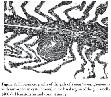
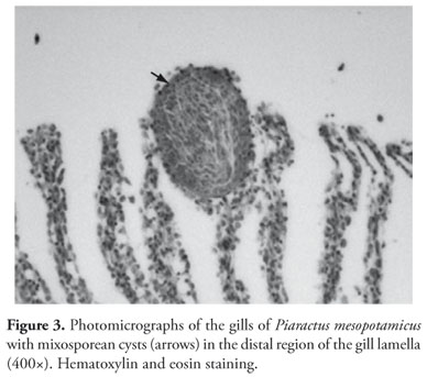
Mixosporean cysts were located inside the capillary in the gills of the intralamellar development type (Figure 1), as defined by McCraren et al. (1975). The presence of cysts inside gill filaments was reported by Martins and Souza (1997) and Martins et al. (1997, 2000). In the samples analyzed in this study, no interlamellar development was observed. Eiras et al. (1999) reported Henneguya piaractus cysts of the intralamellar type present in the epithelium of cultivated pacu gills. Barassa et al. (2003), while performing a histological analysis in the gills of Astyanax altiparanae from natural environment, described the presence of H. chydadea cysts of intralamellar type. This form of infection is considered less pathogenic than the interlamellar infection, which is frequently associated with host death (McCRAREN et al., 1975; EIRAS et al., 1999).
Mixosporean cysts were seen in a variety of sizes, the largest and the smallest cyst had 166.37 and 212.29 µm and 79.22 and 97.81 µm, respectively. Several developmental stages, from early to more mature (Figure 1), were observed, and apparently, unrelated to seasonal distribution since they were found in host collected throughout the months of April, May, June and November.
According to Eiras et al. (1999), this asynchronous development suggests different infection periods. Recently Adriano et al. (2005a) described H. caudalongula parasitizing the gills of P. lineatus cultivated in fish farms in the State of São Paulo and asynchronous parasite development.
In this study, the most common was to find only one mixosporean cyst in the gill lamella in the basal (Figure 2), mid or distal region (Figure 3). Mature cysts containing mature spores v. 20, n. 1, jan.-mar. 2011 Histopathology of gills of Piaractus mesopotamicus (Holmberg, 1887) and Prochilodus lineatus (Valenciennes, 1836) were within the entire lamella and caused it to dilate and compress the epithelial cells that flattened out. In addition, the lamellae infected with parasites pressed the adjacent lamellae, causing them to deform. According to Adriano et al. (2005b) these alterations may partially compromise gill functions and, therefore, diminish the respiratory capacity and ionic exchange.
Henneguya cyst walls were formed by an internal layer of endothelial cells and a thin external layer of connective tissue, as described by Eiras et al. (1999) in cultivated P. mesopotamicus.
Epithelial hyperplasia and deformation of adjacent lamella was described by Ferraz de Lima et al. (1991) while histologically characterizing gills of P. mesopotamicus infected with Ichthyrophthirius multifiliis; Martins and Souza (1997) in their study of gills of the same species infected with Henneguya sp.; Martins et al. (1999), while analyzing Leporinus macrocephalus infected with H. leporinicola, and Barassa et al. (2003), while studying gills of Astyanax altiparanae. Therefore, hyperplasia is a common and unspecific response to parasitic infection, as well as inflammatory and circulatory disturbances seen during massive parasite infections (MARTINS et al., 1997, 2000, 2002).
According to Moraes and Martins (2004) the parasitic fauna of cultivated fish, from sport fishing or fish farms, shows that parasites found in common situations are, with small variations, the same causative agents of severe epizootic epidemics, with high mortality rates when there is an imbalance in the parasite-host-environment system caused by either poor quality environment or inadequate management. In rivers and large reservoirs, this imbalance rarely occurs since the fish are not subject to the same conditions.
In this study, although all fish had some degree of parasite infection, no clinical signs of disease were evident at first. This could be due to the fact that either the intensity of infection was not enough to cause disease manifestations or the fish kept themselves in balance under adequate environmental conditions to the host.
Although no massive infection by Henneguya and monogeneans was found in the species analyzed, the histological alterations here described suggest the pathogenic potential of these parasites.
Acknowledgements
We thank Maria Inês Yamazaki de Campos and Francisca de Assis Ardisson (Pathology Laboratory of the Department of Veterinary Pathology at UNESP, Jaboticabal campus, SP, Brazil) and to Dr. Edson A. Adriano (CEPTA/IBAMA, Pirassununga, SP, Brazil) for specific identification of Henneguya.
Received May 31, 2010
Accepted September 17, 2010
- ADRIANO, E. A.; ARANA, S.; CORDEIRO, N. S. Histophatology and ultrastructure of Henneguya caudalongula sp. n. infecting Prochilodus lineatus (Pisces: Prochilodontidae) cultivated in the State of São Paulo, Brazil. Memórias do Instituto Oswaldo Cruz, v. 100, n. 2, p. 177-181, 2005a.
- ADRIANO, E. A.; ARANA, S.; CORDEIRO, N. S. Histology, ultrastructure and prevalence of Henneguya piaractus (Myxosporea) infecting the gills of Piaractus mesopotamicus (Characidae) cultivated in Brazil. Disease Aquatic Organisms, v. 64, n. 3, p. 229-235, 2005b.
- BARASSA, B.; CORDEIRO, N. S.; ARANA, S. A new species of Henneguya, a gill parasite os Astyanax altiparanae (Pisces: Characidae) from Brazil, with comments on histopathology and seasonality. Memórias do Instituto Oswaldo Cruz, v. 98, n. 6, p. 761-765, 2003.
- EIRAS, J. C. et al. Gill histopathology of Piaractus mesopotamicus (Osteichthyes: Serrasalmidae) infected by Henneguya piaractus Martins et Souza, 1997 (Myxozoa: Myxobolidae). Research and Review in Parasitology, v. 59, n. 3-4, p. 117-120, 1999.
- EIRAS, J. C.; TAKEMOTO, R. M.; PAVANELLI, G. C. Diversidade dos parasitas de peixes de água doce do Brasil Maringá, PR: Ed. Clichetec: NUPÉLIA, 2010. 333 p.
- FERRAZ DE LIMA, C. L. B.; REIS, N. S.; CECCARELLI, P. S. Caracterização histológica da ictiofitiríase em pacu, Piaractus mesopotamicus Holmberg, 1887 (Teleostei, Serrasalminae). Boletim do CEPTA, v. 4, n. 2, p. 39-46, 1991.
- FLORES QUINTANA, C. I.; ROUX, J. P.; DOMITROVIC, H. A. Myxosporidiosis (Henneguya sp.) em brânquias de Serrassalmus sp. (Pisces: Serrassalmídae). Revista de Ictiologia, v. 1, n. 1, p. 11-19, 1992.
- LOM, J.; NOBLE, E. R. Revised classification of the class Myxosporea Bütschli, 1981. Folia Parasitologica, v. 31, n. 3, p. 193-205, 1984.
- MARTINS, M. L. et al. Pathology and behavioral effects associated with Henneguya sp. (Myxozoa: Myxobolidae) infections of captive pacu Piaractus mesopotamicus in Brazil. Journal World Aquaculture Society, v. 28, n. 3, p. 297-300, 1997.
- MARTINS, M. L. et al. Gill infection of Leporinus macrocephalus Garavello & Britski, 1988 (Osteichthyes: Anostomidae) by Henneguya leporinicola n. sp. (Myxozoa: Myxobolidae). Description, histopathology and treatment. Revista Brasileira de Biologia, v. 59, n. 3, p. 527-534, 1999.
- MARTINS, M. L. et al. Parasitic infections in cultivated freshwater fishes a survey of diagnosticated cases from 1993 to 1998. Revista Brasileira de Parasitologia Veterinária, v. 9, n. 1, p. 23-28, 2000.
- MARTINS, M. L. et al. Piscinoodinium pillulare (Schäperclaus, 1954) Lom, 1981 (Dinoflagellida) infection in cultivated freshwater fish from Northeast region of São Paulo State, Brazil. Parasitological and pathological aspects. Revista Brasileira de Biologia, v. 61, n. 4, p. 639-644, 2001.
- MARTINS, M. L.; SOUZA, V. N. Henneguya piaractus n. sp. (Myxozoa: Myxobolidae), a gill parasite of Piaractus mesopotamicus Holmberg, 1887 (Osteichthyes: Characidae), in Brazil. Revista Brasileira de Biologia, v. 57, p. 239-245, 1997.
- McCRAREN, J. P. et al. Variation in response of channel catfish to Henneguya sp. infections (Protozoa: Myxosporidea). Journal Wildlife Diseases, v. 11, n. 1, p. 3-7, 1975.
- MORAES, F. R.; MARTINS, M. L. Condições pré-disponentes e principais enfermidades de teleósteos em piscicultura intensiva. In: CYRINO, J. E. P. et al. (Ed.). Tópicos especiais em piscicultura de água doce tropical intensiva São Paulo: TecArt, 2004. p. 343-386.
- PAVANELLI, G. C.; EIRAS, J. C.; TAKEMOTO, R. M. Doenças de Peixes: profilaxia, diagnóstico e tratamento. 2nd ed. Maringá, PR: EDUEM: NUPÉLIA, 2002. 305 p.
- TAKASHIMA, F.; HIBIYA, T. (Ed.). An atlas of fish histology: normal and pathological features 2nd ed. Tokyi: Kodansha, Stuttgart; NewYork: Fischer, 1995. 195 p.
Corresponding author:
Publication Dates
-
Publication in this collection
20 Apr 2011 -
Date of issue
Mar 2011
History
-
Accepted
18 Sept 2010 -
Received
31 May 2010