Abstracts
Five specimens of Arapaima gigas caught in the Araguaia River (State of Mato Grosso, Brazil) were investigated for helminths in 2004. Numerous adult specimens of the rhapidascarid nematode Goezia spinulosa were found in stomach ulcers in all the specimens of A. gigas and were surrounded by thickening of the mucosa. The gastric glands of all the fish were necrotic and there was a severe and diffuse inflammatory reaction composed of eosinophils (which were predominant), lymphocytes and rare macrophages in the mucosa, submucosa and muscle layer. This is the first report of tissue lesion occurrences in this host, in the presence of G. spinulosa, and it confirms the high pathogenicity of this parasite species.
Pathology; neotropical fish; Araguaia River; Brazil
Cinco espécimens de Arapaima gigas capturados no Rio Araguaia (Estado do Mato Grosso, Brasil) foram investigados para diagnóstico de infecção por helmintos em 2004. Numerosos espécimes adultos do nematóide rafidascarídeo Goezia spinulosa foram encontrados em úlceras do estômago circundadas por um espessamento da mucosa em todos os exemplares de A. gigas. As glândulas gástricas de todos encontravam-se necróticas e havia um acentuado e difuso infiltrado inflamatório composto por eosinófilos, que eram predominantes, linfócitos e raros macrófagos na mucosa, submucosa e camada muscular. As lesões teciduais na presença de nematóide G. spinulosa são relatadas pela primeira vez nesse hospedeiro e confirmam a alta patogenicidade dessa espécie de parasito.
Patologia; peixe neotropical; Rio Araguaia; Brasil
FULL ARTICLE
Tissue alterations in the pirarucu, Arapaima gigas, infected by Goezia spinulosa (Nematoda)
Alterações teciduais em pirarucu, Arapaima gigas, infectado por Goezia spinulosa (Nematoda)
Rodrigo Caldas MenezesI; Sonia Maria Cursino dos SantosII; Paulo Sérgio CeccarelliIII; Luiz Eduardo Roland TavaresIV; Rogério TortellyV; José Luis LuqueII
IClinical Research Laboratory for Domestic Animal Dermatozoonosis, Evandro Chagas Clinical Research Institute, Oswaldo Cruz Foundation - Fiocruz
IIPostgraduate Program on Veterinary Sciences and Department of Animal Parasitology, Veterinary Institute, Federal Rural University of Rio de Janeiro - UFRRJ
IIINational Research and Conservation Center for Continental Fish - CEPTA, Chico Mendes Biodiversity Conservation Institute - ICMBio
IVDepartment of Pathology, Biological and Health Sciences Center, Federal University of Mato Grosso do Sul - UFMS
VDepartment of Pathology and Veterinary Clinical Medicine, Veterinary School, Fluminense Federal University - UFF
Corresponding author Corresponding author: José Luis Luque Curso de Pós-Graduação em Ciências Veterinárias, Departamento de Parasitologia Animal, Instituto de Veterinária, Universidade Federal do Rio de Janeiro - UFRRJ BR-465, Km 7 CEP 23851-970, Seropédica - RJ, Brazil e-mail: jlluque@ufrrj.br
ABSTRACT
Five specimens of Arapaima gigas caught in the Araguaia River (State of Mato Grosso, Brazil) were investigated for helminths in 2004. Numerous adult specimens of the rhapidascarid nematode Goezia spinulosa were found in stomach ulcers in all the specimens of A. gigas and were surrounded by thickening of the mucosa. The gastric glands of all the fish were necrotic and there was a severe and diffuse inflammatory reaction composed of eosinophils (which were predominant), lymphocytes and rare macrophages in the mucosa, submucosa and muscle layer. This is the first report of tissue lesion occurrences in this host, in the presence of G. spinulosa, and it confirms the high pathogenicity of this parasite species.
Keywords: Pathology, neotropical fish, Araguaia River, Brazil.
RESUMO
Cinco espécimens de Arapaima gigas capturados no Rio Araguaia (Estado do Mato Grosso, Brasil) foram investigados para diagnóstico de infecção por helmintos em 2004. Numerosos espécimes adultos do nematóide rafidascarídeo Goezia spinulosa foram encontrados em úlceras do estômago circundadas por um espessamento da mucosa em todos os exemplares de A. gigas. As glândulas gástricas de todos encontravam-se necróticas e havia um acentuado e difuso infiltrado inflamatório composto por eosinófilos, que eram predominantes, linfócitos e raros macrófagos na mucosa, submucosa e camada muscular. As lesões teciduais na presença de nematóide G. spinulosa são relatadas pela primeira vez nesse hospedeiro e confirmam a alta patogenicidade dessa espécie de parasito.
Palavras-chave: Patologia, peixe neotropical, Rio Araguaia, Brasil.
Introduction
The pirarucu, Arapaima gigas (Schinz, 1822), is distributed predominantly in the Amazon basin and has high commercial value as a food resource and ornamental fish (HRBEK et al., 2007). However, over recent decades, the population has been in steady decline, and this fish came under protection through the Convention on International Trade in Endangered Species of Wild Fauna and Flora (CITES) in 1975.
Farming of the pirarucu is limited by parasite infections. The ascarid nematode Goezia spinulosa (Diesing, 1839) was found to be one of the most pathogenic parasites of tank-reared A. gigas on Mexiana Island, Amazon River, Brazil, causing high mortality among cultured fingerlings (SANTOS; MORAVEC, 2009). Santos and Moravec (2009) described gross lesions, characterized by nematodes penetrating deeply into the stomach mucosa and causing ulcers, sometimes with more than 50 parasites involved, but without perforations into the mesentery of large fish. However, nematode larvae and juveniles caused perforations in the stomach wall in fingerlings, thus penetrating the mesentery.
There are no data on microscopic lesions associated with this parasite in this host. Recently, Santos et al. (2008) recorded G. spinulosa in wild pirarucu from the Araguaia River (State of Mato Grosso, Brazil) and provided a quantitative account of this species. Here, we present a full description of the pathological alterations caused by this nematode in the stomach of pirarucu from this locality.
Materials and Methods
In August 2004, five adult specimens of pirarucu (average length of 2.3 m) were caught in the Araguaia River using nylon fishing nets, in the municipality of Cocalinho, State of Mato Grosso, Brazil (13º 23' 07.3" S, 50 º 39' 58.1'' W). These fish were captured and necropsied under permission granted through SISBIO, the Brazilian Biodiversity Information System (License nº 15507-1). Fragments of parasitized stomach were removed and immediately fixed in 10% formalin, and nematode specimens were stored in 70% alcohol. The tissue samples were routinely processed for paraffin embedding (BEHMER et al., 1976). Sections of five micrometers in thickness were stained with hematoxylin and eosin. The parasites were identified based on descriptions by Moravec (1998) and Santos and Moravec (2009). Photomicrographs were taken using a Media Cybernetics Cool Sharp Pro camera connected to a Nikon Eclipse E-800 microscope. The parasitized stomach samples, preserved in 10% formalin, and slides containing histopathological sections through these organs, were deposited in the Helminthological Collection of the Oswaldo Cruz Institute (CHIOC nº 37707-37708, 37315-37317), Rio de Janeiro, RJ, Brazil.
Results
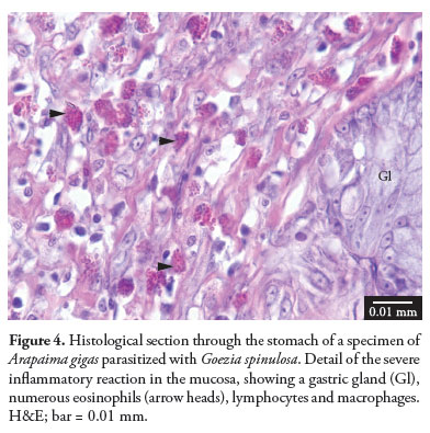
The gross lesions due to G. spinulosa in the stomachs of the five specimens of A. gigas studied here consisted of ulcers at the insertion sites of the nematodes, which were surrounded by thickening of the mucosa (Figure 1). Microscopically, all the fish contained nematodes with cuticular spines in the gastric lumen, and the anterior regions of the nematodes were embedded either superficially or deeply in extensively ulcerated and thickened mucosa, but without reaching the muscularis mucosa and submucosa (Figure 2). At times, nests of bacteria were observed on the ulcer surface (Figure 3). Moreover, there was necrosis in the gastric glands (which were scarce), and a severe and diffuse inflammatory reaction composed of eosinophils (which were predominant), lymphocytes and rare macrophages in the mucosa (Figure 4), submucosa and muscle layers of the five fish studied.
Discussion
The pathological alterations associated with G. spinulosa were severe and were characterized by ulcerative eosinophilic gastritis. According to Martins et al. (2004), fish parasitized with Goezia leporini may present hematological alterations such as anemia, leukocytopenia, neutrophilia and eosinophilia, which can be caused by tissue lesions like those observed in the present study. Moreover, the ulcerative eosinophilic gastritis associated with G. spinulosa may cause digestion disorders and secondary infections, thus worsening the health status of the fish and leading to economic losses in farming this species. Mortality among pirarucu due to parasitism by G. spinulosa has only been reported in fingerlings, which have thin stomach tissue and are highly susceptible to attachment of larvae and juveniles of this nematode, which frequently cause perforations and penetrate into the mesentery (SANTOS; MORAVEC, 2009). The absence of migration of the larvae through the digestive tract wall in the present study may be explained by the fact that the parasitized fish were large and had a thicker stomach wall, which is in agreement with the findings described by Santos and Moravec (2009).
The gross lesions were similar to those described by Santos and Moravec (2009) in the stomach of large specimens of A. gigas parasitized by the same species and similar to those described by Deardorff and Overstreet (1980) in the stomach of Tilapia aurea parasitized by G. sinamora and Rachycentrum canadum parasitized by G. pelagia. Microscopically, however, the fibrotic nodules, collagenous capsules around the parasites, epithelioid tubercles encasing embryonated eggs, penetration of helminths into the body cavity and inflammation only near to the worms with few eosinophils that Deardorff and Overstreet (1980) reported were not seen in the present study. The reason for the different microscopic lesions observed in the present study is likely to be related to the different species of Goezia, as well as the age and species of the host. According to Deardorff and Overstreet (1980), the host response to Goezia species varies between both identical and different hosts. The ulcerative gastritis with numerous eosinophils observed in the present study has also been associated with other anisakid nematodes, such as Anisakis, Contracaecum and Pseudoterranova species, in the stomachs of marine mammals and humans (YOUNG; LOWE, 1969; ORIHEL; ASH, 1995). In contrast with the microscopic lesions associated with these anisakids in mammals, the specimens of G. spinulosa in the present study were only embedded in the mucosa and did not reach the submucosa or muscle layers. Moreover, no abscesses, granulomas or areas of mineralization were observed.
Acknowledgements
We thank the research group of the Instituto Chico Mendes de Conservacão de Biodiversidade (ICMBio), Brazil, for collecting the fish studied. José L. Luque was supported by a research fellowship from Conselho Nacional de Desenvolvimento Científico e Tecnológico do Brasil (CNPq).
Received December 13, 2010
Accepted February 7, 2011
- BEHMER, O. A.; TOLOSA, E. M. C.; FREITAS-NETO, A. G. Manual de técnicas para histologia normal e patológica São Paulo: EDART, 1976. 256 p.
- DEARDORFF, T. L.; OVERSTREET, R. M. Taxonomy and biology of North American species of Goezia (Nematoda: Anisakidae) from fishes, including three new species. Proceedings of Helminthological Society of Washington, v. 47, n. 2, p. 192-217, 1980.
- HRBEK, T.; CROSSA, M.; FARIAS, I. P. Conservation strategies for Arapaima gigas (Schinz, 1822) and the Amazonian várzea ecosystem. Brazilian Journal of Biology, v. 67, p. 909-917, 2007. Suplemento 4.
- MARTINS, M. L. et al. Haematological alterations of Leporinus macrocephalus (Osteichthyes: Anostomidae) naturally infected by Goezia leporini (Nematoda: Anisakidae) in fish pond. Arquivos Brasileiros de Medicina Veterinária e Zootecnia, v. 56, n. 5, p. 640-646, 2004.
- MORAVEC, F. Nematodes of freshwater fishes of the Neotropical region Praga: Academia, 1998. 464 p.
- ORIHEL, T. C.; ASH, L. R. Parasites in human tissues Chicago: ASPC Press, 1995. 386 p.
- SANTOS, C. P.; MORAVEC, F. Goezia spinulosa (Nematoda: Raphidascarididae), a pathogenic parasite of the arapaima Arapaima gigas (Osteichthyes). Folia Parasitologica, v. 56, n. 1, p. 55-63, 2009.
- SANTOS, M. C.; CECCARELLI, P. S.; LUQUE, J. L. Helmintos parasitos do pirarucu, Arapaima gigas (Schinz, 1822) (Osteoglossiformes: Arapaimidae), no rio Araguaia, Estado de Mato Grosso, Brasil. Revista Brasileira de Parasitologia Veterinária, v. 17, n. 3, p. 171-173, 2008.
- YOUNG, P. C.; LOWE, D. Larval nematodes from fish of the subfamily Anisakinae and gastro-intestinal lesions in mammals. Journal of Comparative Pathology, v. 79, n. 3, p. 301-313, 1969. http://dx.doi.org/10.1016/0021-9975(69)90043-7
Corresponding author:
Publication Dates
-
Publication in this collection
14 Oct 2011 -
Date of issue
Sept 2011
History
-
Accepted
07 Feb 2011 -
Received
13 Dec 2010