
Abstract
In the present study, Litomosoides silvai parasitizing Akodon montensis in the southern region of Brazil is reported for the first time. New morphological information is provided for some structures of this nematode species, such as a flattened cephalic extremity, presence of two dorsal cephalic papillae, female tail with a constriction at its tip, “s” shaped vagina, spicules characteristic of the carinii species group and microfilaria tail constricted at the tip. This nematode was found parasitizing the thoracic cavity with a prevalence of 10% (2/20), mean intensity of 4 (6/2), mean abundance of 0.4 (8/20) and range of infection of 2-6 specimens per host, in southern Brazil. This occurrence of L. silvai in A. montensis is a new geographical record for southern Brazil, in the Upper Paraná Atlantic Forest ecoregion of the northwestern region of Rio Grande do Sul, which is part of the Atlantic Forest biome.
Keywords:
Filarioid; Atlantic Forest biome; nematode; rodent; Rio Grande do Sul
Resumo
No presente estudo é relatado pela primeira vez Litomosoides silvai parasitando Akodon montensis coletados na região Sul do Brasil. Foram fornecidas novas informações morfológicas para algumas estruturas desta espécie de nematódeo, tais como extremidade cefálica achatada, a presença de duas papilas cefálicas dorsais, cauda das fêmeas com uma constrição na ponta da cauda, vagina em forma de “s”, espículas de característica do grupo de espécies de carinii e cauda da microfilária com constrição na ponta. Este nematódeo parasitava a cavidade torácica com uma prevalência de 10% (2/20), intensidade média de 4 (8/2) e abundância média de 0,4 (8/20), e intervalo de infecção de 2-6 espécimes por hospedeiro no Sul do Brasil. A ocorrência de L. silvai em A. montensis é um novo registro geográfico, no sul do Brasil, a noroeste do estado do Rio Grande do Sul, na ecorregião da Mata Atlântica do Alto Paraná, parte do bioma da Mata Atlântica.
Palavras-chave:
Filarideo; bioma Mata Atlântica; nematoda; roedor; Rio Grande do Sul
Introduction
Turvo State Park (Parque Estadual do Turvo, PST) is a natural reserve located in the northwest of the state of Rio Grande do Sul, southern Brazil, comprising an area of approximately 17,491 hectares. The park is covered almost entirely by subtropical broadleaf or broadleaf deciduous forest. The mammal fauna of the park consists of carnivores such as Galictis sp., Cerdocyon thous, Nasua nasua, Procyon cancrivorus, Eira barbara, wild cats, herbivorous like deer and tapirs, some species of lagomorphs, primates, didelphimorphia, and xenarthrans, many species of chiropterans and as well as numerous rodents (KASPER et al., 2007Kasper CB, Mazim FD, Soares JBG, Oliveira TG, Fabián ME. Composição e abundância relativa dos mamíferos de médio e grande porte no Parque Estadual do Turvo, Rio Grande do Sul, Brasil. Rev Bras Zool 2007; 24(4): 1087-1100. http://dx.doi.org/10.1590/S0101-81752007000400028.
http://dx.doi.org/10.1590/S0101-81752007...
; MELO et al., 2011Melo GL, Sponchiado J, Machado AF, Cáceres NC. Small-mammal community structure in a South American deciduous Atlantic Forest. Community Ecol 2011; 12(1): 58-66. http://dx.doi.org/10.1556/ComEc.12.2011.1.8.
http://dx.doi.org/10.1556/ComEc.12.2011....
).
One of the genera of Cricetidae rodents that is well represented in Brazil is Akodon Meyen, 1833, with 10 species recorded in this country (SILVEIRA et al., 2013Silveira F, Sbalqueiro IJ, Monteiro-Filho ELA. Identification of the brazilian species of Akodon (Rodentia: Cricetidae: Sigmodontinae) through the microstructure of the hair. Biota Neotrop 2013; 13(1): 339-345. http://dx.doi.org/10.1590/S1676-06032013000100033.
http://dx.doi.org/10.1590/S1676-06032013...
). Among these, Akodon montensis Thomas, 1913 is the only species of this genus found in this reserve (MELO et al., 2011Melo GL, Sponchiado J, Machado AF, Cáceres NC. Small-mammal community structure in a South American deciduous Atlantic Forest. Community Ecol 2011; 12(1): 58-66. http://dx.doi.org/10.1556/ComEc.12.2011.1.8.
http://dx.doi.org/10.1556/ComEc.12.2011....
). This species occurs in Paraguay, northern Argentina and southeastern to southern Brazil, including the states of Minas Gerais, Rio de Janeiro, São Paulo, Paraná, Santa Catarina, and Rio Grande do Sul (MUSSER & CARLETON, 2005Musser GM, Carleton MD. Superfamily Muroidea. In: Wilson DE, Reeder DM, editors. Mammal species of the world: A taxonomic and geographic reference. 3rd ed. Baltimore: Johns Hopkins University Press; 2005. p. 894-1531.).
Filarioids of the genus Litomosoides Chandler, 1931, are parasites of the abdominal and thoracic cavities of bats, marsupials and many rodents. Currently, this genus is represented by 43 known species (NOTARNICOLA et al., 2012Notarnicola J, Jiménez-Ruiz FA, Gardner SL. A new species of Molinema (Nematoda: Onchocercidae) in bolivian rodents and emended description of Litomosoides esslingeri Bain, Petit, and Diagne, 1989. J Parasitol 2012; 98(6): 1200-1208. PMid:22680839. http://dx.doi.org/10.1645/GE-3059.1.
http://dx.doi.org/10.1645/GE-3059.1...
; NOTARNICOLA & de la SANCHA, 2015Notarnicola J, de la Sancha NU. Description of Litomosoides ysoguazu n. sp. (Nematoda, Onchocercidae) a parasite of the tuft-toed rice rat Sooretamys angouya (Fisher) (Rodentia, Cricetidae), and a first record of L. esslingeri Bain, Petit & Berteaux, 1989 in Paraguay. Syst Parasitol 2015; 91(2): 175-184. PMid:25962465. http://dx.doi.org/10.1007/s11230-015-9563-2.
http://dx.doi.org/10.1007/s11230-015-956...
; OVIEDO et al., 2016Oviedo MC, Notarnicola J, Miotti MD, Claps LE. Emended description of Litomosoides molossi (Nematoda: Onchocercidae) and first records of Litomosoides species parasitizing argentinean bats. J Parasitol 2016; 102(4): 440-450. PMid:27098753. http://dx.doi.org/10.1645/15-776.
http://dx.doi.org/10.1645/15-776...
). In Brazil, species of Litomosoides have been found parasitizing marsupials (BAIN et al., 1980Bain O, Petit G, Berteaux S. Description de deux nouvelles Filaires du genre Litomosoides et de leurs stades infestants. Ann Parasitol Hum Comp 1980; 55(2): 225-237. PMid:7458160. http://dx.doi.org/10.1051/parasite/1980552225.
http://dx.doi.org/10.1051/parasite/19805...
; MULLER, 1980Muller R. Litomosoides barretti n. sp. from the ashy opossum in Brazil (Nematoda, Filarioidea). Rev Bras Biol 1980; 40(1): 81-83.), bats (LINS DE ALMEIDA, 1936 apud NOTARNICOLA et al., 2010Notarnicola J, Jiménez-Ruiz FA, Gardner SL. Litomosoides (Nemata: Filarioidea) of Bats from Bolivia with Records for Three Known Species and the Description of a New Species. J Parasitol 2010; 96(4): 775-782. PMid:20738202. http://dx.doi.org/10.1645/GE-2371.1.
http://dx.doi.org/10.1645/GE-2371.1...
; REGO, 1961Rego AA. Sobre algumas espécies do gênero Litomosoides Chandler, 1931 (Nematoda: Filarioidea). Mem Inst Oswaldo Cruz 1961; 59(1): 1-9. PMid:13740379. http://dx.doi.org/10.1590/S0074-02761961000100001.
http://dx.doi.org/10.1590/S0074-02761961...
; GUERRERO et al., 2002Guerrero R, Martin C, Gardner SL, Bain O. New and known species of Litomosoides (Nematoda: Filarioidea): Important adult and larval characters and taxonomic changes. Comp Parasitol 2002; 69(2): 177-195. http://dx.doi.org/10.1654/1525-2647(2002)069[0177:NAKSOL]2.0.CO;2.
http://dx.doi.org/10.1654/1525-2647(2002...
; BAIN et al., 2003Bain O, Guerrero R, Rodriguez B, Babayan S, Jouvenet N. Examination of type material of two species of Litomosoides (Filarioidea: Onchocercidae), parasites from bats; taxonomic consequences. Parasite 2003; 10(3): 211-218. PMid:14535160. http://dx.doi.org/10.1051/parasite/2003103211.
http://dx.doi.org/10.1051/parasite/20031...
) and rodents (TRAVASSOS, 1919 apud PADILHA & FARIA, 1977Padilha TN, Faria MJ. Litomosoides silvai n. sp. proveniente de rato do mato, Akodon arviculoides (Wagner) (Nematoda, Filarioidea). Rev Bras Biol 1977; 37(3): 535-537.; PADILHA & FARIA, 1977Padilha TN, Faria MJ. Litomosoides silvai n. sp. proveniente de rato do mato, Akodon arviculoides (Wagner) (Nematoda, Filarioidea). Rev Bras Biol 1977; 37(3): 535-537.; BAIN et al., 1989Bain O, Petit G, Diagne M. Étude de quelques Litomosoides parasites de rongeurs; coséquences taxonomiques. Ann Parasitol Hum Comp 1989; 64(4): 268-289. PMid:2817694. http://dx.doi.org/10.1051/parasite/1989644268.
http://dx.doi.org/10.1051/parasite/19896...
; MORAES-NETO et al., 1997Moraes-Neto AHA, Lanfredi RM, Souza W. Litomosoides chagasfilhoi sp. nov. (Nematoda: Filarioidea) parasitizing the abdominal cavity of Akodon cursor (Winge, 1887) (Rodentia: Muridae) from Brazil. Parasitol Res 1997; 83(2): 137-143. PMid:9039695. http://dx.doi.org/10.1007/s004360050223.
http://dx.doi.org/10.1007/s004360050223...
; SIMÕES et al., 2012Simões RO, Maldonado-Júnior A, Luque JL. Helminth communities in three sympatric rodents from Brazilian Atlantic Forest: contrasting biomass and numerical abundance. Braz J Biol 2012; 72(4): 909-914. PMid:23295521. http://dx.doi.org/10.1590/S1519-69842012000500018.
http://dx.doi.org/10.1590/S1519-69842012...
). Two species, Litomosoides silvai (PADILHA & FARIA, 1977Padilha TN, Faria MJ. Litomosoides silvai n. sp. proveniente de rato do mato, Akodon arviculoides (Wagner) (Nematoda, Filarioidea). Rev Bras Biol 1977; 37(3): 535-537.) and Litomosoides chagasfilhoi (MORAES-NETO et al., 1997Moraes-Neto AHA, Lanfredi RM, Souza W. Litomosoides chagasfilhoi sp. nov. (Nematoda: Filarioidea) parasitizing the abdominal cavity of Akodon cursor (Winge, 1887) (Rodentia: Muridae) from Brazil. Parasitol Res 1997; 83(2): 137-143. PMid:9039695. http://dx.doi.org/10.1007/s004360050223.
http://dx.doi.org/10.1007/s004360050223...
) have been recorded in the rodent Akodon cursor Winge, 1887, in Itaguaí and Rio Bonito, in the Atlantic Forest region of the state of Rio de Janeiro (PADILHA & FARIA, 1977Padilha TN, Faria MJ. Litomosoides silvai n. sp. proveniente de rato do mato, Akodon arviculoides (Wagner) (Nematoda, Filarioidea). Rev Bras Biol 1977; 37(3): 535-537.; MORAES-NETO et al., 1996Moraes-Neto AHA, Lanfredi RM, Souza W. Emended description of Litomosoides silvai (Nematoda: Filarioidea) of Akodon cursor (Rodentia: Muridade). J Parasitol 1996; 82(6): 988-991. PMid:8973410. http://dx.doi.org/10.2307/3284210.
http://dx.doi.org/10.2307/3284210...
, 1997Moraes-Neto AHA, Lanfredi RM, Souza W. Litomosoides chagasfilhoi sp. nov. (Nematoda: Filarioidea) parasitizing the abdominal cavity of Akodon cursor (Winge, 1887) (Rodentia: Muridae) from Brazil. Parasitol Res 1997; 83(2): 137-143. PMid:9039695. http://dx.doi.org/10.1007/s004360050223.
http://dx.doi.org/10.1007/s004360050223...
; MORAES-NETO & LANFREDI 1997Moraes-Neto AHA, Lanfredi RM. Prevalence of two species of Litomosoides (Nematoda: Filarioidea) parasites of Akodon cursor (Rodentia: Muridae) from southeastern Brazil. Rev Bras Biol 1997; 57(4): 579-582. PMid:9440357.; SIMÕES et al., 2011Simões RO, Souza JGR, Maldonado-Júnior A, Luque JL. Variation in the helminth community structure of three sympatric sigmodontine rodents from the coastal Atlantic Forest of Rio de Janeiro, Brazil. J Helminthol 2011; 85(2): 171-178. PMid:20701831. http://dx.doi.org/10.1017/S0022149X10000398.
http://dx.doi.org/10.1017/S0022149X10000...
, 2012Simões RO, Maldonado-Júnior A, Luque JL. Helminth communities in three sympatric rodents from Brazilian Atlantic Forest: contrasting biomass and numerical abundance. Braz J Biol 2012; 72(4): 909-914. PMid:23295521. http://dx.doi.org/10.1590/S1519-69842012000500018.
http://dx.doi.org/10.1590/S1519-69842012...
).
The aim of the present study was to report on L. silvai parasitizing A. montensis in the Turvo State Park, state of Rio Grande do Sul, Brazil, which is in the Upper Paraná Atlantic Forest ecoregion, part of Atlantic Forest biome. Furthermore, this study provides new morphological data for some structures, such as the cephalic extremity, tail and vagina, spicules and microfilariae tail; and it presents parasitological indices (prevalence, mean intensity and mean abundance) and data on range and infection site.
Materials and Methods
During a survey carried out to study tick-borne diseases (KRAWCZAK, 2016Krawczak FS. Pesquisa de infecção por riquétsias do grupo da febre maculosa em cães, pequenos mamíferos e carrapatos em área endêmica e não endêmica nos biomas Pampa e Mata Atlântica no estado do Rio Grande do Sul. [tese]. São Paulo: Universidade de São Paulo; 2016.), 164 specimens of small mammals were caught: 117 Akodon montensis, 13 Brucepattersonius iheringi, 1 Cryptonanus guahybae, 7 Didelphis aurita, 1 Euryoryzomys russatus, 9 Oligoryzomys nigripes, 7 Oxymycterus judex, 4 Sooretamys angouya and 5 Thaptomys nigrita. They were caught in the Turvo State Park (latitude 27°00’ S to 27°20’ S; longitude 53°40’ W to 54°10’ W), in the northwest of the state of Rio Grande do Sul, Brazil, between June 2013 and January 2014. However, only some of the rodents, which died due to the procedures for anesthesia and blood collection, were used in the present study: 20 specimens of Akodon montensis Thomas, 1913; two Brucepattersonius iheringi Thomas, 1896; one Oxymycterus judex Thomas, 1909; and one Oligoryzomys nigripes Olfers, 1818. Some other rodents that died were preserved in absolute ethanol until arrival at the Parasitic Diseases Laboratory of the School of Veterinary Medicine and Animal Science, Universidade de São Paulo (USP). These were then analyzed for tick presence and were identified in accordance with Bonvicino et al. (2008)Bonvicino CR, Oliveira JA, D’Andrea PS. Guia dos roedores do Brasil, com chaves para gêneros baseadas em caracteres externos. Duque de Caxias: Centro Pan-Americano de Febre Aftosa; 2008. and Melo et al. (2011)Melo GL, Sponchiado J, Machado AF, Cáceres NC. Small-mammal community structure in a South American deciduous Atlantic Forest. Community Ecol 2011; 12(1): 58-66. http://dx.doi.org/10.1556/ComEc.12.2011.1.8.
http://dx.doi.org/10.1556/ComEc.12.2011....
. These animals were deposited in the Rodent Collection of the “Luiz de Queiroz” Higher Agriculture School, USP, state of São Paulo, Brazil.
The animals were caught using a total of 80 live traps (75 Sherman and five Tomahawk) baited with bacon, banana, apple and peanut butter. These were set up for four consecutive nights during each field campaign. Additionally, three pitfall stationary traps each consisting of five buckets of 42.5 cm in diameter and 60 cm in height, which were connected using a plastic fence (at least 30 m long and 50 cm high), were set up for the same period. The animals thus trapped were then anaesthetized using ketamine and xylazine.
Adult nematode specimens were recovered from the thoracic cavity of A. montensis, stored in Railliet & Henry liquid, and were sent to the Veterinary Parasitology Laboratory, Universidade Federal de Santa Maria, state of Rio Grande do Sul, Brazil, for identification. To study their morphological characteristics, they were then cleared in lactophenol, temporarily mounted, examined under a bright-field microscope (Olympus BX-51) and photographed using a Zeiss Axiophot. Microfilariae from the uterus of one fixed female were dissected and examined. Illustrations were made with the aid of a drawing tube. Measurements are given in micrometers unless otherwise stated. The parasite terms used are in accordance with Bush et al. (1997)Bush AO, Lafferty KD, Lotz JM, Shostak AW. Parasitology meets ecology on its own terms: Margolis et al. revisited. J Parasitol 1997; 83(4): 575-583. PMid:9267395. http://dx.doi.org/10.2307/3284227.
http://dx.doi.org/10.2307/3284227...
.
Nematode specimens were deposited in the Helminth Collection of the Museum of La Plata (MLP-he). Moreover, specimens of L. silvai parasitizing A. cursor, from Rio Bonito, state of Rio de Janeiro (MORAES-NETO et al., 1996Moraes-Neto AHA, Lanfredi RM, Souza W. Emended description of Litomosoides silvai (Nematoda: Filarioidea) of Akodon cursor (Rodentia: Muridade). J Parasitol 1996; 82(6): 988-991. PMid:8973410. http://dx.doi.org/10.2307/3284210.
http://dx.doi.org/10.2307/3284210...
) deposited in the Oswaldo Cruz Institute Helminthological Collection (CHIOC) under number 33518 (one female and one male) were studied to improve the diagnosis.
This study was approved by the Ethics Committee on Animal Use of the School of Veterinary Medicine and Animal Science, USP, under protocol number 2908/2013; by the Chico Mendes Institute for Biodiversity Conservation (ICMBio), under license number SISBIO 38502-1; and by the State Environmental Department of Rio Grande do Sul (SEMA, RS).
Results
Three female filarioid specimens, two anterior extremities and three posterior extremities were recovered from the thoracic cavity of two specimens of A. montensis. Morphometric data on the female and microfilarial specimens are showed in Table 1. The species identification was aided through studying male and female specimens deposited in CHIOC (number 33518).
Compared measurements of females from Litomosoides silvai reported in the literature and the present study.
Litomosoides silvai Padilha and Faria, 1977 (Figures 1, 2, 3, 4, 5, 6, 7, 8, 9, 10, 11)

1-4. Female specimens of Litomosoides silvai parasitizing Akodon montensis from Turvo State Park, state of Rio Grande do Sul. 1. Anterior region of female, lateral view. 2. Anterior extremity, lateral view. 3. Detail of the vulva, lateral view. 4. Uterine microfilaria. 5-7. Male specimen of L. silvai parasitizing A. cursor, from Rio Bonito, state of Rio de Janeiro, CHIOC 33518. 5. Posterior extremity showing the spicules and the cloaca, lateral view. 6. Detail of the left spicule, lateral view. 7. Detail of the right spicule, lateral view. Scale bars: 1 = 200 µm; 2 and 4 = 20 µm; 3, 5-7 = 50 µm.
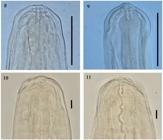
Detail of anterior extremities of Litomosoides silvai. 8-9. Female and male specimens, respectively, of L. silvai parasitizing Akodon cursor, from Rio Bonito, state of Rio de Janeiro, CHIOC 33518. 10-11. Female specimen of L. silvai parasitizing A. montensis from Turvo State Park, state of Rio Grande do Sul. Scale bars: 8-9 = 25 µm; 10-11: 10 µm.
Diagnosis: Adult worms small, males four times smaller than females. Cephalic extremity flattened. In apical view, 4 labial papillae distributed in a stretched dorsoventral rectangle; 2 dorsal cephalic papillae. Buccal capsule slender; anterior segment transparent and posterior chitinous portion with irregular external walls; buccal cavity with 2 short enlargements near the base. Female tail slender, with a constriction at tip of tail. Esophagus muscular, becoming slightly glandular in the posterior portion. Vulva posterior to the esophagus-intestine junction. Vagina “s” shaped; vagina perpendicular to the axis of the vulva with a flattened lumen consisting of a simple “s” shaped tube surrounded by muscular walls. Male tail slender; left spicule with handle longer than blade; blade consisting of a membranous ala that converts into a filament; right spicule is a twisted conduit with a heel that is not strongly cuticularized. Microfilariae slender, with sheath closely applied to the body; nuclei not extending to the tip of tail; tail becomes constricted.
Taxonomic summary
Host:Akodon montensis Thomas, 1913.
Locality: Turvo State Park, state of Rio Grande do Sul, Brazil.
Infection site: thoracic cavity.
Parasitological indices: prevalence (P) = 10% (2/20), mean intensity (MI) = 4 (6/2), mean abundance (MA) = 0.4 (8/20) and range of infection of 2-6 specimens per host.
Specimen deposition numbers: MLP-he 7204; MLP-he 7205.
Remarks
The specimens found in A. montensis correspond to the species L. silvai and are in accordance with the morphology and morphometrical characteristics given by Padilha & Faria (1977)Padilha TN, Faria MJ. Litomosoides silvai n. sp. proveniente de rato do mato, Akodon arviculoides (Wagner) (Nematoda, Filarioidea). Rev Bras Biol 1977; 37(3): 535-537. and Moraes-Neto et al. (1996)Moraes-Neto AHA, Lanfredi RM, Souza W. Emended description of Litomosoides silvai (Nematoda: Filarioidea) of Akodon cursor (Rodentia: Muridade). J Parasitol 1996; 82(6): 988-991. PMid:8973410. http://dx.doi.org/10.2307/3284210.
http://dx.doi.org/10.2307/3284210...
. The identification was confirmed by the shape of the buccal capsule, flattened anterior extremity, vulva located posteriorly to the esophagus-intestine junction, non-globular shape of the vulva, tail tip with a constriction, shape of the spicules corresponding to the carinii species group, and constricted tail tip of the microfilariae. In the present study, female specimens were compared with previous specimens deposited in CHIOC, which confirmed the identity of the species.
Discussion
Litomosoides silvai was originally described by Padilha & Faria (1977)Padilha TN, Faria MJ. Litomosoides silvai n. sp. proveniente de rato do mato, Akodon arviculoides (Wagner) (Nematoda, Filarioidea). Rev Bras Biol 1977; 37(3): 535-537., as a parasite of A. cursor. Later on, Moraes-Neto et al. (1996)Moraes-Neto AHA, Lanfredi RM, Souza W. Emended description of Litomosoides silvai (Nematoda: Filarioidea) of Akodon cursor (Rodentia: Muridade). J Parasitol 1996; 82(6): 988-991. PMid:8973410. http://dx.doi.org/10.2307/3284210.
http://dx.doi.org/10.2307/3284210...
redescribed the species including new morphological data and scanning electron microphotographs. These authors showed the distribution of the head papillae, the rugose area, the tip of the tail and the structure of the cuticle at the vulva. However, the position of the head papillae was not stated.
Here, four labial papillae distributed in a stretched dorsoventral rectangle and two dorsal cephalic papillae were observed; amphids were located ventrally in relation to the cephalic papillae. Additionally, the vulva was “s” shaped and differed from the globular shape of other Litomosoides species (NOTARNICOLA et al., 2000Notarnicola J, Bain O, Navone GT. Two new species of Litomosoides (Nematoda: Filarioidea) in sigmodontines (Rodentia: Muridae) from Rio de La Plata marshland, Argentina. J Parasitol 2000; 86(6): 1318-1325. PMid:11191910. http://dx.doi.org/10.1645/0022-3395(2000)086[1318:TNSOLN]2.0.CO;2.
http://dx.doi.org/10.1645/0022-3395(2000...
; OVIEDO et al., 2016Oviedo MC, Notarnicola J, Miotti MD, Claps LE. Emended description of Litomosoides molossi (Nematoda: Onchocercidae) and first records of Litomosoides species parasitizing argentinean bats. J Parasitol 2016; 102(4): 440-450. PMid:27098753. http://dx.doi.org/10.1645/15-776.
http://dx.doi.org/10.1645/15-776...
).
Recently, Simões et al. (2011Simões RO, Souza JGR, Maldonado-Júnior A, Luque JL. Variation in the helminth community structure of three sympatric sigmodontine rodents from the coastal Atlantic Forest of Rio de Janeiro, Brazil. J Helminthol 2011; 85(2): 171-178. PMid:20701831. http://dx.doi.org/10.1017/S0022149X10000398.
http://dx.doi.org/10.1017/S0022149X10000...
, 2012Simões RO, Maldonado-Júnior A, Luque JL. Helminth communities in three sympatric rodents from Brazilian Atlantic Forest: contrasting biomass and numerical abundance. Braz J Biol 2012; 72(4): 909-914. PMid:23295521. http://dx.doi.org/10.1590/S1519-69842012000500018.
http://dx.doi.org/10.1590/S1519-69842012...
) reported the presence of L. silvai parasitizing the sympatric rodents A. cursor and A. montensis in Teresópolis, state of Rio de Janeiro (RJ). All previous reports of L. silvai are from the Atlantic Forest ecoregion, in the state of Rio de Janeiro. Therefore, in the present study, the occurrence of L. silvai in A. montensis in southern Brazil is a new geographical record, in the Upper Paraná Atlantic Forest ecoregion of the northwest of the state of Rio Grande do Sul, which is part of the Atlantic Forest biome.
According to Moraes-Neto & Lanfredi (1997)Moraes-Neto AHA, Lanfredi RM. Prevalence of two species of Litomosoides (Nematoda: Filarioidea) parasites of Akodon cursor (Rodentia: Muridae) from southeastern Brazil. Rev Bras Biol 1997; 57(4): 579-582. PMid:9440357., L. silvai parasitizing A. cursor in Rio Bonito, RJ (Serra do Mar Coastal Atlantic Forest ecoregion), presented P = 20.7% (12/58), MI = 18.6 (223/12) and MA = 3.8 (223/58); while Simões et al. (2011)Simões RO, Souza JGR, Maldonado-Júnior A, Luque JL. Variation in the helminth community structure of three sympatric sigmodontine rodents from the coastal Atlantic Forest of Rio de Janeiro, Brazil. J Helminthol 2011; 85(2): 171-178. PMid:20701831. http://dx.doi.org/10.1017/S0022149X10000398.
http://dx.doi.org/10.1017/S0022149X10000...
reported P = 12%, MI = 2 and MA = 0.2, in Teresópolis, RJ, which is a nearby locality. Moreover, these last authors also found L. silvai parasitizing the sympatric rodent A. montensis (P = 2%, MI = 26 and MA = 0.5). The results from the present study show intermediate values that may be related to a different ecoregion or that may show that environmental factors are involved. Interestingly, in Rio Bonito A. cursor was also found to be parasitized with L. chagasfilhoi, with a prevalence of 12%, and both species of filarioids were concurrent in 4 out of the 58 hosts sampled (MORAES-NETO & LANFREDI, 1997Moraes-Neto AHA, Lanfredi RM. Prevalence of two species of Litomosoides (Nematoda: Filarioidea) parasites of Akodon cursor (Rodentia: Muridae) from southeastern Brazil. Rev Bras Biol 1997; 57(4): 579-582. PMid:9440357.).
It seems that L. silvai always is located in the thoracic cavity when it parasitizes A. cursor or A. montensis, while L. chagasfilhoi is located in the abdominal cavity. Both species of filarioid are present in Rio Bonito, while only L. silvai is present in the other localities (Teresópolis and Turvo State Park). Thus, L. silvai is present in both Akodon species, but because of the different southern distribution of their hosts, the filarioid only follows A. montensis, which is the only species of this genus that has been trapped in the study area (MELO et al., 2011Melo GL, Sponchiado J, Machado AF, Cáceres NC. Small-mammal community structure in a South American deciduous Atlantic Forest. Community Ecol 2011; 12(1): 58-66. http://dx.doi.org/10.1556/ComEc.12.2011.1.8.
http://dx.doi.org/10.1556/ComEc.12.2011....
). It is interesting to note that among more than 236 specimens of A. montensis that were sampled by one of the present authors (JN, unpublished data) in different localities in Misiones province (Argentina), which is close to Turvo State Park, no filarioid specimens were detected.
Acknowledgements
This work was supported financially by the Fundação de Amparo a Pesquisa do Estado de São Paulo (FAPESP) (processes nº 2012/21915-4 and 2016/07241-1), Conselho Nacional de Desenvolvimento Científico e Tecnológico (CNPq) and Coordenadoria de Apoio a Pesquisa e Desenvolvimento (CAPES), and was authorized by the Chico Mendes Institute for Biodiversity Conservation (authorization SISBIO 38502-1) and the Ethics Committee on Animal Research of the School of Veterinary Medicine of the University of São Paulo (protocol 2908/2013).
References
- Bain O, Guerrero R, Rodriguez B, Babayan S, Jouvenet N. Examination of type material of two species of Litomosoides (Filarioidea: Onchocercidae), parasites from bats; taxonomic consequences. Parasite 2003; 10(3): 211-218. PMid:14535160. http://dx.doi.org/10.1051/parasite/2003103211
» http://dx.doi.org/10.1051/parasite/2003103211 - Bain O, Petit G, Berteaux S. Description de deux nouvelles Filaires du genre Litomosoides et de leurs stades infestants. Ann Parasitol Hum Comp 1980; 55(2): 225-237. PMid:7458160. http://dx.doi.org/10.1051/parasite/1980552225
» http://dx.doi.org/10.1051/parasite/1980552225 - Bain O, Petit G, Diagne M. Étude de quelques Litomosoides parasites de rongeurs; coséquences taxonomiques. Ann Parasitol Hum Comp 1989; 64(4): 268-289. PMid:2817694. http://dx.doi.org/10.1051/parasite/1989644268
» http://dx.doi.org/10.1051/parasite/1989644268 - Bonvicino CR, Oliveira JA, D’Andrea PS. Guia dos roedores do Brasil, com chaves para gêneros baseadas em caracteres externos. Duque de Caxias: Centro Pan-Americano de Febre Aftosa; 2008.
- Bush AO, Lafferty KD, Lotz JM, Shostak AW. Parasitology meets ecology on its own terms: Margolis et al. revisited. J Parasitol 1997; 83(4): 575-583. PMid:9267395. http://dx.doi.org/10.2307/3284227
» http://dx.doi.org/10.2307/3284227 - Guerrero R, Martin C, Gardner SL, Bain O. New and known species of Litomosoides (Nematoda: Filarioidea): Important adult and larval characters and taxonomic changes. Comp Parasitol 2002; 69(2): 177-195. http://dx.doi.org/10.1654/1525-2647(2002)069[0177:NAKSOL]2.0.CO;2
» http://dx.doi.org/10.1654/1525-2647(2002)069[0177:NAKSOL]2.0.CO;2 - Kasper CB, Mazim FD, Soares JBG, Oliveira TG, Fabián ME. Composição e abundância relativa dos mamíferos de médio e grande porte no Parque Estadual do Turvo, Rio Grande do Sul, Brasil. Rev Bras Zool 2007; 24(4): 1087-1100. http://dx.doi.org/10.1590/S0101-81752007000400028
» http://dx.doi.org/10.1590/S0101-81752007000400028 - Krawczak FS. Pesquisa de infecção por riquétsias do grupo da febre maculosa em cães, pequenos mamíferos e carrapatos em área endêmica e não endêmica nos biomas Pampa e Mata Atlântica no estado do Rio Grande do Sul [tese]. São Paulo: Universidade de São Paulo; 2016.
- Melo GL, Sponchiado J, Machado AF, Cáceres NC. Small-mammal community structure in a South American deciduous Atlantic Forest. Community Ecol 2011; 12(1): 58-66. http://dx.doi.org/10.1556/ComEc.12.2011.1.8
» http://dx.doi.org/10.1556/ComEc.12.2011.1.8 - Moraes-Neto AHA, Lanfredi RM, Souza W. Emended description of Litomosoides silvai (Nematoda: Filarioidea) of Akodon cursor (Rodentia: Muridade). J Parasitol 1996; 82(6): 988-991. PMid:8973410. http://dx.doi.org/10.2307/3284210
» http://dx.doi.org/10.2307/3284210 - Moraes-Neto AHA, Lanfredi RM, Souza W. Litomosoides chagasfilhoi sp. nov. (Nematoda: Filarioidea) parasitizing the abdominal cavity of Akodon cursor (Winge, 1887) (Rodentia: Muridae) from Brazil. Parasitol Res 1997; 83(2): 137-143. PMid:9039695. http://dx.doi.org/10.1007/s004360050223
» http://dx.doi.org/10.1007/s004360050223 - Moraes-Neto AHA, Lanfredi RM. Prevalence of two species of Litomosoides (Nematoda: Filarioidea) parasites of Akodon cursor (Rodentia: Muridae) from southeastern Brazil. Rev Bras Biol 1997; 57(4): 579-582. PMid:9440357.
- Muller R. Litomosoides barretti n. sp. from the ashy opossum in Brazil (Nematoda, Filarioidea). Rev Bras Biol 1980; 40(1): 81-83.
- Musser GM, Carleton MD. Superfamily Muroidea. In: Wilson DE, Reeder DM, editors. Mammal species of the world: A taxonomic and geographic reference 3rd ed. Baltimore: Johns Hopkins University Press; 2005. p. 894-1531.
- Notarnicola J, Bain O, Navone GT. Two new species of Litomosoides (Nematoda: Filarioidea) in sigmodontines (Rodentia: Muridae) from Rio de La Plata marshland, Argentina. J Parasitol 2000; 86(6): 1318-1325. PMid:11191910. http://dx.doi.org/10.1645/0022-3395(2000)086[1318:TNSOLN]2.0.CO;2
» http://dx.doi.org/10.1645/0022-3395(2000)086[1318:TNSOLN]2.0.CO;2 - Notarnicola J, de la Sancha NU. Description of Litomosoides ysoguazu n. sp. (Nematoda, Onchocercidae) a parasite of the tuft-toed rice rat Sooretamys angouya (Fisher) (Rodentia, Cricetidae), and a first record of L. esslingeri Bain, Petit & Berteaux, 1989 in Paraguay. Syst Parasitol 2015; 91(2): 175-184. PMid:25962465. http://dx.doi.org/10.1007/s11230-015-9563-2
» http://dx.doi.org/10.1007/s11230-015-9563-2 - Notarnicola J, Jiménez-Ruiz FA, Gardner SL. A new species of Molinema (Nematoda: Onchocercidae) in bolivian rodents and emended description of Litomosoides esslingeri Bain, Petit, and Diagne, 1989. J Parasitol 2012; 98(6): 1200-1208. PMid:22680839. http://dx.doi.org/10.1645/GE-3059.1
» http://dx.doi.org/10.1645/GE-3059.1 - Notarnicola J, Jiménez-Ruiz FA, Gardner SL. Litomosoides (Nemata: Filarioidea) of Bats from Bolivia with Records for Three Known Species and the Description of a New Species. J Parasitol 2010; 96(4): 775-782. PMid:20738202. http://dx.doi.org/10.1645/GE-2371.1
» http://dx.doi.org/10.1645/GE-2371.1 - Oviedo MC, Notarnicola J, Miotti MD, Claps LE. Emended description of Litomosoides molossi (Nematoda: Onchocercidae) and first records of Litomosoides species parasitizing argentinean bats. J Parasitol 2016; 102(4): 440-450. PMid:27098753. http://dx.doi.org/10.1645/15-776
» http://dx.doi.org/10.1645/15-776 - Padilha TN, Faria MJ. Litomosoides silvai n. sp. proveniente de rato do mato, Akodon arviculoides (Wagner) (Nematoda, Filarioidea). Rev Bras Biol 1977; 37(3): 535-537.
- Rego AA. Sobre algumas espécies do gênero Litomosoides Chandler, 1931 (Nematoda: Filarioidea). Mem Inst Oswaldo Cruz 1961; 59(1): 1-9. PMid:13740379. http://dx.doi.org/10.1590/S0074-02761961000100001
» http://dx.doi.org/10.1590/S0074-02761961000100001 - Silveira F, Sbalqueiro IJ, Monteiro-Filho ELA. Identification of the brazilian species of Akodon (Rodentia: Cricetidae: Sigmodontinae) through the microstructure of the hair. Biota Neotrop 2013; 13(1): 339-345. http://dx.doi.org/10.1590/S1676-06032013000100033
» http://dx.doi.org/10.1590/S1676-06032013000100033 - Simões RO, Maldonado-Júnior A, Luque JL. Helminth communities in three sympatric rodents from Brazilian Atlantic Forest: contrasting biomass and numerical abundance. Braz J Biol 2012; 72(4): 909-914. PMid:23295521. http://dx.doi.org/10.1590/S1519-69842012000500018
» http://dx.doi.org/10.1590/S1519-69842012000500018 - Simões RO, Souza JGR, Maldonado-Júnior A, Luque JL. Variation in the helminth community structure of three sympatric sigmodontine rodents from the coastal Atlantic Forest of Rio de Janeiro, Brazil. J Helminthol 2011; 85(2): 171-178. PMid:20701831. http://dx.doi.org/10.1017/S0022149X10000398
» http://dx.doi.org/10.1017/S0022149X10000398
Publication Dates
-
Publication in this collection
23 Oct 2017 -
Date of issue
Oct-Dec 2017
History
-
Received
27 June 2017 -
Accepted
15 Sept 2017


