
Abstract
Humiria balsamifera is used in traditional medicine as anthelmintic, expectorant, to treat hepatitis, diarrhea, hemorrhoids; to cure chronic wounds; and to alleviate toothaches. This species occurs in Jurubatiba shoal, Rio de Janeiro state-Brazil, a rich region which offers a variety of promising bioactive product sources. The present study focuses on the chemical and pharmacological evaluation of H. balsamifera. The n-hexane, dichloromethane and ethyl acetate leaf fractions exhibited higher inhibitory potential on NO production. Friedelin (1), quercetin (2) and quercetin-3-α-O-arabinopyranoside (3) were isolated and characterized; the latter is described for the first time for H. balsamifera. Quercetin (2) showed the best inhibitory activity on NO production and moderate inhibition of TNF-α production. These results contribute to the knowledge of Humiria balsamifera as a source of anti-inflammatory compounds. Furthermore, the identification of the terpenes ß-amyrone, betulin, citronellol, eremophillene, dihydroactinolide and borneol, and the isolation of quercetin-3-α-O-arabinopyranoside are being reported for the first time for this species.
Key words
inflammatory; Friedelin; Humiriaceae; arabinopyranoside; α
Resumo
Humiria balsamifera é utilizada na medicina tradicional como antihelmintico, expectorante, para tratar hepatite, diarreia, hemorroidas, na cura de feridas crônicas, e no alívio da dor de dente. Esta espécie ocorre na Restinga de Jurubatiba, no estado do Rio de Janeiro-Brasil, uma região rica que oferece uma variedade de fontes promissoras de produtos bioativos. O presente estudo foca na avaliação química e farmacológica da espécie H. balsamifera. As frações de folhas em n-hexano, diclorometano e acetato de etila apresentaram maior potencial inibitório na produção de NO. Friedelina (1), quercetina (2) e quercetina-3-O-α-arabinopiranosídeo (3) foram isolados e caracterizados; o último está sendo descrito pela primeira vez para H. balsamifera. A quercetina (2) apresentou a melhor atividade inibitória na produção de NO e moderada inibição da produção de TNF-α. Estes resultados contribuem para o conhecimento da espécie Humiria balsamifera como potencial fonte de substâncias anti-inflamatórias. E, ainda, a identificação dos terpenos ß-amirona, betulina, citronelol, eremofileno, dihidroactinolideo e borneol, e o isolamento da quercetina-3-α-O-arabinopiranosideo estão sendo descritos pela primeira vez para esta espécie.
Palavras-chave
inflamatório; Friedelina; Humiriaceae; arabinopiranosideo; α
Introduction
nflammation is part of the survival strategy of the organism, however, there may be an excessive or inappropriate inflammatory response causing injuries to the body (Dinarello 2010Dinarello CA (2010). Anti-inflammatory agents: present and future. Cell 140: 935-950. ). Steroidal and non-steroidal anti-inflammatory drugs (NSAIDs) have been commonly used for treating inflammation. However, the use of these drugs are associated with several adverse effects (Harirforoosh et al. 2013Harirforoosh S, Asghar W & Jamali F (2013) Adverse effects of nonsteroidal antiinflammatory drugs: an update of gastrointestinal, cardiovascular and renal complications. Journal of Pharmaceutical Sciences 16: 821-47. ; Ronchetti et al. 2018Ronchetti S, Migliorati G, Bruscoli S & Riccardi C (2018) Defining the role of glucocorticoids in inflammation. Clinical Science 132: 1529-1543.).
Considering the importance of plants as alternative sources for obtaining new anti-inflammatory bioactive constituents (Bernstein et al. 2018Bernstein N, Akram M, Daniyal M, Koltai H, Fridlender M & Gorelick J (2018) Antiinflammatory potential of medicinal plants: a source for therapeutic secondary metabolites. Advances in Agronomy 150: 131-183. ), this paper proposes to study Humiria balsamifera Aubl. This species also knows as “umiri-de-cheiro”, “umiri-do-pará”, “umirizeiro”, “umiri” e “muréua” (Lorenzi & Matos 2008Lorenzi H & Matos FJA (2008) Plantas medicinais no Brasil: nativas e exóticas. 2nd ed. Plantarum, Nova Odessa. 296p.) occurs in the North, Northeast, Midwest and Southeast regions of Brazil (BFG 2018BFG - The Brazil Flora Group (2018) Brazilian Flora 2020: innovation and collaboration to meet Target 1 of the Global Strategy for Plant Conservation (GSPC). Rodriguésia 69: 1513-1527.).
Humiria balsamifera is popularly used in the Amazon as a perfume and in the treatment of diseases such as hepatitis, diarrhea and hemorrhoids (Coelho-Ferreira 2009Coelho-Ferreira M (2009) Medicinal knowledge and plant utilization in an Amazonian coastal community of Marudá, Pará state (Brazil). Journal of Ethnopharmacology 126: 159-175.). Its use has also been described as anthelmintic, expectorant, healing action against toothache and chronic wounds (Lorenzi & Matos 2008Lorenzi H & Matos FJA (2008) Plantas medicinais no Brasil: nativas e exóticas. 2nd ed. Plantarum, Nova Odessa. 296p.). Considering some of these popular applications, such as for the treatment of hemorrhoids and toothache, it may be directly related to the modulation of the inflammatory process, so, the evaluation of this species in terms of its anti-inflammatory potential became relevant.
This species can be found in Jurubatiba shoal, RJ, Brazil. The shoal is a complex ecosystem in very delicate balance that have a typical flora, well adapted to characteristic conditions. The combination of several physical and chemical factors of this region, such as high temperature, salinity and high exposure to light (Cogliatti-Carvalho et al. 2001Cogliatti-Carvalho L, Freitas AFN, Rocha CFD & Van Sluys M (2001) Variação na estrutura e na composição de Bromeliaceae em cinco zonas de restinga no Parque Nacional da Restinga de Jurubatiba, Macaé, RJ. Revista Brasileira de Botânica 24: 1-9.; Kelecom et al. 2002Kelecom A, Reis GL, Fevereiro PC, Silva JG, Santos MG, Mello Neto CB, Gonzalez MS, Gouvea RC & Almeida GS (2002) A multidisciplinary approach to the study of the fluminense vegetation. Anais da Academia Brasileira de Ciências 74: 171-181.) makes it a promising source in the search for new bioactive products and evidencing the importance of its preservation.
Therefore, this study aims to investigate the chemical composition of this species, to evaluate the inhibitory activity on pro-inflammatory mediators produced by LPS-stimulated RAW 264.7 macrophages for the first time, and to isolate and identify compounds which can be related to this activity. This study intends to contribute to the knowledge related to the popular use of H. balsamifera associated with inflammation.
Materials and Methods
Chemicals and reagents
Lipopolysaccharide (Escherichia coli O111:B4) (LPS), dimethyl sulfoxide (DMSO), phosphate buffered saline (PBS), Griess reagent (p-aminobenzenosulphonamide 1% + 0.1% naphthylethylenediamine dihydrochloride in 5% phosphoric acid), NG-monomethyl-L-arginine (L-NMMA), 3-(4,5-dimethyl-thiazol-2-yl)-2,5-diphenyl tetrazolium bromide (MTT) and actinomycin D were purchased from Sigma-chemicals. Dulbecco Modified Eagle’s Medium DMEM/F-12 and fetal bovine serum (FBS) were purchased from Gibco BRL®. The other chemicals used were: Triton X-100 (Vetec Chemical®), LDH KIT (Doles®), gentamicin (Invitrogen®), and murine recombinant TNF-α (Biosource®).
Plant material
The plant material was collected from: Rio de Janeiro, Brazil: Quissamã, Jurubatiba Shoal National Park, 22.19970°S, 41.4712°W, 20.IX.2010, fl., T.U.P. Konno (RFA33650), (University Federal Rio de Janeiro Herbarium). The botanical material was identified by the botanist T.U.P. Konno.
Preparation of the crude extracts
The plant material was properly separated into leaves and bark and then dried in an oven with air circulation for 72 h at 45 ºC. Dried leaves (224 g) and bark (66.6 g) were macerated at room temperature with ethanol for 7 days, and the resulting extractive alcoholic solutions were subsequently evaporated under vacuum to provide the extracts (39.80 g and 3.55 g, respectively). Fresh leaves (50 g) were also extracted with boiling distilled water (2 L) for 15 min. This material was then subjected to a second extraction step at low temperature (40 °C) for 30 min with magnetic stirring. The aqueous extract (AqEx, 4.2 g) was lyophilized for quantification (Santos et al. 2020Santos FM, Malafaia CA, Simas DLR, Paulino AB, Muzitano MF, Simas NK, Da-Rocha EAC, Amaral ACF, Leal ICR (2020) Phenolic compounds from Tocoyena bullata mart (Rubiaceae) with inhibitory activity in mast cells degranulation. Natural Product Research 34: 3295-3298.).
Fractions obtained by liquid-liquid partition
The leaf ethanol extract (LE) was suspended (36.25 g) in MeOH:H2O (9:1, 250 mL) and initially submitted to partition with n-hexane (HEX) (4 × 150 mL, 4.39 g). The separated aqueous MeOH layer was evaporated under reduced pressure and then resuspended in water. The aqueous solution was extracted by increasing polarity solvents, as follows: dichloromethane (DCL) (4 × 150 mL, 0.35 g), ethyl acetate (EtOAc) (4 × 150 mL, 2.13 g) and butanol (BuOH) (5 × 150 mL, 3.41 g) (Santos et al. 2020Santos FM, Malafaia CA, Simas DLR, Paulino AB, Muzitano MF, Simas NK, Da-Rocha EAC, Amaral ACF, Leal ICR (2020) Phenolic compounds from Tocoyena bullata mart (Rubiaceae) with inhibitory activity in mast cells degranulation. Natural Product Research 34: 3295-3298.). The CH2Cl2:H2O mixture led to forming an emulsion which reduced according to dichloromethane renewal. The final residual emulsion was separated and named as the residual dichloromethane fraction (RDCL, 0.35 g). The ethyl acetate fraction spontaneously precipitated resulting in ethyl acetate supernatant (EtOAc Sup.) and precipitate (EtOAc Ppt., 107.4 mg). The partitions (hexane, dichloromethane, ethyl acetate and butanol) were dried under reduced pressure, while the aqueous residue was freezed and liophylized. All samples were resuspended in DMSO (20 mg/mL) for later biologicals assays.
General experimental conditions GC-MS
The fractions were analyzed by GC-MS in a gas chromatograph (Shimadzu 2010) with a GCMS-QP2010 interface with electronic ionization energy of 70 eV, split ratio 1/20; mass range of m/z 30–500 D, and a scanning time of 1 s. We used a RTx-5MS column (30 m × 0.25 mm × 0.25 µm film; Restek Corporation, USA). The following conditions were adopted: helium as the carrier gas (1.1 mL min-1); temperature setting from 60 to 280 °C (15 °C min-1); injector temperature (250 °C) and interface (300 °C). The samples were prepared by adding 200 μL MSTFA in 2 mg of fraction. Each sample was dissolved in 2 ml of chloroform after 15 minutes, and 1 μl was then injected with an autosampler. Compound identification was based on comparing the mass spectra and the NIST spectrometer data bank together with a literature data comparison.
NMR
1D (1H-, APT) and 2D (HSQC, HMBC, COSY) NMR spectra acquisitions were performed in a Varian Inova 500 or Varian Mercury-VX 400 (500 or 400 MHz for 1H and 125 MHz for 13C NMR) NMR spectrometer using CDCl3 or CD3OH as deuterated solvents. Chemical shifts (δ) were referenced to internal TMS standards (δ=0,1H), being expressed in ppm units and the coupling constants (Ј) in Hertz (Hz).
HPLC-DAD
HPLC-DAD analysis was performed using a 250 × 4.6 mm NUCLEOSIL® C18 5 µm column (SHIMADZU® LC-20AT). The adopted mobile phase was (A): H2O adjusted to pH 3.0 by H3PO4, and (B): CH3CN, with a gradient elution constituted by acetonitrile from 0 to 18% over 10 min, 18–20% over 20 min, 20–21% over 40 min, 21–22% over 22 min, 22–100% over 50 min, 100–0% over 55 min, and 0% over 60 min at a flow rate of 1 mL/min. The detection was registered in a diode array detector (DAD) (Shimadzu SPD -M20A). The samples were injected at 2 mg/mL in acetonitrile with a volume injection of 20 µL by an autosampler (Shimadzu SIL-20A).
Isolation of the compounds
Compound 1 (Sub-fraction 65–71) (11.4 mg) was isolated from the HEX fraction. The hexane fraction (2.07 g) was resuspended in a minimun amount of ethyl acetate and, then, a portion of silica gel (8.12 g) was added into this solution. The mixture was taken to the rotary evaporator until dry and produce a pastille that was later applied in the top of an open column chromatography (CC) (silica gel, 40–63 micras SILICYCLE®) (h = 60 cm; i.d. = 3.4 cm). It was adopted the following elution gradient protocol: dichloromethane:hexane (1:9) (3:7) (1:1); ethyl acetate:hexane (1:9), (1:4), (3:7), (2:3), (1:1), (3:2), (7:3), (4:1), (9:1) to pure ethyl acetate, methanol:ethyl acetate (1:9), (1:4), (3:7), (2:3), (1:1), (3:2), (7:3), (4:1), (9:1) to pure methanol. The samples (1–390) (20 mL) were grouped according TLC comparison, using a combination of HEX:DCL and HEX:EtOAc as eluents in different proportions. Sulfuric anisaldehyde spray under heating was adopted as the chromogenic reagent (Wagner & Bladt 1996Wagner H & Bladt S (1996). Plant drug analysis: athin layer chromatography atlas. 2nd ed. Springer, Berlim. 359p.). Sub-fractions of 65 to 71 with high purity were eluted at the proportion of ethyl acetate:hexane (1:9), and were grouped for further spectroscopic analysis.
Compounds 2 (Sub-fraction 105-107) (4.3 mg) and 3 (Sub-fraction 96) (4.5 mg) were isolated from EtOAc Ppt. (101.1 mg) by a CC (silica C18, 40–63 µm, SILICYCLE®) (h = 38 cm; i.d. = 1.7 cm) by adopting a gradient elution varying from water, methanol:water (1:9), (1:4), (2:3), (3:2), (4:1), methanol and methanol:acetonitrile (1:1). The samples (1–115) (20 mL) were grouped according to TLC comparison using a combination of butanol:acetic acid:water as eluent in different proportions. Furthermore, 2-aminoethyl diphenylborinate/polyethylene glycol 400 were used as chromogenic reagents for detection (Wagner & Bladt 1996Wagner H & Bladt S (1996). Plant drug analysis: athin layer chromatography atlas. 2nd ed. Springer, Berlim. 359p.). Sub-fractions 105 to 107 (compound 2) and 96 (compound 3) with high purities eluted at methanol:water (3:2) and (4:1), respectively, were concentrated and quantified.
Biological assay Inhibition of NO production by LPS-stimulated macrophages
The murine macrophage cell line RAW 264.7 was obtained from the American Type Culture Collection (ATCC), grown at 37 °C and 5% CO2 in DMEM/F-12 supplemented with 10% FBS. Macrophages (105 cells/mL) were seeded in 96-well plates and incubated for 24 h under 5% CO2 37 °C. Cells were treated with samples (0.8, 4, 20 and 100 µg/mL) and stimulated with 1 µg/mL LPS. Culture supernatants were collected for NO and TNF-α assays after 24 h (TNF-α assay as described below). Nitrite concentration was determined as an indicator of NO production, according to the Griess test (Griess 1879Griess JP (1879) Bemerkungen zu der Abhandlung der HH: Wesely und Benedikt. “Uber einige Azoverbindungen”. Berichte der Deutschen Chemischen Gesellschaft 12: 426-428.). Absorbance was spectrophotometrically measured at 540 nm. Nitrite concentration was calculated by comparison with a sodium nitrite standard curve.
Cytoxicity
The cytotoxic effects of samples on RAW 264.7 stimulated with LPS were determined using the lactate dehydrogenase (LDH) release assay (for extacts and fractions) and using the MTT assay (for isolated compounds). In both cases, the cytotoxicity percentage was calculated in relation to the negative control (untreated and LPS-stimulated macrophages), and to the positive control (stimulated macrophages treated with 1% (v/v) Triton X-100).
LDH assay
Cytoplasmic enzyme lactate dehydrogenase (LDH) release was determined using 50 µL of culture supernatant collected at the end of the assay (Muzitano et al. 2006Muzitano MF, Cruz EA, Almeida AP, Silva SAG, Kaiser CR, Guette C, Rossi-Bergmann B & Costa SS (2006) Quercitrin: an antileishmanial flavonoid glycoside from Kalanchoe pinnata. Planta Médica 72: 81-83.). The LDH amount was colorimetrically verified using a commercial kit (Doles®).
MTT assay
3-(4,5-dimethylthiazol-2yl-)-2,5-diphenyl tetrazolium bromide (MTT) (Mosmann 1983Mosmann T (1983) Rapid colorimetric assay for cellular growth and survival: application to proliferation and cytotoxicity assays. Journal of Immunological Methods 65: 55-63. ) was used to evaluate the viability of the cells exposed to the isolated substances. The adherent macrophages remaining were treated with 10 µL of MTT (5 mg/mL), and further incubated for 2 h at 37 °C and 5% CO2. The viable cells reduced the MTT into formazan crystals, which were dissolved in HCl (4 mM) in isopropanol. This reduction was spectrophotometrically estimated at 570 nm to determine cell viability.
TNF-α assay
TNF-α was measured by a L929 fibroblast bioassay (Shiau et al. 2001Shiau M, Chiou H & Lee Y (2001) Establishment of a consistent L929 bioassay system for TNF-α quantitation to evaluate the effect of lipopolysaccharide, phytomitogens and cytodifferentiation agents on cytotoxicity of TNF-α secreted by adherent human mononuclear cells. Mediators of Inflammation 10: 199-208. ), based on the sensitivity of L929 cells to the cytotoxic effect of TNF-α. First, L929 cells were seeded at a density of 2.5×10⁵ cells/mL in a 96-well plate and incubated for 24 h in DMEM/F-12 with 10% FBS and 20 µg/mL of gentamicin at 37 °C and 5% CO2. The resulting cell monolayers were treated with the macrophage culture supernatants in the presence of actinomycin D (2 μg/mL) after 24 h of incubation. L929 viability was determined by the MTT method (Mosmann 1983Mosmann T (1983) Rapid colorimetric assay for cellular growth and survival: application to proliferation and cytotoxicity assays. Journal of Immunological Methods 65: 55-63. ). The reading was performed at 570 nm on a spectrophotometer plate. The cell viability percentage was determined using the culture supernatant of non-stimulated macrophages (negative control TNF-α production) and the culture supernatant of stimulated macrophages (positive control TNF-α production) as controls. A standard curve with recombinant mouse TNF-α was used to measure the TNF-α concentration found in the samples.
Statistical analysis
All experiments were performed in triplicate and the results were expressed as mean±standard error (M±SEM). The results were representative of three independent experiments. Statistical analyses were performed by one-way ANOVA followed by a Tukey’s post-test. The results were considered statistically significant for p < 0.05. The IC50 was determined by non-linear regression based on the results of the concentration-response curve.
Results and Discussion
Extracts and fractions analysis by chromatographic techniques and corresponding activities
No significant differences (P > 0.05) were observed between the leaf and bark ethanolic extracts, nor for the AqEx (decoction), concerning NO and TNF-α inhibition, as well as cytotoxicity in LPS-stimulated RAW 264.7 cells (IC50 >100 µg/mL). The HEX, DCL and EtOAc Ppt leaf fractions exhibited the highest NO inhibition, with IC50 of 9.26±0.83, 15.43±0.63 and 19.34±0.70 µg/mL, respectively. The DCL fraction and EtOAc Ppt. displayed the greatest promising TNF-α inhibition, with IC50 equal to 44.92±0.84 and 52.52 ± 0.96 µg/mL, respectively. Among the most active fractions, a cytotoxic effect was also verified (78.79±1.03 µg/ml) for the DCL fraction, indicating that the activity described above is probably non-specific (Tab. 1). These results are being described for the first time to extracts and fractions from this species, adding an important pharmacological interest.
Effect of the extracts and fractions of Humiria balsamifera on the inhibition of NO and TNF-α production, as well as cytotoxicity in LPS-stimulated RAW 264.7 macrophage cells.
The following crude extracts EBC, EBF and AqEx were evaluated by HPLC-DAD and showed similar chromatographic profiles (Fig. 1), with compounds in TR less than 15.0 min (a - TR 10.2 min and b - TR 14.5 min) with UV suggestive of phenolic compounds such as bergenin (approx. 215 and 270 nm) (Qin et al. 2010Qin X, Yang Y, Fan TT, Gong T, Zhang XN & Huang Y (2010) Preparation, characterization and in vivo evaluation of bergenin-phospholipid complex. Acta Pharmacologica Sinica 31: 127-136.), already described for this specie (Silva et al. 2004Silva TBC, Alves VL, Mendonça LVH, Conserva LM, Rocha EMM, Andrade EHA & Lemos RPL (2004) Chemical constituents and preliminary antimalarial activity of Humiria balsamifera. Pharmaceutical Biology 42: 94-97.). There were also observed compounds with TR between 20.0 and 27.0 min (highlighting c - TR 24.9 min) showing to be compatible to flavonoid derivatives (approx. 205, 254 and 354 nm) (De Rijke et al. 2006De Rijke E, Out P, Niessen WMA, Ariese F, Gooijer C & Brinkman UAT (2006) Analytical separation and detection methods for flavonoids. Journal of Chromatography A 1112: 31-63.). These results can justify the fact that the described extracts showed similar behavior when evaluated in NO and TNF-α inhibition, and in cytotoxicity test in LPS-stimulated RAW 264.7 cells. The ethyl acetate supernatant (EtOAc Sup.) when analyzed by HPLC-DAD also showed bergenin (TR 14.5 min) and the same flavonoid derivatives (TR 20.0 to 27.0 min) in a considerable higher amount compared to the extracts. While in the ethyl acetate precipitate (EtOAc Ppt.) it was observed two major compounds with TR 14.5 (a) and 24.9 min (b) (Fig. 1), compatible to bergenin and a flavonoid derivative, respectively.
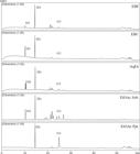
HPLC chromatogram (254 nm) of EBF, EBC, AqEx extracts and EtOAc Sob. and EtOAc Ppt. fractions of Humiria balsamifera. [a = TR 10.2 min; b = TR 14.5 min; c = TR 24.9 min. UV spectra (a) and (b) approx. 215 and 270 nm and (c) approx. 205, 254 and 354 nm].
The analysis of the hexane fraction by GC-MS showed, based on the characteristic mass spectrum, the majority presence of hexadecanoic acid, tetratetracontane and terpenes derivatives (betulin, friedelin, ß-amyrone) (Figure S1, available on supplementary material <https://doi.org/10.6084/m9.figshare.16569393.v1>; Tab. 2).
In the dichloromethane fraction, other major compounds were identified in addition to friedelin and hexadecanoic acid (Figure S2, available on supplementary material <https://doi.org/10.6084/m9.figshare.16569393.v1>; Tab. 3). Among these compounds, the presence of some terpenes is highlighted, such as citronellol, eremophillene, dihydroactinolide and borneol, the last as the majority compound of this fraction (38.57% peak purity). This factor may be relevant to justify the observed activity since this compound has already been described for its anti-inflammatory potential (Zhong et al. 2014Zhong W, Cui Y, Yu Q, Xie X, Liu Y, Wei M, Ci X & Peng L (2014) Modulation of LPS-stimulated pulmonary inflammation by borneol in murine acute lung injury model. Inflammation 37: 1148-1157.; Zou et al. 2017Zou L, Zhang Y, Li W, Zhang J, Wang D, Fu J & Wang P (2017) Comparison of chemical profiles, anti-inflammatory activity, and UPLC-Q-TOF/MS-Based metabolomics in endotoxic fever rats between synthetic borneol and natural borneol. Molecules 22: 1446.). In addition, the ability of borneol to increase cell uptake of other substances is also reported, which would lead to increased apoptosis in tumor cells (Su et al. 2013Su J, Lai H, Chen J, Li L, Wong YS, Chen T & Li X (2013) Natural borneol, a monoterpenoid compound, potentiates selenocystine-induced apoptosis in human hepatocellular carcinoma cells by enhancement of cellular uptake and activation of ROS-mediated DNA damage. PLoS One 8.). This could contribute to understand the cytotoxic profile presented by this fraction against Raw 264.7 macrophages, which could be associated with an increase in cellular uptake of the other compounds present in this fraction.
To sum up, the EtOAc Sup. and the HEX fractions showed to be the most actives, with less citotoxicity, which are in accordance with the literature, that has been described the anti-inflammatory potential of some of the derivatives found in those fractions, such as the hexadecanoic acid (Aparna et al. 2012Aparna V, Dileep KV, Mandal PK, Karthe P, Sadasivan C & Haridas M (2012) Anti-Inflammatory property of n-hexadecanoic acid: structural evidence and kinetic assessment. Chemical Biology & Drug Design 80: 434-439.), phenolic (Gao et al. 2015Gao X jiao, Guo M yao, Zhang Z cai, Wang T cheng, Cao Y guo & Zhang N sheng (2015) Bergenin plays an anti-inflammatory role via the modulation of MAPK and NF-κB signaling pathways in a mouse model of LPS-induced mastitis. Inflammation 38: 1142-1150.; Maleki et al. 2019Maleki SJ, Crespo JF & Cabanillas B (2019) Anti-inflammatory effects of flavonoids. Food Chemistry 299: 125124.) and terpenes derivatives, including betulin (Reyes et al. 2006Reyes CP, Núñez MJ, Jiménez IA, Busserolles J, Alcaraz MJ & Bazzocchi IL (2006) Activity of lupane triterpenoids from Maytenus species as inhibitors of nitric oxide and prostaglandin E2. Bioorganic & Medicinal Chemistry 14: 1573-1579.; Liu et al. 2019Liu D, Yin Q, Zhang Q, Xiang J, Ruan C, Liu H, Li B, Zhu W, Yin C & Fang J (2019) New NO production inhibitors from Hyssopus cuspidatus in LPS-induced RAW264.7 cells. Phytochemistry Letters 34: 91-95.), friedelin (Jin et al. 2018Jin M, Zhou W, Jin C, Jiang Z, Diao S, Jin Z & Li G (2018) Anti-inflammatory activities of the chemical constituents isolated from Trametes versicolor. Natural Product Research 33: 2422-2425.) and amyrone (Almeida et al. 2015Almeida PDO, Boleti APA, Rudiger AL, Lourenço GA, Veiga Junior, VF & Lima ES (2015) Anti-Inflammatory Activity of Triterpenes Isolated from Protium paniculatum Oil-Resins. Evidence-Based Complementary and Alternative Medicine 2015:293768.). Some of the identified compounds have already been described for species from the same family Humiriaceae, such as bergenin in Sacoglottis gabonensis (Ogan 1971Ogan AU (1971) An isocoumarin from the bark of Sacoglottis gabonensis. Phytochemistry 10: 2832-2833.), and hexadecanoic acid, friedelin, betulin and bergenin identified in Endopleura uchi (Marx et al. 2002Marx F, Andrade EHA, Zoghbi MDGB & Maia JGS (2002) Studies of edible Amazonian plants. Part 5: chemical characterisation of Amazonian Endopleura uchi fruits. European Food Research and Technology 214: 331-334.; Abreu et al. 2013Abreu VGC, Correa GM, Lagos IAS, Silva RR & Alcântara AFC (2013) Pentacyclic triterpenes and steroids from the stem bark of uchi (Sacoglottis uchi, Humiriaceae ). Acta Amazônica 43: 525-528.), being bergenin associated to the anti-inflammatory activitiy of the plant (Nunomura et al. 2009Nunomura RCS, Oliveira VG & Nunomura SM (2009) Characterization of Bergenin in Endopleura uchi bark and its anti-inflammatory activity. Journal of the Brazilian Chemical Society 20: 1060-1064.). It is important to highlight that it is the first time that tetratetracontane, ß-amyrone, betulin, citronellol, eremophillene, dihydroactinolide and borneol are being reported for this species and also for the genus Humiria.
Compound identification
Compound 1 (85%) presented by GC-MS m/z 426 [M+] is compatible with a triterpene skeleton (Figure S3, available on supplementary material <https://doi.org/10.6084/m9.figshare.16569393.v1>). The loss of a methyl group is shown by the fragment m/z 411. Other important fragmentations were observed at m/z 341, 273, 205 and 123, assigned to the A, B, C and D rings, respectively (Shiojima et al. 1992Shiojima K, Arai Y, Masuda K, Takase Y, Ageta T & Ageta H (1992) Mass spectra of pentacyclic triterpenoids. Chemical and Pharmaceutical Bulletin 40: 1683-1690. ). 1H-NMR (500 MHz, CDCl3) showed signals between 0.72 and 2.49 ppm characteristic of the triterpene friedelin, which had already been earlier isolated from H. balsamifera (Silva et al. 2004Silva TBC, Alves VL, Mendonça LVH, Conserva LM, Rocha EMM, Andrade EHA & Lemos RPL (2004) Chemical constituents and preliminary antimalarial activity of Humiria balsamifera. Pharmaceutical Biology 42: 94-97.). The following methyl hydrogens were observed: δH 0.82-0.90 (3H, m, H-23); 0.72 (3H, s, H-24); 0.82-0,90 (3H, m, H-25); 1.01 (3H, s, H-26); 1.05 (3H, s, H-27); 1.18 (3H, s, H-28); 1.00 (3H, s, H-29); and 0.95 (3H, s, H-30). This analysis also indicated the presence of metilenic hydrogens between δH 1.25-2.40, and metinic hydrogens δH 2.24 (1H, m, H-4), 1.39 (1H, m, H-8), 1.54 (1H, m, H-10), and 1.56 (1H, m, H-18). HSQC (500 MHz, CDCl3) indicated a characteristic correlation of a friedelane ring, a H-23 methyl group with C-23 in the most protected region of the spectra (δc 6.9) due to effect caused by the carbonyl group (C-3, δc 213.1). HMBC (500 MHz, CDCl3) showed seven quaternary carbons δC 213.1 (C-3), 42.0 (C-5), 37.6 (C-9), 39.7 (C-13), 38.3 (C-14), 29.9 (C-17), 28.2 (C-20). Characteristic correlations to the friedelan ring as H-23/C-3 and H-4/C-3 were also observed in this spectrum. The correlation between the H-4 and H-23 was verified in COSY spectrum (500 MHz, CDCl3). These data are in agreement with literature (Queiroga et al. 2000Queiroga CL, Silva GF, Dias PC, Possenti A & De Carvalho JE (2000) Evaluation of the antiulcerogenic activity of friedelan-3β-ol and friedelin isolated from Maytenus ilicifolia (Celastraceae). Journal of Ethnopharmacology 72: 465-468.; Almeida et al. 2011Almeida MFO, Melo ACR, Pinheiro MLB, Silva JRA, de Souza ADL, Barison A, Campos FR, Amaral ACF, Machado GMC & Leon LLP (2011) Constituintes químicos e atividade leishmanicida de Gustavia elliptica (LECYTHIDACEAE). Química Nova 34: 1182-1187.).
Compound 2 (91%): HPLC/UV-DAD: tR 47.06 min and λmáx 201, 254 and 369 nm in the UV spectra (characteristic of flavonoids) (Fig. 2). 1H-NMR (500 MHz, CD3OD): 6.16 (1H, s H-6), 6.37 (1H, s, H-8), 7.72 (1H, m, H-2’), 6.89 (1H, d, J = 8.5 Hz, H-5’), and 7.62 (1H, d, J = 8.5 Hz, H-6’). HSQC (500 MHz, CDCl3) analysis allowed to observe δC 98.0 (C-6) and 92.9 (C-8) coupling to δH 6.16 (H-6) and 6.37 (H-8), referring to the A ring. In addition, the δC 114.6 (C-5’), 120.1 (C-6’) and 147.2 (C-2’) coupling to 6.89 (H-5’) and 7.62 (H-2’,6’), respectively, relative to B ring. HMBC (500 MHz, CDCl3) showed nine quaternary carbons δC 147.2 (C-2), 175.5 (C-4), 161.0 (C-5), 164.8 (C-7), 156.8 (C-9), 102.9 (C-10), 122.6 (C-1’), 147.2 (C-3’) and 144.8 (C-4’). COSY (500 MHz, CDCl3) analysis allowed to observe the H-5’ and H-6’ correlation of the B ring. These data are characteristic of flavonol quercetin (Fossen et al. 1998Fossen T, Pedersen AT & Andersen ØM (1998) Flavonoids from red onion (Allium cepa). Phytochemistry 47: 281-285.; Saaby et al. 2009Saaby L, Rasmussen HB & Jäger AK (2009) MAO-A inhibitory activity of quercetin from Calluna vulgaris (L.) Hull. Journal of Ethnopharmacology 121: 178-181.), which had already been isolated earlier from H. balsamifera (Silva et al. 2004Silva TBC, Alves VL, Mendonça LVH, Conserva LM, Rocha EMM, Andrade EHA & Lemos RPL (2004) Chemical constituents and preliminary antimalarial activity of Humiria balsamifera. Pharmaceutical Biology 42: 94-97.).

Compound 3 (70%): HPLC/UV-DAD 25.49 min and λmáx 202, 255 and 355 nm in the UV spectra (Fig. 3). Spectroscopy data showed signals characteristic of quercetin-3-O-α-arabinopyranoside (Wollenweber et al. 1997Wollenweber E, Stüber A & Kraut L (1997) Flavonoid aglycones and flavonol glycosides in the lipophilic leaf exudate of Nothofagus antarctica. Phytochemistry 44: 1399-1400.; Ahmadu et al. 2007Ahmadu AA, Hassan HS, Abubakar MU & Akpulu IN (2007) Flavonoid glycosides from Byrsocarpus coccineus leaves. Schum and Thonn (connaraceae). African Journal of Traditional, Complementary and Alternative Medicines 4: 257-260.), described for the first time for this species. The 1H-NMR (500 MHz, CD3OD) displayed characteristic signals of the aglycone quercetin δH 6.21 (1H, d, J = 2.0, H-6), 6.41 (1H, d, J = 2.0, H-8), 7.74 (1H, d, J = 2.2, H-2’), 6.90 (1H, d, J = 8.5, H-5’), 7.57 (1H, dd, J = 2.2 and 8.5, H-6’) and sugar signals δH 5.10 (1H, d, J = 6.7, H-1’’), 3.90 (1H, dd, J = 6.6 and 8.5, H-2’’), 3.65 (1H, dd, J = 3.1 and 8.4, H-3’’), 3.83 (1H, s, J = 8.5, H-4’’), 3.80 (1H, d, J = 3.5, H-5a’’), and 3.43 (1H, d, J = 10.8, H-5b’’). APT (100 MHz, CD3OD) data indicated the presence of nine methine carbons δC 100.3 (C-6) and 95.1 (C-8) of ring A, 116.3 (C-5’), 123.1 (C-6’) e 117.5 (C-2’) of ring B and 104.4 (C-1’’), 72.8 (C-2’’), 69.1 (C-3’’), and 74.0 (C-4’’), and one methylene carbon 67.0 (C-5’’) of sugar portion. It was possible to identify 10 quaternary carbons from HMBC (500 MHz, CD3OD), namely: 158.2 (C-2), 135.2 (C-3), 179.7 (C-4), 162.4 (C-5), 166.6 (C-7), 158.2 (C-9), 105.0 (C-10), 122.8 (C-1’), 145.8 (C-3’) and 149.7 (C-4’). This spectra alowed to confirm the existence of a glycoside moiety attached to the quercetin through the OH of C-3 of the C ring by the H-1’’ (5.10 ppm) and C-3 (135.2 ppm) correlation and enable confirmation of this structure by other observed correlations.

HPLC Chromatogram (254 nm) and UV spectra of the compound 3 (quercetin-3-O-α-arabinopyranoside).
NO and TNF-α inhibition and cytotoxicity of isolated compounds in LPS-stimulated RAW 264.7 cells
Quercetin (2) presented the highest potential in NO (IC50 4.58±0.95 µg/mL) and TNF-α (IC50 33.25±0.88 µg/mL) inhibition. In addition, this compound was more active than the L-NMMA inhibitor and showed no cytotoxic effect up to a concentration of 20 µg/mL, in which excellent inhibitory activity was observed for NO production (84.87±1.47%). Friedelin (1) and quercetin-3-O-α-arabinopyranosyl (3) demonstrated no significant NO and TNF-α inhibition (IC50 > 100 µg/mL). Besides, we observed low cytotoxicity for compounds 1 and 3 (Tab. 4).
Effect of the isolated compounds on the inhibition of nitric oxide, TNF-α production and cytotoxicity in LPS-stimulated RAW 264.7 macrophage cells.a = samples were different from the positive control MΦ + LPS (0.00 ± 0.40%), except Quercetin-3-O-α-arabinopyranoside at 0.8 µg/mL; b = samples were different from the positive control MΦ + LPS (0.00 ± 0.27%), except Quercetin at 0.8 µg/mL and Quercetin-3-O-α-arabinopyranoside at 0.8-20 µg/mL; c = all samples were different from the positive control Triton (100 ± 0.01%); *** = P < 0.001; ** = P < 0.01; * = P < 0.05. L-NMMA at 20 µg/mL (51.36 ± 0.54% NO inhibition).
Friedelin (1) has already been briefly described in the literature for its anti-inflammatory activity. Friedelin isolated from the stems of Heritiera littoralis Aiton. (Malvaceae) exhibited IC50 > 100 μM (approximately 42.67 μg/mL) in the NO inhibition (Tewtrakul et al. 2010Tewtrakul S, Tansakul P, Daengrot C, Ponglimanont C & Karalai C (2010) Anti-inflammatory principles from Heritiera littoralis bark. Phytomedicine 17: 851-855.), a result which can be considered compatible with the observed in this study (at 100 μg/mL; 24.23 ± 2.41% inhibition). This substance was isolated from the leaves and twigs of Acer mandshuricum Maxim. (Aceraceae), being evaluated at 100 nM (approximately 0.04 μg/mL) and inhibiting 23.5% of TNF-α release (Ding et al. 2010Ding Y, Liang C, Kim JH, Lee YM, Hyun JH, Kang HK, Kim JA, Min BS & Kim YH (2010) Triterpene compounds isolated from Acer mandshuricum and their anti-inflammatory activity. Bioorganic & Medicinal Chemistry Letters 20: 1528-1531. ). A slightly reduced TNF-α inhibition percentage (14.98 ± 0.20%) was verified in this study at 0.8 μg/mL and, in addition to that, it was also observed that even with the increase of the evaluated concentrations, no increase in the inhibitory effect was noted, suggesting that it may be the maximum effect caused by friedelin.
Quercetin (2), as well as friedelin (1), already earlier isolated from H. balsamifera (Silva et al. 2004Silva TBC, Alves VL, Mendonça LVH, Conserva LM, Rocha EMM, Andrade EHA & Lemos RPL (2004) Chemical constituents and preliminary antimalarial activity of Humiria balsamifera. Pharmaceutical Biology 42: 94-97.), has been widely described in the literature as having anti-inflammatory potential. Quercetin at 20 and 100 μg/mL was able to inhibit approximately 85% of NO production (IC50 4.58 ± 0.95 μg/mL) in this study. These data agree with the literature since this compound inhibited between 75–80% of NO production at 20 μM (6.04 μg/mL) and 100 μM (30.2 μg/mL) (Manjeet & Ghosh 1999Manjeet KR & Ghosh B (1999). Quercetin inhibits LPS-induced nitric oxide and tumor necrosis factor-α production in murine macrophages. International Immunopharmacology 21: 435-443.; Shen et al. 2002Shen SC, Lee WR, Lin HY, Huang HC, Ko CH, Yang LL & Chen YC (2002) In vitro and in vivo inhibitory activities of rutin, wogonin, and qyercetin on lipopolysaccharide-induced nitricnoxide and prostaglandin E2 production. European Journal of Pharmacology 1-3: 187-194.; Ho et al. 2017Ho GTT, Wangensteen H & Barsett H (2017) Elderberry and elderflower extracts, phenolic compounds, and metabolites and their effect on complement, RAW 264.7 macrophages and dendritic cells. International Journal of Molecular Sciences 18: 584.) showing IC50 4.08 μg/mL (Wang et al. 2019Wang L, Zhang K, Han S, Bai H, Bao F, Zheng Y, Wang J, Du H, Liu Y & Yang Z (2019) Constituents isolated from the leaves of Glycyrrhiza uralansis and their anti-inflammatory activities on LPS-induced RAW264.7 cells. Molecules 24: 1923.). Quercetin was also described for having the ability to inhibit the release of TNF-α, presenting IC50 < 200 μg/mL (667 μM) (Calixto et al. 2004Calixto JB, Campos MM, Otuki MF & Santos ARS (2004) Anti-inflammatory compounds of plant origin. Part II. Modulation of pro-inflammatory cytokines, chemokines and adhesion molecules. Planta Médica 70: 93-103.). A recent study evaluated the activity of quercetin from 6.25 to 25 μM in the release of TNF-α, showing no relevant effect on the production of TNFα at the tested concentrations (Lee et al. 2018Lee HN, Shin SA, Choo GS, Kim HJ, Park YS, Kim BS, Kim SK, Cho SD, Nam JS, Choi CS, Che JH, Park BK & Jung JY (2018) Anti-inflammatory effect of quercetin and galangin in LPS-stimulated RAW264.7 macrophages and DNCB-induced atopic dermatitis animal models. International Journal of Molecular Medicine 41: 888-898.).
Quercetin-3-O-α-arabinopyranoside (3) is herein described for the first time in H. balsamifera extracts. The non-expressive effects of this compound on NO and TNF-α production compared with the correspondent aglicone quercetin agree with literature data. Quercetin-3-O-α-arabinopyranoside isolated from guava leaves (Kim et al. 2015Kim MH, Kim JN, Han SN & Kim HK (2015) Ursolic acid isolated from guava leaves inhibits inflammatory mediators and reactive oxygen species in LPS-stimulated macrophages. Immunopharmacology and Immunotoxicology 37: 228-235. ) and Acer tegmentosum Maxim. (Aceraceae) (Lee et al. 2014Lee KJ, Song NY, Oh YC, Cho WK & Ma JY (2014) Isolation and bioactivity analysis of ethyl acetate extract from acer tegmentosum using in vitro assay and on-line screening HPLC-ABTS+ system. Journal of Analytical Methods in Chemistry 2014: 150509.) evaluated at 10–100 μM demonstrated no significant NO and TNF-α inhibition in RAW 264.7 cells. This compound similarly isolated from Psidium acutangulum DC. (Myrtaceae) did not inhibit NO when tested at 50 μg/mL (Houël et al. 2016Houël E, Nardella F, Jullian V, Valentin A, Vonthron-Sénécheau C, Villa P, Obrecht A, Kaiser M, Bourreau E, Odonne G, Fleury M, Bourdy G, Eparvier V, Deharo E & Stien D (2016) Wayanin and guaijaverin, two active metabolites found in a Psidium acutangulum Mart. ex DC (syn. P. persoonii McVaugh) (Myrtaceae) antimalarial decoction from the Wayana Amerindians. Journal of Ethnopharmacology 187: 241-248. ). A higher inhibitory activity of pro-inflammatory mediators for quercetin (2) compared to quercetin-3-O-α-arabinopyranoside (3) was also observed in the present study. Therefore, it can be suggested that glycosylation leads to an increase in the polarity and molecular size, and hence may alter the interaction with the target molecule and its passage through the phospholipidic membrane, modifying the possible activity on intracellular sites.
Other activities previously described in the literature for these compounds such as analgesic, antipyretic, antimicrobial, and wound healing (Antonisamy et al. 2011Antonisamy P, Duraipandiyan V & Ignacimuthu S (2011) Anti-inflammatory, analgesic and antipyretic effects of friedelin isolated from Azima tetracantha Lam. in mouse and rat models. Journal of Pharmacy and Pharmacology 63: 1070-1077.; Maalik et al. 2014Maalik A, Khan FA, Mumtaz A, Mehmood A, Azhar S, Atif M, Karim S, Altaf Y & Tariq I (2014) Pharmacological applications of quercetin and its derivatives: a short review. Tropical Journal of Pharmaceutical Research 13: 1561-1566.; Hatahet et al. 2016Hatahet T, Morille M, Hommoss A, Devoisselle JM, Müller RH & Bégu S (2016) Quercetin topical application, from conventional dosage forms to nanodosage forms. European Journal of Pharmaceutics and Biopharmaceutics 108: 41-53. ; Noundou et al. 2016Noundou XS, Krause RWM, Van Vuuren SF, Ndinteh DT & Olivier DK (2016) Antibacterial effects of Alchornea cordifolia (Schumach. and Thonn.) Müll. Arg extracts and compounds on gastrointestinal, skin, respiratory and urinary tract pathogens. Jounal of Ethnopharmacology 179: 76-82. ; Sa et al. 2017Sa FAS, Paula JAM, Santos PA, Oliveira LDAR, Oliveira GDAR, Liao LM, Paula JR & Silva MDRR (2017) Phytochemical analysis and antimicrobial activity of Myrcia tomentosa (Aubl.) DC. leaves. Molecules 22: 1-10.; Özbilgin et al. 2018Özbilgin S, Acıkara ÖB, Akkol EK, Süntar I, Keleş H & İşcan GS (2018) In vivo wound-healing activity of Euphorbia characias subsp. wulfenii: isolation and quantification of quercetin glycosides as bioactive compounds. Journal of Ethnopharmacology 224: 400-408.; Singh et al. 2018Singh AK, Kumar S & Vinayak M (2018) Recent development in antihyperalgesic effect of phytochemicals: anti-inflammatory and neuro-modulatory actions. Inflammation Research 67: 633-654. ) may contribute to justify the use of this species in traditional medicine to treat hepatitis, diarrhea, hemorrhoids, in curing chronic wounds, and alleviating toothaches.
In summary, this study describes the NO and TNF-α inhibition by H. balsamifera, the isolation of quercetin-3-O-α-arabinopyranoside and identification of terpenes ß-amyrone, betulin, citronellol, eremophillene, dihydroactinolide and borneol for the first time in this species. Thus, promising results for a species of Jurubatiba shoal are demonstrated in this work, evidencing this region as being a relevant source in the search for new bioactive products. These aspects reinforce the importance of biodiversity preservation of Jurubatiba shoal. The results also strengthen the proposal that inhibition of NO and TNF-α production might be a useful screening strategy in searching for new anti-inflammatory compounds, especially by using medicinal plant extracts.
Acknowledgements
This work was supported by a CAPES scholarship, by funding from CNPq: Universal 2011 (486565/2011-4) and Produtividade em Pesquisa (312045/2014-0); and also from FAPERJ: Emergentes (E-26/110.127/2014) and BIOTA (E-26/111.396/2012).
References
- Abreu VGC, Correa GM, Lagos IAS, Silva RR & Alcântara AFC (2013) Pentacyclic triterpenes and steroids from the stem bark of uchi (Sacoglottis uchi, Humiriaceae ). Acta Amazônica 43: 525-528.
- Ahmadu AA, Hassan HS, Abubakar MU & Akpulu IN (2007) Flavonoid glycosides from Byrsocarpus coccineus leaves. Schum and Thonn (connaraceae). African Journal of Traditional, Complementary and Alternative Medicines 4: 257-260.
- Almeida MFO, Melo ACR, Pinheiro MLB, Silva JRA, de Souza ADL, Barison A, Campos FR, Amaral ACF, Machado GMC & Leon LLP (2011) Constituintes químicos e atividade leishmanicida de Gustavia elliptica (LECYTHIDACEAE). Química Nova 34: 1182-1187.
- Almeida PDO, Boleti APA, Rudiger AL, Lourenço GA, Veiga Junior, VF & Lima ES (2015) Anti-Inflammatory Activity of Triterpenes Isolated from Protium paniculatum Oil-Resins. Evidence-Based Complementary and Alternative Medicine 2015:293768.
- Antonisamy P, Duraipandiyan V & Ignacimuthu S (2011) Anti-inflammatory, analgesic and antipyretic effects of friedelin isolated from Azima tetracantha Lam. in mouse and rat models. Journal of Pharmacy and Pharmacology 63: 1070-1077.
- Aparna V, Dileep KV, Mandal PK, Karthe P, Sadasivan C & Haridas M (2012) Anti-Inflammatory property of n-hexadecanoic acid: structural evidence and kinetic assessment. Chemical Biology & Drug Design 80: 434-439.
- Bernstein N, Akram M, Daniyal M, Koltai H, Fridlender M & Gorelick J (2018) Antiinflammatory potential of medicinal plants: a source for therapeutic secondary metabolites. Advances in Agronomy 150: 131-183.
- BFG - The Brazil Flora Group (2018) Brazilian Flora 2020: innovation and collaboration to meet Target 1 of the Global Strategy for Plant Conservation (GSPC). Rodriguésia 69: 1513-1527.
- Calixto JB, Campos MM, Otuki MF & Santos ARS (2004) Anti-inflammatory compounds of plant origin. Part II. Modulation of pro-inflammatory cytokines, chemokines and adhesion molecules. Planta Médica 70: 93-103.
- Coelho-Ferreira M (2009) Medicinal knowledge and plant utilization in an Amazonian coastal community of Marudá, Pará state (Brazil). Journal of Ethnopharmacology 126: 159-175.
- Cogliatti-Carvalho L, Freitas AFN, Rocha CFD & Van Sluys M (2001) Variação na estrutura e na composição de Bromeliaceae em cinco zonas de restinga no Parque Nacional da Restinga de Jurubatiba, Macaé, RJ. Revista Brasileira de Botânica 24: 1-9.
- De Rijke E, Out P, Niessen WMA, Ariese F, Gooijer C & Brinkman UAT (2006) Analytical separation and detection methods for flavonoids. Journal of Chromatography A 1112: 31-63.
- Dinarello CA (2010). Anti-inflammatory agents: present and future. Cell 140: 935-950.
- Ding Y, Liang C, Kim JH, Lee YM, Hyun JH, Kang HK, Kim JA, Min BS & Kim YH (2010) Triterpene compounds isolated from Acer mandshuricum and their anti-inflammatory activity. Bioorganic & Medicinal Chemistry Letters 20: 1528-1531.
- Fossen T, Pedersen AT & Andersen ØM (1998) Flavonoids from red onion (Allium cepa). Phytochemistry 47: 281-285.
- Gao X jiao, Guo M yao, Zhang Z cai, Wang T cheng, Cao Y guo & Zhang N sheng (2015) Bergenin plays an anti-inflammatory role via the modulation of MAPK and NF-κB signaling pathways in a mouse model of LPS-induced mastitis. Inflammation 38: 1142-1150.
- Griess JP (1879) Bemerkungen zu der Abhandlung der HH: Wesely und Benedikt. “Uber einige Azoverbindungen”. Berichte der Deutschen Chemischen Gesellschaft 12: 426-428.
- Harirforoosh S, Asghar W & Jamali F (2013) Adverse effects of nonsteroidal antiinflammatory drugs: an update of gastrointestinal, cardiovascular and renal complications. Journal of Pharmaceutical Sciences 16: 821-47.
- Hatahet T, Morille M, Hommoss A, Devoisselle JM, Müller RH & Bégu S (2016) Quercetin topical application, from conventional dosage forms to nanodosage forms. European Journal of Pharmaceutics and Biopharmaceutics 108: 41-53.
- Ho GTT, Wangensteen H & Barsett H (2017) Elderberry and elderflower extracts, phenolic compounds, and metabolites and their effect on complement, RAW 264.7 macrophages and dendritic cells. International Journal of Molecular Sciences 18: 584.
- Houël E, Nardella F, Jullian V, Valentin A, Vonthron-Sénécheau C, Villa P, Obrecht A, Kaiser M, Bourreau E, Odonne G, Fleury M, Bourdy G, Eparvier V, Deharo E & Stien D (2016) Wayanin and guaijaverin, two active metabolites found in a Psidium acutangulum Mart. ex DC (syn. P. persoonii McVaugh) (Myrtaceae) antimalarial decoction from the Wayana Amerindians. Journal of Ethnopharmacology 187: 241-248.
- Jin M, Zhou W, Jin C, Jiang Z, Diao S, Jin Z & Li G (2018) Anti-inflammatory activities of the chemical constituents isolated from Trametes versicolor Natural Product Research 33: 2422-2425.
- Kelecom A, Reis GL, Fevereiro PC, Silva JG, Santos MG, Mello Neto CB, Gonzalez MS, Gouvea RC & Almeida GS (2002) A multidisciplinary approach to the study of the fluminense vegetation. Anais da Academia Brasileira de Ciências 74: 171-181.
- Kim MH, Kim JN, Han SN & Kim HK (2015) Ursolic acid isolated from guava leaves inhibits inflammatory mediators and reactive oxygen species in LPS-stimulated macrophages. Immunopharmacology and Immunotoxicology 37: 228-235.
- Lee HN, Shin SA, Choo GS, Kim HJ, Park YS, Kim BS, Kim SK, Cho SD, Nam JS, Choi CS, Che JH, Park BK & Jung JY (2018) Anti-inflammatory effect of quercetin and galangin in LPS-stimulated RAW264.7 macrophages and DNCB-induced atopic dermatitis animal models. International Journal of Molecular Medicine 41: 888-898.
- Lee KJ, Song NY, Oh YC, Cho WK & Ma JY (2014) Isolation and bioactivity analysis of ethyl acetate extract from acer tegmentosum using in vitro assay and on-line screening HPLC-ABTS+ system. Journal of Analytical Methods in Chemistry 2014: 150509.
- Liu D, Yin Q, Zhang Q, Xiang J, Ruan C, Liu H, Li B, Zhu W, Yin C & Fang J (2019) New NO production inhibitors from Hyssopus cuspidatus in LPS-induced RAW264.7 cells. Phytochemistry Letters 34: 91-95.
- Lorenzi H & Matos FJA (2008) Plantas medicinais no Brasil: nativas e exóticas. 2nd ed. Plantarum, Nova Odessa. 296p.
- Maalik A, Khan FA, Mumtaz A, Mehmood A, Azhar S, Atif M, Karim S, Altaf Y & Tariq I (2014) Pharmacological applications of quercetin and its derivatives: a short review. Tropical Journal of Pharmaceutical Research 13: 1561-1566.
- Maleki SJ, Crespo JF & Cabanillas B (2019) Anti-inflammatory effects of flavonoids. Food Chemistry 299: 125124.
- Manjeet KR & Ghosh B (1999). Quercetin inhibits LPS-induced nitric oxide and tumor necrosis factor-α production in murine macrophages. International Immunopharmacology 21: 435-443.
- Marx F, Andrade EHA, Zoghbi MDGB & Maia JGS (2002) Studies of edible Amazonian plants. Part 5: chemical characterisation of Amazonian Endopleura uchi fruits. European Food Research and Technology 214: 331-334.
- Mosmann T (1983) Rapid colorimetric assay for cellular growth and survival: application to proliferation and cytotoxicity assays. Journal of Immunological Methods 65: 55-63.
- Muzitano MF, Cruz EA, Almeida AP, Silva SAG, Kaiser CR, Guette C, Rossi-Bergmann B & Costa SS (2006) Quercitrin: an antileishmanial flavonoid glycoside from Kalanchoe pinnata. Planta Médica 72: 81-83.
- Noundou XS, Krause RWM, Van Vuuren SF, Ndinteh DT & Olivier DK (2016) Antibacterial effects of Alchornea cordifolia (Schumach. and Thonn.) Müll. Arg extracts and compounds on gastrointestinal, skin, respiratory and urinary tract pathogens. Jounal of Ethnopharmacology 179: 76-82.
- Nunomura RCS, Oliveira VG & Nunomura SM (2009) Characterization of Bergenin in Endopleura uchi bark and its anti-inflammatory activity. Journal of the Brazilian Chemical Society 20: 1060-1064.
- Ogan AU (1971) An isocoumarin from the bark of Sacoglottis gabonensis Phytochemistry 10: 2832-2833.
- Özbilgin S, Acıkara ÖB, Akkol EK, Süntar I, Keleş H & İşcan GS (2018) In vivo wound-healing activity of Euphorbia characias subsp. wulfenii: isolation and quantification of quercetin glycosides as bioactive compounds. Journal of Ethnopharmacology 224: 400-408.
- Qin X, Yang Y, Fan TT, Gong T, Zhang XN & Huang Y (2010) Preparation, characterization and in vivo evaluation of bergenin-phospholipid complex. Acta Pharmacologica Sinica 31: 127-136.
- Queiroga CL, Silva GF, Dias PC, Possenti A & De Carvalho JE (2000) Evaluation of the antiulcerogenic activity of friedelan-3β-ol and friedelin isolated from Maytenus ilicifolia (Celastraceae). Journal of Ethnopharmacology 72: 465-468.
- Reyes CP, Núñez MJ, Jiménez IA, Busserolles J, Alcaraz MJ & Bazzocchi IL (2006) Activity of lupane triterpenoids from Maytenus species as inhibitors of nitric oxide and prostaglandin E2. Bioorganic & Medicinal Chemistry 14: 1573-1579.
- Ronchetti S, Migliorati G, Bruscoli S & Riccardi C (2018) Defining the role of glucocorticoids in inflammation. Clinical Science 132: 1529-1543.
- Sa FAS, Paula JAM, Santos PA, Oliveira LDAR, Oliveira GDAR, Liao LM, Paula JR & Silva MDRR (2017) Phytochemical analysis and antimicrobial activity of Myrcia tomentosa (Aubl.) DC. leaves. Molecules 22: 1-10.
- Saaby L, Rasmussen HB & Jäger AK (2009) MAO-A inhibitory activity of quercetin from Calluna vulgaris (L.) Hull. Journal of Ethnopharmacology 121: 178-181.
- Santos FM, Malafaia CA, Simas DLR, Paulino AB, Muzitano MF, Simas NK, Da-Rocha EAC, Amaral ACF, Leal ICR (2020) Phenolic compounds from Tocoyena bullata mart (Rubiaceae) with inhibitory activity in mast cells degranulation. Natural Product Research 34: 3295-3298.
- Shiau M, Chiou H & Lee Y (2001) Establishment of a consistent L929 bioassay system for TNF-α quantitation to evaluate the effect of lipopolysaccharide, phytomitogens and cytodifferentiation agents on cytotoxicity of TNF-α secreted by adherent human mononuclear cells. Mediators of Inflammation 10: 199-208.
- Shen SC, Lee WR, Lin HY, Huang HC, Ko CH, Yang LL & Chen YC (2002) In vitro and in vivo inhibitory activities of rutin, wogonin, and qyercetin on lipopolysaccharide-induced nitricnoxide and prostaglandin E2 production. European Journal of Pharmacology 1-3: 187-194.
- Shiojima K, Arai Y, Masuda K, Takase Y, Ageta T & Ageta H (1992) Mass spectra of pentacyclic triterpenoids. Chemical and Pharmaceutical Bulletin 40: 1683-1690.
- Silva TBC, Alves VL, Mendonça LVH, Conserva LM, Rocha EMM, Andrade EHA & Lemos RPL (2004) Chemical constituents and preliminary antimalarial activity of Humiria balsamifera Pharmaceutical Biology 42: 94-97.
- Singh AK, Kumar S & Vinayak M (2018) Recent development in antihyperalgesic effect of phytochemicals: anti-inflammatory and neuro-modulatory actions. Inflammation Research 67: 633-654.
- Su J, Lai H, Chen J, Li L, Wong YS, Chen T & Li X (2013) Natural borneol, a monoterpenoid compound, potentiates selenocystine-induced apoptosis in human hepatocellular carcinoma cells by enhancement of cellular uptake and activation of ROS-mediated DNA damage. PLoS One 8.
- Tewtrakul S, Tansakul P, Daengrot C, Ponglimanont C & Karalai C (2010) Anti-inflammatory principles from Heritiera littoralis bark. Phytomedicine 17: 851-855.
- Wagner H & Bladt S (1996). Plant drug analysis: athin layer chromatography atlas. 2nd ed. Springer, Berlim. 359p.
- Wang L, Zhang K, Han S, Bai H, Bao F, Zheng Y, Wang J, Du H, Liu Y & Yang Z (2019) Constituents isolated from the leaves of Glycyrrhiza uralansis and their anti-inflammatory activities on LPS-induced RAW264.7 cells. Molecules 24: 1923.
- Wollenweber E, Stüber A & Kraut L (1997) Flavonoid aglycones and flavonol glycosides in the lipophilic leaf exudate of Nothofagus antarctica Phytochemistry 44: 1399-1400.
- Zhong W, Cui Y, Yu Q, Xie X, Liu Y, Wei M, Ci X & Peng L (2014) Modulation of LPS-stimulated pulmonary inflammation by borneol in murine acute lung injury model. Inflammation 37: 1148-1157.
- Zou L, Zhang Y, Li W, Zhang J, Wang D, Fu J & Wang P (2017) Comparison of chemical profiles, anti-inflammatory activity, and UPLC-Q-TOF/MS-Based metabolomics in endotoxic fever rats between synthetic borneol and natural borneol. Molecules 22: 1446.
Supplementary Material
See supplementary material at <https://doi.org/10.6084/m9.figshare.16569393.v1>
Edited by
Publication Dates
-
Publication in this collection
27 Sept 2021 -
Date of issue
2021
History
-
Received
16 Mar 2020 -
Accepted
20 May 2020


