Abstracts
pyoderma gangrenosum is a rare inflammatory skin condition characterized by progressive and recurrent skin ulceration of destructive course. It is usually associated with rheumatoid arthritis, paraproteinemia, myeloproliferative diseases and inflammatory bowel diseases, especially non-specific ulcerative proctocolitis. In these situations, skin lesions are described as concurrent with the intestinal condition. However, reports on pyoderma gangrenosum preceding intestinal findings are less frequent. The authors describe a case of a woman with febrile condition associated with skin lesions diagnosed by biopsy as pyoderma gangrenosum. Two weeks later, she developed diarrhea, arthralgia and sepsis, being diagnosed as ulcerative proctocolitis. After the administration of the treatment for ulcerative proctocolitis, she showed improvements in sepsis care, remission of diarrhea and regression of skin lesions. This case highlights the importance of considering pyoderma gangrenosum as a manifestation associated with inflammatory bowel disease, regardless of its timing in relation to intestinal symptoms.
pyoderma gangrenosum; proctocolitis; pyoderma; colitis
Pioderma gangrenoso é uma forma de inflamação cutânea, caracterizada por ulceração progressiva e recorrente da pele, com curso destrutivo. Geralmente é associada à artrite reumatoide, paraproteinemia, doenças mieloproliferativas e doença inflamatória intestinal, em especial retocolite ulcerativa inespecífica. Em tais casos, as lesões cutâneas são descritas concomitantes ao quadro intestinal, porém, relatos com descrição de pioderma gangrenoso precedendo achados intestinais são menos frequentes. Os autores relatam caso de mulher com quadro febril associado a lesões cutâneas diagnosticadas por biópsia como pioderma gangrenoso. Duas semanas depois, apresentou diarreia, artralgia e sepse sendo diagnosticada retocolite ulcerativa. Com o tratamento para retocolite ulcerativa apresentou melhora do quadro séptico, remissão da diarreia e regressão das lesões cutâneas. Este caso enfatiza a importância em considerar o pioderma gangrenoso como manifestação associada à doença inflamatória intestinal, independente de sua temporalidade em relação aos sintomas intestinais.
pioderma gangrenoso; proctocolite; pioderma; colite
CASE REPORT
Pyoderma gangrenosum as a initial manifestation of ulcerative proctocolitis
Carla Bortolin FonsecaI,II; Guilherme Lang MottaI,II; Alexandre RampazzoI; João Carlos Cantarelli JuniorI,II; Renato Borges FagundesII,III
IUniversity Hospital of Universidade Federal de Santa Maria (UFSM) Santa Maria (RS), Brazil
IIDepartament of Medical Clinic/Center of Health Sciences of UFSM Santa Maria (RS), Brazil
IIIPost Graduation Program: Gastroenterology Sciences of the Medical School at Universidade Federal do Rio Grande do Sul (UFRGS) Porto Alegre (RS), Brazil
Correspondence Correspondence: Renato B. Fagundes, MD, PhD Avenida Grécia 1000, ap. 1002 B Passo d'areia CEP: 91350-070 Porto Alegre (RS), Brazil. E-mail: fagundesrb@gmail.com
ABSTRACT
pyoderma gangrenosum is a rare inflammatory skin condition characterized by progressive and recurrent skin ulceration of destructive course. It is usually associated with rheumatoid arthritis, paraproteinemia, myeloproliferative diseases and inflammatory bowel diseases, especially non-specific ulcerative proctocolitis. In these situations, skin lesions are described as concurrent with the intestinal condition. However, reports on pyoderma gangrenosum preceding intestinal findings are less frequent. The authors describe a case of a woman with febrile condition associated with skin lesions diagnosed by biopsy as pyoderma gangrenosum. Two weeks later, she developed diarrhea, arthralgia and sepsis, being diagnosed as ulcerative proctocolitis. After the administration of the treatment for ulcerative proctocolitis, she showed improvements in sepsis care, remission of diarrhea and regression of skin lesions. This case highlights the importance of considering pyoderma gangrenosum as a manifestation associated with inflammatory bowel disease, regardless of its timing in relation to intestinal symptoms.
Keywords: pyoderma gangrenosum; proctocolitis; pyoderma; colitis.
RESUMO
Pioderma gangrenoso é uma forma de inflamação cutânea, caracterizada por ulceração progressiva e recorrente da pele, com curso destrutivo. Geralmente é associada à artrite reumatoide, paraproteinemia, doenças mieloproliferativas e doença inflamatória intestinal, em especial retocolite ulcerativa inespecífica. Em tais casos, as lesões cutâneas são descritas concomitantes ao quadro intestinal, porém, relatos com descrição de pioderma gangrenoso precedendo achados intestinais são menos frequentes. Os autores relatam caso de mulher com quadro febril associado a lesões cutâneas diagnosticadas por biópsia como pioderma gangrenoso. Duas semanas depois, apresentou diarreia, artralgia e sepse sendo diagnosticada retocolite ulcerativa. Com o tratamento para retocolite ulcerativa apresentou melhora do quadro séptico, remissão da diarreia e regressão das lesões cutâneas. Este caso enfatiza a importância em considerar o pioderma gangrenoso como manifestação associada à doença inflamatória intestinal, independente de sua temporalidade em relação aos sintomas intestinais.
Palavras-chave: pioderma gangrenoso; proctocolite; pioderma; colite.
INTRODUCTION
Pyoderma gangrenosum (PG) is a type of inflammatory skin condition of unknown origin characterized by progressive and recurrent skin ulceration of destructive course1-3. Dermatosis is usually unpredictable, sudden and aggressive, but it can also be chronic, slow and insidious, presenting skin ulcers that expand centrifugally4. The aggressive form may cause painful ulcerative lesions, with necrotic and hemorrhagic base5. It is associated with systemic disease in about 50% of the cases, such as rheumatoid arthritis, inflammatory bowel disease, paraproteinemia and myeloproliferative disease6,7. It is described as one of the extraintestinal manifestations for the patients who have inflammatory bowel disease (IBD), especially the severe forms of unspecified ulcerative proctocolitis (UUP)8,9. PG affects people at any age group, especially young women with proctocolitis and diffuse compromise of the entire colon10,11. It can appear in any part of the body, but is mostly described in the inferior limbs, especially the lower third of the legs. The skin lesion usually manifests during the two first years of inflammatory bowel disease, being more prevalent during the periods of UUP clinical exacerbation12. Skin lesions that precede the onset of the intestinal picture are a less frequent situation13. The authors report the case of a patient with severe UUP whose initial manifestation was the pyoderma gangrenosum, preceding diarrhea in two weeks.
CLINICAL CASE
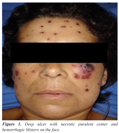
A white 46-year-old female patient, who was previously healthy, presented with a 38.5ºC fever and skin lesions in the face, limbs and vulva, characterized as deep and painful ulcers, with necrotic purulent center and small hemorrhagic blisters (Figures 1 and 2), five days prior to the first visit. At the doctor's appointment, the patient was given antithermic drugs; skin lesions were biopsied. The anatomopathological examination of the lesions showed superficial and deep diffuse dermatitis with the prevalence of neutrophils (Figure 3) and epidermal ulceration, which are compatible with pyoderma gangrenosum and evidence of deep abscesses in the subcutaneous tissue (Figure 4).
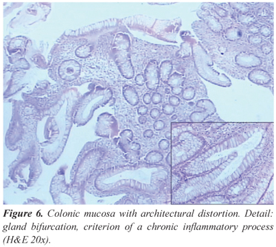
Two weeks after, she started presenting liquid diarrhea without mucus, suppuration or blood, followed by vomit and persistent fever. She reported the ingestion of untreated water, but denied having contact with sick people, trips, previous medications, insect bites and similar cases in the family. She was admitted at the hospital and submitted to complementary examinations, with the following results: hemoglobin 7.4 g/dL, leukocytes 22500/mm3, band cells 7232/mm3, segmented 12,430/mm3; parasitological examination of stools without evidence of intestinal parasites or their evolutive forms. Fecal leukocytes test was positive (++). At coproculture, gram positive cocci were prevalente. The Widal reaction was negative. Right after admission, diarrhea had mucus and blood, and the patient started reporting arthralgia of the knees. On the seventh day after admission, she evolved to a septic picture and was transferred to the intensive care unit. Then, antibiotic therapy and support measurements were performed, including oxacilin and imipenem. The abdominal computed tomography showed a general distension of loops and thickening of the right colonic wall. Colonoscopy showed edema, hyperemia, erosions and pseudopolyps throughout the colon, compatible with inflammatory bowel disease (Figures 5 and 6). The treatment also consisted of prednisone, mesalamine and azathioprine, with the respective daily doses: 40 mg, 3 g and 100 mg. The patient showed improvements, reduction in the number of evacuations, no more blood and mucus in the stool and gradual regression of skin lesions. She was discharged from the hospital 30 days after the treatment for UUP started. Seven months after discharge, on mesalamine and azathioprine, the patient had regular intestinal habits and full remission of the skin lesions.
DISCUSSION
Pyoderma gangrenosum is believed to be the skin manifestation of several systemic diseases. It is associated with: rheumatoid arthritis, myeloproliferative diseases, liver disease, monoclonal gammopathy, Wegener's granulomatosis, diabetes mellitus and inflammatory bowel disease14. In approximately 50% of the cases, no associated disease can be identified, thus being called idiopathic pyoderma. Among the systemic diseases, IBD is the most frequently found, corresponding to 27% of the cases15. About 20% of the patients who present with skin lesions suggestive of PG can have IBD16. According to other data, the established relation is 0.5 to 5%8,9. The relation between between PG and the extension, length and severity of IBD is controversial. PG is a relatively rare extraintestinal presentation of ulcerative proctocolitis, and its incidence ranges from 2 and 12%17,18,19, affecting both genders and all age groups. It is also associated with Chron's disease, however, the prevalence of such association is lower than the one observed for ulcerative proctocolitis20,21.
It is believed that PG occurs due to a reaction against antigens of bowel disease. The presence of bacterial antigens in the bowel lumen and their absorption through the affected colonic mucosa could trigger and continue a local and systemic inflammatory reaction, which would be a result of the stimulation of cells in the immune system and of the production of proinflammatory cytokines22,23. The existence of an antigenic relation between bacterial antigens and the colonic mucosa, biliary tract, skin and/or joints would turn these organs into real "target antigens", which would explain the different manifestations24,25. Deficiencies in immunoglobulin synthesis, the production of an inhibitory factor for mast cells, neutrophil dysfunction and skin allergies can also be involved8,5. In most cases described in literature, PG presents during the active bowel disease12. Bowel symptoms precede or are concomitant with PG, and exacerbations of the disease can be usually related with worse skin lesions26. However, PG can occur in any stage of the disease, at the absence of active inflammation, even after total colectomy24,27,28. In the described report, skin manifestations preceded the diagnosis of ulcerative proctocolitis in two weeks. PG preceded the intestinal picture in the described report, which reinforces how important it is to correlate both pathologies in order to conduct an early diagnosis.
Clinically, PG may be presented in four types: classic, pustulous, bullous and vegetative29. The patient had the classic type, characterized by deep and painful ulcer, with violaceous border and necrotic purulent center. This type usually affects the legs, but it can also reach the head, neck and the genitalia13, which was the case for this patient.
When PG is associated with IBD, the therapy should be directed to the bowel disease, whose remission is followed by the clinical improvement of the skin lesion30. It is also important to have extra local hygiene care to avoid the secondary infection15.
CONCLUSION
It is essential to consider the presence of IBD in patients with pyoderna gangrenosum, even at the absence of gastrointestinal symptoms, so that it is possible to have an early diagnosis. Thus, the treatment can be rapidly administered to avoid the development of the disease and further complications.
Submitted on: 22/12/2010
Accepted on: 16/02/2011
Financing source: none
Conflict of interest: nothing to declare
Study carried out at the University Hospital of Universidade Federal de Santa Maria, Santa Maria (RS), Brazil
- 1. Schwaegerle SM, Bergfeld WF, Senitzer D, Tidrick RT. Pyoderma gangrenosum: a review. J Am Acad Dermatol 1988;18(3):559-68.
- 2. Owell FC, Su WP, Perry HO. Pyoderma gangrenosum: classification and management. J Am Acad Dermatol 1996;34(3):395-409.
- 3. Tromm A, May D, Almus E, Voigt E, Greving I, Schwegler U,Griga T. Cutaneous manifestations in inflammatory bowel disease. Z Gastroenterol 2001;39(2):137-44.
- 4. Brunsting LA, Goeckerman WH, O'Leary PA. Pyoderma (echtyma) gangrenosum. Clinical and experimental observations in five cases occurring in adults. Arch Derm Syphilol 1930;22:655-80.
- 5. Houli J, Netto, Gumercindo M. Retocolite ulcerativa inespecífica. Bras Colo-Proct 1984;4(4):191-205.
- 6. Powell FC, O'Kane M. Management of pyoderma gangrenosum. Dermatol Clin 2002;20(2):347-55.
- 7. Su WP, Davis MD, Weenig RH, Powell FC, Perry HO. Pyoderma gangrenosum: clinico-pathologic correlation and proposed diagnostic criteria. Int J Dermatol 2004;43(11):790-800.
- 8. Futami H, Kodaira M, Furuta T, Hanai H, Kaniko E. Pyoderma gangrenosum complicating ulcerative colitis: Successful treatment with methylprednisolone pulse therapy and cyclosporine. J Gastroenterol 1998;33(3):408-11.
- 9. Mir-Madjlessi SH, Taylor JS, Farmer RG. Clinical course and evolution of erythema nodosum and pioderma gangrenosum in chronic ulcerative colitis: A study of 42 patients. Am J Gastroenterol 1985;80(8):615-20.
- 10. Menachem Y, Gotsman I. Clinical manifestations of pyoderma gangrenosum associated with inflammatory bowel disease. Isr Med Assoc J 2004;6(2):88-90.
- 11. Tromm A, May D, Almus E, Voigt E, Greving I, Schwegler U, et al. Cutaneous manifestations in inflammatory bowel disease. Z Gastroenterol 2001;39(2):137-44.
- 12. Restrepo AJ, Farfán YQ, Angarita O, Cifuentes S, Hormaza N, Marulanda JC, et al. Ulcerative colitis associated with cutaneous manifestations. Rev Colomb Gastroenterol 2006;21(4):300-5.
- 13. Callen JP. Pyoderma gangrenosum. Lancet 1998;351(9102):5815.
- 14. Fiocchi C. Inflammatory bowel disease: etiology and pathogenesis. Gastroenterology 1998;115(1):182-205.
- 15. Martinez CAR, Priolli DG, Ramos RFB, Nonose R, Schmidt KH. Complete remission of gangrenous pyoderma after total colectomy in patient with ulcerative colitis. Arq Méd ABC 2005;30(2):106-10.
- 16. Powell FC, Schroeter AL, Su WP, Perry HO. Pyoderma gangrenosum: a review of 86 patients. Q J Med 1985;55(217):173-86.
- 17. Bennett ML, Jackson JM, Jorizzo JL, Fleischer Jr AB, White WL, Callen JP. Pyoderma gangrenosum. A comparison of typical and atypical forms with an emphasis on time to remission. Case review of 86 patients from 2 institutions. Medicine (Baltimore) 2000;79(1):37-46.
- 18. Veloso FT, Carvalho J, Magro F. Immune-related systemic manifestations of inflammatory bowel disease. A prospective study of 792 patients. J Clin Gastroenterol 1996;23(1):29-34.
- 19. López San Román A, Bermejo F, Aldanondo I, Carrera E, Boixeda D, Muñoz Zato E. Pyoderma gangrenosum associated with ulcerative colitis: response to infliximab. Rev Esp Enferm Dig 2004;96(6):420-2; 422-4.
- 20. Weiner SR, Clarke J, Taggart NA, Utsinger PD. Rheumatic manifestations of inflammatory bowel disease. Semin Arth Rheum 1991;20:35366.
- 21. Bernstein CN, Blanchard JF, Rawsthorne P, Yu N. The prevalence of extraintestinal diseases in inflammatory bowel disease: a population-based study. Am J Gastroenterol 2001;96(4):1116-22.
- 22. Barbieri D. Inflammatory bowel disease. J Pediatr 2000;76(Supl.2):s173-s80.
- 23. Veloso FT. Review article: skin complications associated with inflammatory bowel disease. Aliment Pharmacol Ther 2004;20(Suppl. 4):503.
- 24. Cabral VLR, Miszputen SJ, Catapani WR. Antineutrophil cytoplasmic antibody (ANCA) in pyoderma gangrenosum, a serologic marker for associated systemic diseases: a study of eight cases. An Bras Dermatol 2004;79(1).
- 25. Chowdhury SMZ, Broomhead V, Spickett GP, Wilkinson R. Pitfalls of formalin fixation for determination of antineutrophil cytoplasmic antibodies. J Clin Pathol 1999;52(6):475-7.
- 26. Souza CS, Chiossi MPV, Takada MH, Foss NT, Roselino AMF. Pioderma gangrenoso: casuística e revisão de aspectos clínico-laboratoriais e terapêuticos. Ann Bras Dermatol 1999;74(5):465-72.
- 27. Sheldon DG, Sawchuk LL, Kozarek RA, Thirlby RC. Twenty cases of peristomal pyoderma gangrenosum diagnostic implications and management. Arch Surg 2000;135(5):564-8.
- 28. Levitt MD, Ritchie JK, Lennard-Jones JE, Phillips RKS. Pyoderma gangrenosum in inflammatory bowel disease. Br J Surg 1991;78(6):676-8.
- 29. Ruocco E, Sangiuliano S, Gravina AG, Miranda A, Nicoletti G. Pyoderma gangrenosum: an updated review. JEADV 2009;23(9):100817.
- 30. Souza CS, Chiossi MPV, Takada MH, Foss NT, Roselino AMF. Pioderma gangrenoso: casuística e revisão de aspectos clínico-laboratoriais e terapêuticos. An Bras Dermatol 1999;74(5):465-72.
Publication Dates
-
Publication in this collection
07 May 2012 -
Date of issue
Sept 2011
History
-
Received
22 Dec 2010 -
Accepted
16 Feb 2011