Abstracts
The presence of cutaneous metastases (CM) from a digestive tract cancer is infrequent, appearing in less than 5% of the cases. Given the low incidence of metastasis from colon adenocarcinoma, an unusual case that occurred in the Colorectal Surgery Department of Santa Casa de São Paulo is described in this report, and a brief review of literature is presented. A 78-year-old female patient who underwent right hemicolectomy, developed a tumor with approximately 8 cm in diameter in her gluteus eight months later, which was surgically resected, and the histological diagnosis was metastatic adenocarcinoma of the right colon. Although the literature estimated short survival in cases of CM, the patient developed no signs of tumor recurrence five months after the onset of metastasis in the gluteus, with significant improvement in her quality of life.
colonic neoplasms; adenocarcinoma; neoplasm metastasis; skin; neovascularization; pathologic
A presença de metástase cutânea (MC) de neoplasia de trato digestivo é um evento pouco frequente, presente em menos de 5% dos casos da doença. Tendo em vista a baixa incidência da metástase cutânea de adenocarcinoma de cólon, será descrito no presente relato um caso incomum que ocorreu no Serviço de Coloproctologia do Departamento de Cirurgia da Irmandade da Santa Casa de São Paulo e será feita uma breve revisão de literatura. Uma paciente do sexo feminino, 78 anos, submetida à hemicolectomia direita, desenvolveu após oito meses tumoração de aproximadamente 8 cm de diâmetro em sua nádega, que foi ressecada cirurgicamente, tendo diagnóstico histológico de metástase de adenocarcinoma de cólon direito. Apesar de ser estimada pela literatura uma sobrevida curta nos casos de MC, a paciente evolui sem sinais de recidiva do tumor após cinco meses do surgimento da metástase em glúteo, com melhora significativa em sua qualidade de vida.
neoplasias do colo; adenocarcinoma; metástase neoplásica; pele; neovascularização patológica
CASE REPORT
Cutaneous metastases from colonic adenocarcinoma case report
Metástase cutânea de adenocarcinoma de cólon relato de caso
Caroline Merci Caliari de Neves GomesI; Fang Chia BinII; Wilmar Artur KlugIII; Tiago ValoisIV; Marilia Tassinari Gonçalves PrezaIV
IResident doctor at the Coloproctology discipline in Santa Casa de Misericórdia de São Paulo São Paulo (SP), Brazil
IIAdjunct Professor at the Coloproctology Discipline in Santa Casa de Misericórdia de São Paulo São Paulo (SP), Brazil
IIIHead of the Coloproctology Discipline in Santa Casa de Misericórdia de São Paulo São Paulo (SP), Brazil
IVStudent at the Medical School of Santa Casa de Misericórdia de São Paulo São Paulo (SP), Brazil
Correspondence to Correspondence to: Caroline Merci Caliari de Neves Gomes Rua Dona Veridiana, 484, ap. 72 A 01238-010 São Paulo (SP), Brazil E-mail: caliaricarol@hotmail.com
ABSTRACT
The presence of cutaneous metastases (CM) from a digestive tract cancer is infrequent, appearing in less than 5% of the cases. Given the low incidence of metastasis from colon adenocarcinoma, an unusual case that occurred in the Colorectal Surgery Department of Santa Casa de São Paulo is described in this report, and a brief review of literature is presented. A 78-year-old female patient who underwent right hemicolectomy, developed a tumor with approximately 8 cm in diameter in her gluteus eight months later, which was surgically resected, and the histological diagnosis was metastatic adenocarcinoma of the right colon. Although the literature estimated short survival in cases of CM, the patient developed no signs of tumor recurrence five months after the onset of metastasis in the gluteus, with significant improvement in her quality of life.
Keywords: colonic neoplasms; adenocarcinoma; neoplasm metastasis; skin; neovascularization, pathologic.
RESUMO
A presença de metástase cutânea (MC) de neoplasia de trato digestivo é um evento pouco frequente, presente em menos de 5% dos casos da doença. Tendo em vista a baixa incidência da metástase cutânea de adenocarcinoma de cólon, será descrito no presente relato um caso incomum que ocorreu no Serviço de Coloproctologia do Departamento de Cirurgia da Irmandade da Santa Casa de São Paulo e será feita uma breve revisão de literatura. Uma paciente do sexo feminino, 78 anos, submetida à hemicolectomia direita, desenvolveu após oito meses tumoração de aproximadamente 8 cm de diâmetro em sua nádega, que foi ressecada cirurgicamente, tendo diagnóstico histológico de metástase de adenocarcinoma de cólon direito. Apesar de ser estimada pela literatura uma sobrevida curta nos casos de MC, a paciente evolui sem sinais de recidiva do tumor após cinco meses do surgimento da metástase em glúteo, com melhora significativa em sua qualidade de vida.
Palavras-chave: neoplasias do colo; adenocarcinoma; metástase neoplásica; pele; neovascularização patológica.
INTRODUCTION
Colorectal cancer (CRC) is the fourth most common malignant neoplasm in the world, and also the most frequent in the digestive tract, presenting with high mortality rates about half a million deaths per year1,2. According to Andrade and Pereira, the adenocarcinoma responds for 90% of the CRC cases1.
The general incidence of cutaneous metastasis (CM) of gastrointestinal neoplasms is 5.3%3, and most of them appearing near the site of the primary tumor. Therefore, the CM of colonic tumors is usually in the pelvis and abdomen4. Frequently, CM means that the disease is at an advanced stage, with poor prognosis5.
The mechanisms for the cutaneous metastases to occur are yet to be established. However, the speculation is that they can appear due to continuity through the peritoneal cavity or by the interconnected routes of the lymphatic and hematologic systems. The iatrogenic implantation during the surgical primary tumor resection is described as rare, but accounted for in the literature5.
Considering the rare occurrence of cutaneous metastasis from colonic adenocarcinoma, an unusual case that took place at the Coloproctology Service in the Surgery Department of Santa Casa de São Paulo was described.
CASE REPORT
E.G.C.R., a 78-year-old female white patient, widower, was born in Portugal and had lived in São Paulo for 50 years. She weighed 46 kilos, her height was 1.58 m, body mass index of 18.47 and personal history of hypertension. She attended the service on October 23, 2009, complaining of changes in the bowel habit, mesogastric pain and anemia. Colonoscopy with biopsy and a computerized tomography of the abdomen and chest were performed, and the diagnosis was a right colon adenocarcinoma. On February 10, 2010, the patient underwent right hemicolectomy and resection of a tumor implant in the gastric wall. There were no signs of cutaneous metastasis.
The result of the anatomopathologic study of the hemicolectomy specimen came as a poorly differentiated adenocarcinoma of 12x10 cm, which invaded up to the pericolic fat (meso). Angiolymphatic invasion was present, and perineural neoplastic invasion was absent. Proximal and distal surgical margins were free of neoplastic compromise, and metastasis was present in 1 of the 10 dissected lymph nodes. The result also showed that the fiber conjunctive tissue of the gastric wall was compromised. Therefore, the patient was staged as pT3N1M1.
She was referred to chemotherapy with capecitabine, however, she only underwent two sections due to side-effect intolerance.
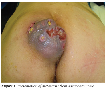
On October 5, 2010, the patient attended an outpatient clinic appointment and reported the appearance of a painful and fast-growing tumor on the left buttock (Figure 1). At the physical examination, an approximately 3cm x 8cm tumor was observed. It was purple, hard, with central ulcerations, draining serosanguinolent secretion and located in the lower left buttock.
A pelvic nuclear magnetic resonance (NMR) was performed and showed a solid and large formation at the left gluteal region, adjacent to the intergluteal line, presenting contact with the gluteus maximus and the elevator of the anus, measuring 5.3x4.7x4.7 cm (Figure 2). An incisional biopsy of the tumor was also performed.
The anatomopathological study showed metastatic adenocarcinoma negative for CK7 and positive for CK20 in the skin of the gluteal region, with primary location most likely being the colorectum.
Treatment with complete tumor exeresis was performed (Figure 3). The patient was discharged from the hospital in good conditions. Five months after the onset of metastasis in the gluteus, the patient evolved with no signs of recurrence.
DISCUSSION
The most common form of CRC dissemination is the lymphohematogenous pathway, affecting mainly the liver (75%), lungs (15%), bones (5%) and the central nervous system (5%). However, it can also be disseminated by contiguity to the peritoneum or intra-abdominal organs1; there are many reports of metastasis to unusual locations.
Balsamo and Formiga described colorectal adenocarcinoma metastasis to the jaws6, while Contu et al. described colonic metastasis to endotracheal and endobronchial sites after the primary treatment7. Torosian et al. described colorectal tumor metastasis to the thigh skeletal muscles8. Costa also reported vaginal metastasis, isolated from the colon cancer, which is extremely rare9. The reports of CRC metastases to the skin are unusual and poorly described in literature4,7.
The general incidence of cutaneous metastasis is 5.3%3, and most occur near the site of the primary tumor4. Therefore, the metastasis of the colonic tumor is usually to the pelvis and abdomen4. Another study with 724 patients showed that the neoplasms that mostly cause cutaneous metastases in women are: breast (69%), colon (9%), melanoma (5%), ovarian (4%) and pulmonary (4%)4. Among men, these metastases come from the lungs (24%), the colon (19%), melanoma (13%) and the oral cavity (12%)4.
A retrospective study by Lookingbill demonstrated that skin involvement caused by colorectal tumor at the time of diagnosis was present in 0.05% of the patients with cutaneous metastasis. This study included 7,316 patients with cancer, and found 367 with skin involvement (5%)10. In a sample of 413 patients with metastatic colorectal cancer, 18 (4.4%) presented with cutaneous metastases, and most were in the abdominal incision5,10.
As described in literature, the adenocarcinoma metastasis to the skin is uncommon, and usually happens near the primary site due to continuity or surgical implant10. However, this case reports isolated metastasis from the right colon to the skin, with no signs of the dissemination pathway. Also, the access pathway of the primary tumor resection was distant from the place of metastasis implantation.
The mechanisms for the cutaneous metastases to occur are yet to be estabished. However, the speculation is that they may occur due to continuity through the peritoneal cavity or by the interconnected routes of the lymphatic and hematologic systems. The iatrogenic implantation during the surgical primary tumor resection is rare, but considered in literature4.
With the current knowledge on molecular biology, there is not only the possibility of passive dissemination pathway, known as "tumor embolization", but also an easier way through the processes of angiogenesis and angiolymphatic growth factors that help to increase the total surface of blood vessels for tumor cell invasion. Following the blood vessel invasion, tumor cells that resist to the mechanic stress of circulation and to the immune system "colonize" the subcutaneous tissues until they acquire the necessary angiogenic properties and a favorable microenvironment to leave the state of "numbness"1.
The estimated survival rate is short, from 1 to 34 months, after the diagnosis of cutaneous metastasis. The mean of 18 months has been described for patients with cutaneous metastasis from colorectal tumor5,10. The cutaneous metastasis from a visceral neoplasm predicts the systemic compromise of the tumor with multiple metastases and poor prognosis5,10,11. In our case, no evidence of hepatic, lung or bone compromise has been found, and these are common metastatic dissemination pathways of colorectal neoplasm.
Both in the primary tumor and the metastasis, the patient was submitted to the treatment recommended in literature, that is, the surgical tumor resection. Chemotherapy was indicated due to the lymph node involvement observed in the right colon tumor resection, and due to the presence of resected neoplastic implant in the gastric wall; however, the patient refused to take the treatment because of the side effects.
Regarding the diagnosis of metastatic colorectal cancer to the skin, the treatment can be based on the relief of symptoms and comfort for the patient, but the local treatment, with radiotherapy or surgery, should be evaluated.
CONCLUSION
Even though the presence of cutaneous metastasis from colorectal cancer is associated with poor prognosis, the surgical resection of these metastases is a good treatment option when associated with low morbidity and improvement in the quality of life.
Submitted on: 03/21/2011
Approved on: 05/12/2011
Study carried out at the Coloproctology Discipline in Santa Casa de Misericórdia de São Paulo São Paulo (SP), Brazil.
Financing source: none.
Conflict of interest: nothing to declare.
- 1. Andrade SMS, Pereira FL. Câncer colorretal sincrônico: relato de caso e revisão de literatura. Rev bras Coloproct 2007;27(1):69-79.
- 2. Freitas AHA, Nunes TA, Wainstein AJA, Barroso AA, Ricardo-Filho OP, Dias MA, et al. Pesquisa de linfonodo-sentinela em pacientes com adenocarcinoma de cólon. Rev Bras Coloproct 2008;28(2):170-7.
- 3. Krathen RA, Orengo IF, Rosen T. Cutaneous metastasis: a meta-analysis of data. South Med J 2003. 96:164-7.
- 4. Helm TN, Lee TC. Metastatic carcinoma of the skin: treatment & medication [serial on the Internet] 2010 [cited 2010 Dez 18]. Available from: URL: http://emedicine.medscape.com/Article/1101058-Treatment
- 5. Lookingbill DP, Spangler N, Helm KF. Cutaneous metastases in patients with metastatic carcinoma: a retrospective study of 4020 patients. J Am Acad Dermatol 1993;29(2 Pt 1):228-36.
- 6. Balsamo F, Formiga GJS. Adenocarcinoma de reto com metástase para mandíbula: relato de caso. Rev bras Coloproct 2009;29(4):493-6.
- 7. Contu PC, Tarta C, Damin DC, Duarte IF, Contu SS, Moreira FF. Metástase endotraqueal e endobrônquica de adenocarcinoma de cólon. Rev bras Coloproct 2006;26(1):54-6.
- 8. Torosian MH, Botet JF, Paglia M. Colon carcinoma metastatic to the thigh - an unusual site of metastasis. Report of a case. Dis Colon Rectum 1987;30(10):805-8.
- 9. Costa SRP, Antunes RCP, Abraão AT, Silva RM, Paula RP, Lupinacci RA. Metástase vaginal isolada de câncer de cólon direito: relato de um caso. Einstein 2009;7(2):219-21.
- 10. Lookingbill DP, Spangler N, Sexton FM. Skin involvement as the presenting sign of internal carcinoma. A retrospective study of 7316 cancer patients. J Am Acad Dermatol 1990;22(1):19-26.
- 11. Llaguna OH, Desai P, Ferder AB, Zedek DC, Meyers MO, O'Neil BH, et al. Subcutaneous Metastatic Adenocarcinoma: An Unusual Presentation of Colon Cancer Case Report and Literature Review. Case Rep Oncol 2010;3(3):386-90.
Publication Dates
-
Publication in this collection
09 Jan 2013 -
Date of issue
Sept 2012
History
-
Received
21 Mar 2011 -
Accepted
12 May 2011