Abstracts
Colorectal cancer is a common malignancy in our clinical practice and it often evolves to a metastatic stage. Cutaneous dissemination, however, is a rare form of presentation of this disease. This study reports the case of a 38-year-old female patient that, even after neoadjuvant chemotherapy, presented cutaneous metastases of signet-ring cell colorectal adenocarcinoma. The malignancy proved to be extremely aggressive, without response to clinical therapy nor allowing surgical management, leading the patient to death about six months after the diagnosis.
colorectal neoplasms; neoplastic metastasis; adenocarcinoma; colorectal neoplasm
O câncer colorretal é uma neoplasia comum em nossa prática clínica e frequentemente evolui ao estágio metastático. Disseminação cutânea, entretanto, é uma forma rara de manifestação da doença. Apresentaremos aqui o caso de uma paciente de 38 anos que, mesmo após a realização de quimioterapia neoadjuvante, apresentou metástases cutâneas de um adenocarcinoma colorretal com células em anel de sinete. A neoplasia demonstrou-se extremamente agressiva, não respondendo ao tratamento medicamentoso nem permitindo o tratamento cirúrgico, levando a paciente ao óbito cerca de seis meses após o diagnóstico.
neoplasias colorretais; metástase neoplásica; adenocarcinoma; câncer colorretal
CASE REPORT
Cutaneous metastases of signet-ring cell colorectal adenocarcinoma
André Petter RodriguesI; Gustavo Becker PereiraII; Rafael Felix SchlindweinII; José Vinícius CruzIII; Lucas Cogo FurquimIV
IPhysician at the Universidade Federal de Ciências da Saúde de Porto Alegre (UFCSPA) Porto Alegre (RS), Brazil
IIColoproctologist, UFCSPA da Irmandade da Santa Casa de Misericórdia de Porto Alegre (ISCMPA) Porto Alegre (RS), Brazil
IIIFull professor of Coloproctology at UFCSPA; Head of Service of Coloproctology at ISCMPA Porto Alegre (RS), Brazil
IVPhysician at the UFCSPA Porto Alegre (RS), Brazil
Correspondence to Correspondence to: André Petter Rodrigues Rua Cel. Corte Real, 913/202 90630-080 Porto Alegre (RS), Brazil E-mail: andre.ufcspa@gmail.com
ABSTRACT
Colorectal cancer is a common malignancy in our clinical practice and it often evolves to a metastatic stage. Cutaneous dissemination, however, is a rare form of presentation of this disease. This study reports the case of a 38-year-old female patient that, even after neoadjuvant chemotherapy, presented cutaneous metastases of signet-ring cell colorectal adenocarcinoma. The malignancy proved to be extremely aggressive, without response to clinical therapy nor allowing surgical management, leading the patient to death about six months after the diagnosis.
Keywords: colorectal neoplasms; neoplastic metastasis; adenocarcinoma; colorectal neoplasm.
RESUMO
O câncer colorretal é uma neoplasia comum em nossa prática clínica e frequentemente evolui ao estágio metastático. Disseminação cutânea, entretanto, é uma forma rara de manifestação da doença. Apresentaremos aqui o caso de uma paciente de 38 anos que, mesmo após a realização de quimioterapia neoadjuvante, apresentou metástases cutâneas de um adenocarcinoma colorretal com células em anel de sinete. A neoplasia demonstrou-se extremamente agressiva, não respondendo ao tratamento medicamentoso nem permitindo o tratamento cirúrgico, levando a paciente ao óbito cerca de seis meses após o diagnóstico.
Palavras-chave: neoplasias colorretais; metástase neoplásica; adenocarcinoma; câncer colorretal.
INTRODUCTION
Colorectal adenocarcinoma is one of the most frequent neoplasms in our practice, the third most common form of cancer in men and the fourth in women1. Progressive knowledge of the disease led to great improvement in the prognosis; however, some more agressive forms of tumors persist as challenges to therapy. We will present here the case of a young patient who, during the course of a disseminated disease, evolved to cutaneous metastases of a rectal cancer.
CASE REPORT
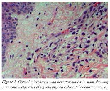
A 38-year-old-female patient, without comorbidities or family history of neoplasms, was admitted with obstructive acute abdomen. Abdominal x-ray showed bowel distension with abrupt interruption at the distal sigmoid colon level. As the clinical status worsened, she was took to the operating room for exploratory laparotomy. During surgery, a voluminous lesion was identified in the rectum, with sacral invasion, showing no possibility of resection, so loop sigmoidostomy was performed. The cavity inventory did not present findings that suggested secondary implants. After the postoperative recovery, colonoscopy was performed, which showed a stenosing tumor 9 cm from the anal margin (the anatomopathological analysis showed poor-differentiated signet-ring cell adenocarcinoma). Then, the patient was evaluated by the service of clinical oncology for neoadjuvant therapy, and was submitted to chemotherapy protocol with 5-fluorouracil and radiotherapy at the dose of 45 Gy for five weeks. Eight weeks later, the patient was admitted again for re-staging, complaining of eventual vomiting and the presence of subcutaneous nodules under the right costal margin. Computed tomography (CT) showed pelvic and retroperitoneal lymphadenomegaly, colon wall thickening, especially at the splenic flexure, infiltration of perirectal fat and tumor in the upper rectum. The patient presented subocclusion during the hospitalization period, with vomiting and interrupted eliminations via sigmoidostomy. A palliative surgery was planned, aiming at relieving obstructive symptoms by a new transit bypass or a tumor resection. At laparotomy, extensive peritoneal carcinomatosis was observed, with adhesions and complete immobility of loops, making it impossible to perform a new bypass. Peritoneal and right hypochondium cutaneous biopsies were performed and after surgical recovery the patient was sent for outpatient palliative therapy. After the patient was discharged from hospital, we received the anatomopathological analyses of the cutaneous lesion, which showed poor-differentiated signet-ring cell adenocarcinoma in both samples (Figures 1 and 2). The patient was supposed to come back within seven days to start chemotherapy, but she died at home on the fifth day after discharge. About six months elapsed between clinical presentation and death.
DISCUSSION
Cutaneous metastases of colorectal tumors are infrequent. Some case series report this occurrence in around 4 to 5% of patients with colorectal cancer2, most cases at the site of surgical incision for tumor resection. Cutaneous metastases far from the surgical site are even more unusual, corresponding to only one third of skin metastatic lesions, i.e., around 1.3% of all metastases; in the case reported here, we performed midline incisions, while the cutaneous lesion was in the right hypochondrium. The most common site of cutaneous metastases from the surgical wound is usually the abdominal region, followed by the pelvis and the back3.
The aspect of cutaneous metastatic lesions is completely unspecific. In general, they are small lesion, smaller than 2 cm, hardened and painless2, although occurrences of ulcerated lesions have been reported4,5 and even lesions with inflammatory characteristics6. Our patient presented two hardened and mobile lesions of around 1 cm, causing skin raise, similar to small lipomas.
The presence of cutaneous metastatic lesions usually represents a widely spread disease, and its presentation as initial manifestation of the disease is extremely uncommon3, being typically a follow-up finding. Lookingbill7 observed that survival in these cases may achieve up to 18 months, although the most common is a shorter period. Long survival after surgical treatment of cutaneous lesions with curative purpose, as the one reported by Sarid2, are exceptions.
Another aspect to be pointed out in this case is the tumor histology. Signet-ring cells are more common at other sites of gastrointestinal tract, an atypical occurrence in colorectal tumors around 0.5 to 2.0% of the cases. Such patients tend to be younger many of them under 50 when compared to those with other histological types. Also frequent is the presentation in late stages, with TNM classification usually III or IV8,9, many of them with peritoneal carcinomatosis at diagnosis or during the disease progress. Early diagnosis is rare, usually occuring at routine screening colonoscopy10.
CONCLUSION
The presence of cutaneous metastases of colorectal tumors is a rare occurrence, representing an extremely advanced disease. This study reported a case of fast clinical progress, with no response to the proposed treatment, in which the cutaneous metastases appeared when the disease was already widely spread. Anyway, the assistant physician should be alert to complaints of cutaneous lesions in patients being treated for such neoplasms, aiming at a proper staging.
Submitted on: 03/27/2011
Approved on: 04/14/2011
Study carried out at the Department of Surgery, Discipline of Coloproctology at the Universidade Federal de Ciências da Saúde de Porto Alegre (UFCSPA) Porto Alegre (RS), Brazil.
Financing source: none.
Conflict of interest: nothing to declare.
- 1. Estimativas 2008: Incidência do câncer no Brasil. Rio de Janeiro: INCA, 2007.
- 2. Sarid D, Wigler N, Gutkin Z, Merimsky O, Leider-Trejo L, Ron IG. Cutaneous and subcutaneous metastases of colorectal cancer. Int J Clin Oncol 2004;9(3):202-5.
- 3. Brownstein MH, Helwig EG. Metastatic tumors of the skin. Cancer 1972;29(5):1298-307.
- 4. Attili VSS, Rama Chandra C, Dadhich HK, Sahoo TP, Anupama G, Bapsy PP. Unusual metastasis in colorectal cancer. Indian J Cancer 2006;43(2):93-5.
- 5. Moonda A, Fatteh S. Metastatic colorectal carcinoma: an unusual presentation. J Cutan Pathol 2009;36(1):64-6.
- 6. Tan KY, Ho KS, Lai JH, Lim JF, Ooi BS, Tang Cl, et al. Cutaneous and subcutaneous metastases of adenocarcinoma of the colon and rectum. Ann Acad Med Singapore 2006;35(8):585-7.
- 7. Lookingbill DP, Spangler N, Helm KF. Cutaneous metastases in patients with metastatic carcinoma: a retrospective study of 4020 patients. J Am Acad Dermatol 1993;29(2 Pt 1):228-36.
- 8. Chen JS, Hsieh PS, Hung SY, Tang R, Tsai WS, Changchien CR, et al. Clinical significance of signet ring cell rectal carcinoma. Int J Colorectal Dis 2004;19(2):102-7.
- 9. Chew MH, Yeo SA, Ng ZP, Lim KH, Koh PK, Ng KH, et al. Critical analysis of mucin and signet ring cell as prognostic factors in an Asian population of 2,764 sporadic colorectal cancers. Int J Colorectal Dis 2010;25(10):1221-9.
- 10. Fu KI, Sano Y, Kato S, Saito H, Ochiai A, Fujimori T, et al. Primary signet-ring cell carcinoma of the colon at early stage: a case report and a review of the literature. World J Gastroenterol 2006;12(21):3446-9.
Publication Dates
-
Publication in this collection
08 Jan 2013 -
Date of issue
Sept 2012
History
-
Received
27 Mar 2011 -
Accepted
14 Apr 2011