OBJECTIVE:
Investigation of standard intensities of physical exercise is important to better comprehend and develop rehabilitation programs for emphysema. We aimed to evaluate the effects of different intensities (moderate and high-intensity) of physical exercise on the development of a protease-induced (papain intratracheal instillation) emphysema in rats.
METHODS:
Male Wistar rats were randomly separated into five groups that received intratracheal instillation of papain solution or vehicle: (i) papain high intensity exercise, (ii) papain moderate exercise, (iii) saline high intensity exercise, (iv) saline sedentary and (v) papain sedentary. Forty days after intratracheal instillation, the exercise groups were submitted to an exercise-training protocol on a treadmill during 10 weeks, 5 days/week, at 0.9 km/h (Papain and saline high exercise), or at 0.6 km/h (papain moderate exercise).We measured respiratory system elastance and resistance, the collagen fiber lung parenchyma, and the pulmonary mean linear intercept.
RESULTS:
All animal groups that received papain instillation presented higher alveolar wall destruction compared to animals that received only saline solution. The papain high intensity exercise group presented higher values of mean linear intercept compared to emphysema groups that were trained at a moderate intensity or not submitted to exercise.
CONCLUSION:
High intensity exercise training worsened alveolar destruction in an experimental model of emphysema in rats when compared to moderate intensity exercise, or to no exercise.
KEYWORDS:
Emphysema; Pulmonary Disease; Chronic Obstructive; Exercise; Rats
RESUMO
OBJETIVO:
A investigação de intensidades padrão de exercício físico é importante para melhor compreender e desenvolver programas de reabilitação para enfisema. Nosso objetivo foi avaliar os efeitos de diferentes intensidades (moderada e alta) de exercício físico sobre o desenvolvimento de enfisema induzido por protease (instilação endotraqueal de papaína) em ratos.
MÉTODOS:
Ratos Wistar machos foram divididos aleatoriamente em cinco grupos que receberam instilação intra-traqueal de solução de papaína ou veículo: (i) papaína, exercício de alta intensidade, (ii) papaína, exercício moderado, (iii) soro fisiológico, exercício de alta intensidade, (iv) soro fisiológico, sedentário e (v) papaína, sedentário. Quarenta dias após a instilação intra-traqueal os grupos de exercício foram submetidos a um protocolo de treinamento físico em esteira durante 10 semanas, 5 dias/ semana, a 0.9 km/h (papaína e solucão salina exercício de alta intensidade), ou em 0.6km/h (papaína exercício moderado). Medimos elastância do sistema respiratório e resistência, as fibras de colágeno no parênquima pulmonar, e a intercepção linear média pulmonar.
RESULTADOS:
Todos os grupos de animais que receberam instilação de papaína apresentaram maior destruição da parede alveolar em comparação com animais que receberam apenas solução salina. O grupo papaína exercício de alta intensidade apresentou maiores valores de intercepto linear médio em comparação com grupos de enfisema que foram treinados em uma intensidade moderada ou não submetidos ao exercício.
CONCLUSÃO:
A alta intensidade de treinamento físico produziu mais destruição alveolar em um modelo experimental de enfisema em ratos quando comparada ao exercício de intensidade moderada, ou a nenhum exercício.
INTRODUCTION
Chronic obstructive pulmonary disease (COPD) is a pulmonary disorder that affects respiratory and cardiovascular parameters11 Reis MS, Deus AP, Simões RP, Aniceto IA, Catai AM, Borghi-Silva A. Autonomic control of heart rate in patients with chronic cardiorespiratory disease and in healthy participants at rest and during a respiratory sinus arrhythmia maneuver. Rev Bras Fisioter. 2010;14(2):106-13.; it is characterized by alveolar wall destruction with permanent airspace enlargement22 Mahadeva R, Shapiro SD. Chronic obstructive pulmonary disease * 3: Experimental animal models of pulmonary emphysema. Thorax. 2002;57(10):908-1014..
Animal models have been used to elucidate the possible mechanisms that contribute to emphysema development and worsening. Instillation of proteases have been extensively used to induce emphysema, since emphysema was induced in rats after a single intratracheal administration of papain22 Mahadeva R, Shapiro SD. Chronic obstructive pulmonary disease * 3: Experimental animal models of pulmonary emphysema. Thorax. 2002;57(10):908-1014.. Instillation of proteases such as papain and porcine pancreatic elastase (PPE) result in rapid and expressive airspace enlargement that resembles human emphysema22 Mahadeva R, Shapiro SD. Chronic obstructive pulmonary disease * 3: Experimental animal models of pulmonary emphysema. Thorax. 2002;57(10):908-1014..
In this context, exercise training has been considered to be an important component of pulmonary rehabilitation in patients with severe COPD33 Pulmonary rehabilitation. American Thoracic Society. Am J Respir Crit Care Med. 1999;159(5 P1 1):1666-82., resulting in positive effects on dyspnea and exercise tolerance44 American College of Sports Medicine Position stand. The recommended quantity and quality of exercise for developing and maintaining cardiorespiratory and muscular fitness in healthy adults. Med Sci Sports Exerc. 1990;22(2):265-74.,55 Casaburi R, Porszasz J, Burns MR, Chang RSY, Cooper CB. Physiologic benefits of exercise training in rehabilitation of patients with severe chronic obstructive pulmonary disease. Am J Respir Crit Care Med. 1997;155(5):1541-51.. However, some experimental studies suggested that exercise training performed during the development of emphysema could actually aggravate it.
In a previous study in our laboratory, Flo et al. observed more severe emphysema in rats that received papain instillation and were submitted to exercise training compared to the group that received this protease but was not submitted to exercise66 Fló C, Lopes FD, Kasahara DI, Silva AC, Jesus RC, Rivero DH, Saldiva PH, Martins MA, Jacob-Filho W. Effects of exercise training on papaininduced pulmonary emphysema in Wistar rats. J Appl Physiol (1985). 2006;100(1):281-5.. In this study rats were exposed to high intensity exercise. It is possible that the deleterious effect of physical exercise in the development of experimental emphysema depends on the intensity of exercise.
Sahebjami et al. observed that physical exercise in rats that received papain inhalation resulted in a reduction in tissue elasticity in the lungs77 Sahebjami H, Vassallo CL. Exercise stress and enzyme-induced emphysema. J Appl Physiol. 1976;41(3):332-5.. Martorana et al. induced emphysema in hamsters by PPE instillation and then submitted the animals to exercise training88 Martorana PA, Vaneven P, Schaper J. The effect of lung growth on the evolution of elastase-induced emphysema in the hamster. Lung. 1982;160(1):19-27.. They observed that physical activity did influence the progression of emphysema, but was responsible for the structural right-ventricular changes associated with emphysema.
In clinical studies, lung function parameters, specific symptoms, functional status, and exacerbations were some of outcomes considered to establish a rehabilitation program, respecting the individual disorder's severity99 Cooper CB. The Connection Between Chronic Obstructive Pulmonary Disease Symptoms and Hyperinflation and Its Impact on Exercise and Function. Am J Med. 2006;119(10 Suppl 1):21-31.. The evaluation of different intensities of exercise activity could elucidate the role of exercise training in emphysema.
In the present study we evaluated different intensities (moderate and high-intensity) of physical exercise and their effects on the development of a protease-induced emphysema in rats. The high-intensity exercise training employed here was based on Vrabas et al. and Zonderland et al. protocols., which were based on an increase in speed and time compared to the moderate exercise.
METHODS
Study design
In this prospective study, we used specific and validated methods to induce emphysema and evaluate changes in different intensities of exercise activity. This study was approved by the Institutional Review Board of the School of Medicine of the University of São Paulo. All animals received humane care in compliance with the "Principles of Laboratory Animal Care" published by the National Institutes of Health (NIH publication 86-23, revised 1985).1010 Principles of Laboratory Animal Care published by the National Institutes of Health (NIH publication 86-23, revised 1985).
Eight-week-old male Wistar rats weighing 294 ± 23 g (mean ± SD) were used in the study. The rats were anesthetized with a mixture of 1% isoflurane (Baxter Healthcare, Puerto Rico), diluted in 100% oxygen, at a flow rate of 2 L/min with the use of a gas nebulizer (1224 K, Takaoka, Brazil). Before that, a polyethylene cannula was inserted into the trachea of the animal to connect to a rodent ventilator (flexiVent, Scireq, Canada) with a tidal volume of 10 ml/kg and a respiratory rate of 90 breaths/min. Emphysema was induced by a single intratracheal instillation of papain. Six milligrams of papain (10-20U/mg protein, Sigma Chemical, USA) were diluted in 1 ml of saline (0.9% NaCl) and infused into the tracheal cannula. Saline groups (SS and SHE) received intratracheal instillation of 1 ml of saline (0.9% NaCl).
Experimental groups. Rats were randomly separated into five groups (n ¼10 per group): 1) rats that received intratracheal instillation of papain solution and were submitted to a protocol of high intensity exercise (papain high-intensity exercise - PHE); 2) rats receiving intratracheal instillation of papain solution and submitted to a protocol of moderate intensity exercise (papain moderate-intensity exercise - PME); 3) rats receiving intratracheal papain instillation and remaining sedentary (papain sedentary - PS); 4) rats receiving intratracheal instillation of saline solution and submitted to high intensity exercise (saline high-intensity exercise - SHE); and 5) rats receiving intratracheal instillation of saline solution and remaining sedentary (saline sedentary - SS).
Exercise training protocol. Forty days after intratracheal instillation of either papain or saline solution, PHE, PME and SHE groups were submitted to an exercise-training protocol. Animals were initially adapted to the treadmill for 3 days (5 min, 0% inclination, 0.3 km/h). Exercise training was performed on a human treadmill (Imbramed® , Brazil) adapted with eight chambers (10 × 10 × 50 cm each) with Plexiglas covers. Animals were exercised during 10 weeks (5 days/week) between 5:30 PM and 7:30 PM.
Moderate Exercise. Forty days after papain instillation, the selected animals were submitted to the moderate exercise protocol, based on Navarro et al1111 Navarro A, Gomez C, López-Cepero JM, Boveris A. Beneficial effects of moderate exercise on mice aging: survival, behavior, oxidative stress, and mitochondrial electron transfer. Am J Physiol Regul Integr Comp Physiol. 2004;286(3):R505-11. and Hashimoto et al. protocols1212 Hashimoto T, Kambara N, Nohara R, Yazawa M, Taguchi S. Expression of MHC-beta and MCT1 in cardiac muscle after exercise training in myocardial-infarcted rats. J Appl Physiol. 2004;97(3):843-51.. Briefly, in the four initial weeks, animals ran at 0.3 km/h for a gradual increase in duration from 15 to 40 min. Starting at the beginning of the fifth week we started to raise the speed from 0.4 km/h (fifth week) to 0.6 km/h (tenth week) and duration from 40 to 60 min.
High intensity exercise. The high intensity protocol was based on Vrabas et al. and Zonderland et al. protocols.1313 Vrabas IS, Dodd SL, Powers SK, Hughes M, Coombes J, Fletcher L, et al. Endurance training reduces the rate of diaphragm fatigue in vitro. Med Sci Sports Exerc. 1999;31(11):1605-12.,1414 Zonderland ML, Bär PR, Reijneveld JC, Spruijt BM, Keizer HA, Glatz JF. Different metabolic adaptation of heart and skeletal muscles to moderateintensity treadmill training in the rat. Eur J Appl Physiol Occup Physiol. 1999;79(5):391-6. For the first four weeks the same protocol of moderate intensity exercise was maintained. From the beginning of the fifth week onward, we raised the speed from 0.4 km/h (fifth week) to 0.9 km/h (tenth week) and the duration from 40 min to 60 min.
Assessment of respiratory system mechanics. Forty-eight hours after the last training session, rats were anesthetized with Thiopental (50 mg/kg), tracheostomized and connected to a ventilator for small animals (flexiVent, Scireq, Canada) to measure Rrs (respiratory resistance) and Ers (respiratory elastance) according to the linear equation of motion of the respiratory system, as follows:

Where Ptr is tracheal pressure, is airflow, V is volume an Po is a constant to correct for errors in estimating functional residual capacity.1515 Wagers S, Lundblad L, Moriya HT, Bates JH, Irvin CG. Nonlinearity of respiratory mechanics during bronchoconstriction in mice with airway inflammation. J Appl Physiol (1985). 2002;92(5):1802-7.
Lung morphological evaluation. After measurements of respiratory mechanics, and still under anesthesia, the animals were euthanized by exsanguination. The lungs were removed and fixed in formalin 10% for 24 h at the constant pressure of 20 cmH2O. Thereafter, lungs were cut and paraffin embedded; sections for histological analysis were prepared. Five-micrometer tissue sections were stained with hematoxylin and eosin (H&E) for lung structure analysis, and Sirius Red for collagen fiber analysis (Direct Red 80, C.I. 35780; Aldrich, USA).1616 Mauad T, Xavier AC, Saldiva PH, Dolhnikoff M. Elastosis and fragmentation of fibers of the elastic system in fatal asthma. Am J Respir Crit Care Med. 1999;160(3):968-75.
The mean linear intercept (Lm), an indicator of mean diameter of airspaces1717 Margraf LR, Tomashefski JF Jr, Bruce MC, Dahms BB. Morphometric analysis of the lung in bronchopulmonary dysplasia. Am Rev Respir Dis. 1991;143(2):391-400., was assessed employing an integrating eyepiece with a coherent system of 50 lines and 100 points with a known area (1044 American College of Sports Medicine Position stand. The recommended quantity and quality of exercise for developing and maintaining cardiorespiratory and muscular fitness in healthy adults. Med Sci Sports Exerc. 1990;22(2):265-74. µm22 Mahadeva R, Shapiro SD. Chronic obstructive pulmonary disease * 3: Experimental animal models of pulmonary emphysema. Thorax. 2002;57(10):908-1014. of total area) attached to the ocular of the microscope. The number of intercepts of the integrating eyepiece falling on tissue areas was counted in 20 non-overlapping fields of lung parenchyma per animal, at 200 × magnification, and results were expressed in micrometers.
To evaluate the collagen fiber lung parenchyma content, we used an image analysis system (Image Pro Plus 4.0). The collagen and elastic fiber content in lung parenchyma were expressed as the relationship between the total area of lung tissue in a randomized chosen frame and the collagen or elastic fiber area (µm22 Mahadeva R, Shapiro SD. Chronic obstructive pulmonary disease * 3: Experimental animal models of pulmonary emphysema. Thorax. 2002;57(10):908-1014.) of that frame. Twenty fields were analyzed per animal, at a 400 × magnification.1818 Prado CM, Leick-Maldonado EA, Yano L, Leme AS, Capelozzi VL, Martins MA, et al. Effects of nitric oxide synthases in chronic allergic airway inflammation and remodeling. Am J Respir Cell Mol Biol. 2006;35(4):457-65.
Statistical analysis. Statistical analysis was performed using Sigma Stat software (SPSS Inc., Chicago, IL). All values are expressed as means ± SD. Values of Lm were compared using Two Way analysis test followed by Holm-Sidak for multiple comparison procedures. Student t-test was used to compare papain groups that were submitted to different intensities of exercise training and used to compare initial and final values of respiratory elastance for each experimental group. One Way analysis of variance was used to compare mean values of weight and respiratory elastance among the five groups.
RESULTS
Mean values of body weight (±SD) were measured before the beginning of the experimental protocol and at the end of the protocol (Figure 1). There was a significant increase in body weight for all groups, comparing the beginning of the experiments (time 0) to the end of the experimental protocol, after 10 weeks (p = 0.001). We did not observe any statistically significant difference in body weight among the experimental groups.
Body weight values (means ± SD) of the five experimental groups. There was an increase in body weight comparing time 0 (beginning of the experiments) to time 10 (10 weeks of experimental protocol) (p = 0.001) for all groups. There were no statistically significant differences among the groups.
Figure 2 shows mean values (±SD) of Ers obtained before the beginning of the experimental protocol and at the end of the experimental protocol. A significant decrease in the values of Ers was observed in the groups instilled with papain (PS, PME and PHE) compared to the groups instilled with saline solution (SS and SHE) (p < 0.001). There was a significant decrease in Ers between the initial and final values for all groups (p < 0.05), except for the saline high exercise group.
Respiratory system elastance (Ers) measured at the beginning of the experimental protocol (initial) and 10 weeks after, at the end of the experimental protocol (final). *p , 0.001 compared to initial values. **p , 0.05 compared to groups that did not receive papain instillation. Values are means and SD.
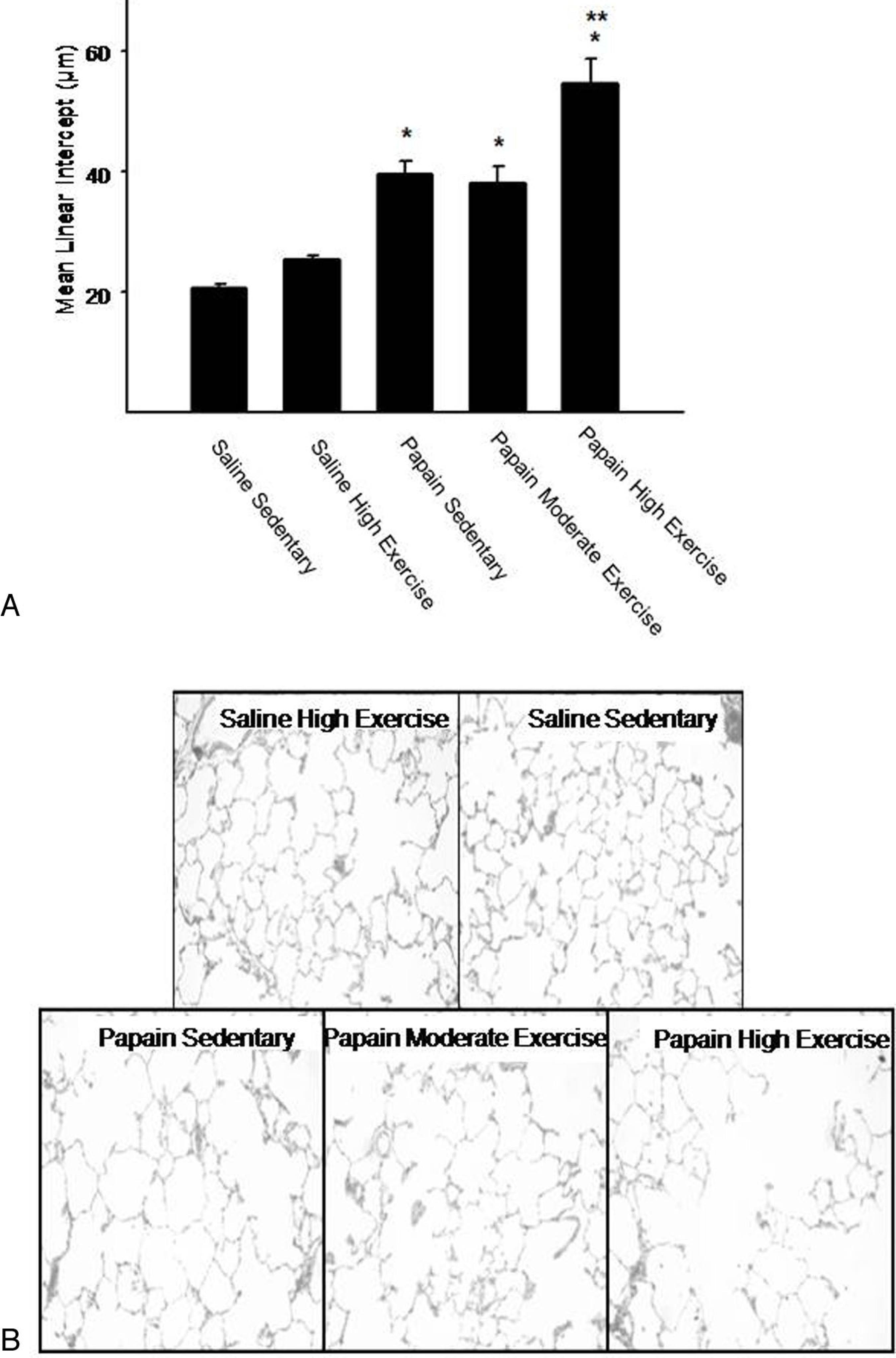
Rats that received intratracheal instillation of papain (PS; PME and PHE) showed a statistically significant increase in mean values of Lm (p ≤ 0.001) compared to the animals that received intratracheal saline instillation (SS and SE) (Figure 3A). In addition, PHE group showed higher values of Lm compared to PME group (p = 0.001). We did not observe significant differences between PS and PME groups (p = 0.239). Figure 3B shows photomicrographs of lung parenchyma 10 weeks after intratracheal instillation of saline (SS and SHE groups) and papain solution (PS; PME; PHE groups). It can be seen that alveolar wall integrity was preserved in saline groups, whereas and the presence of alveolar wall destruction and marked enlargement of distal airway spaces is evident in the papain groups.
(A) Mean linear intercept values measured in the five experimental groups. Values are means and SD. *p < 0.001 compared to the groups that did not receive papain instillation; **p = 0.001 compared to papain-sedentary and papain moderate-intensity exercise groups. (B) Representative photomicrographs of lung parenchyma of rats of the five experimental groups (original magnification × 400, H&E staining).
The volume proportion of collagen fibers in alveolar tissue is shown in Figure 4. Groups that received papain solution showed a significant increased in volume proportion of collagen fibers compared to animals that received saline solution (SS and SHE) (p = 0.049). There was no significant difference in volume proportion of collagen fibers among the groups that received papain instillation.
Volume proportion of collagen in alveolar tissue of the five experimental groups. Values are means and SD. * p < 0.05 compared to groups that did not receive papain instillation.
DISCUSSION
In the present study, after ten weeks of training, we observed a higher alveolar wall destruction, with a significant increase in Lm values, in all animal groups that received papain instillation compared to animals that received only saline solution. The papain group that was trained with high intensity showed higher values of Lm compared to emphysema groups that were trained with moderate intensity or not submitted to exercise. These results showed that exercise training in high intensity worsened the parenchyma destruction in this animal model of emphysema and that exercise training with moderate intensity does not present this deleterious effect.
The purpose of rehabilitation programs is to submit the respiratory muscles to the correction of the multiple factors that impair their structure and function, depending on the presence and severity of COPD.1919 Cambach W, Chadwick-Straver RVM, Wagenaar RC, van Keinpema ARJ, Kemper HCG. The effects of a community-based pulmonary rehabilitation programme on exercise tolerance and quality of life: a randomized controlled trial. Eur Respir J. 1997;10(1):104-13. Furthermore, it is essential that any rehabilitation program should aim to make every patient reach the highest possible level of exercise capability. COPD patients often show weakness and reduced endurance in respiratory muscles, which is correlated with a difficulty in maintaining the level of respiratory power output.2020 Orozco-Levi M. Structure and function of the respiratory muscles in patients with COPD: impairment or adaptation. Eur Respir J Suppl. 2003;46:41s-51s. Clinical studies report that patients with CPOD who are submitted to rehabilitation programs present improvement in quality of life and exercise tolerance and that this is considered the major objectives of pulmonary rehabilitation.2121 Ries AL, Kaplan RM, Limberg TM, Prewitt LM. Effects of pulmonary rehabilitation on physiologic and psychosocial outcomes in patients with chronic obstructive pulmonary disease. Ann Intern Med. 1995;122 (11):823-32.
Animal models have been extensively used to closely reproduce human respiratory diseases such as emphysema. Protease-induced emphysema reproduces many aspects of the disease, promoting alveolar wall destruction and lung repair.2222 Shapiro SD. Animal models for COPD. Chest. 2000;117(5 Suppl 1):223S-7S.
Papain is a proteolitic enzyme that has been used for many years, and it is able to induce emphysema in animals. Although many recent studies used elastase to induce emphysema, there are no published studies comparing both experimental models. Previous studies by our research group55 Casaburi R, Porszasz J, Burns MR, Chang RSY, Cooper CB. Physiologic benefits of exercise training in rehabilitation of patients with severe chronic obstructive pulmonary disease. Am J Respir Crit Care Med. 1997;155(5):1541-51.,2323 Lopes FD, Pinto TS, Arantes-Costa FM, Moriya HT, Biselli PJ, Ferraz LF, et al. Exposure to ambient levels of particles emitted by traffic worsens emphysema in mice. Environ Res. 2009;109(5):544-51. and others2424 Pastor LM, Sánchez-Gascón F, Girona JC, Bernal-Mañas CM, Morales E, Beltrán-Frutos E, et al. Morphogenesis of rat experimental pulmonary emphysema induced by intratracheally administered papain: changes in elastic fibres. Histol Histopathol. 2006;21(12):1309-19. have shown that papain is efficient to induce lung parenchyma destruction in different animals species and that this effect can be quantified by the increase in mean linear intercept associated with lung remodeling, characterized by deposition of collagen and elastic fibers in alveolar septa. These models provide rapid, significant and reproducible results.2525 Dawkins PA, Stockley RA. Animal models of chronic obstructive pulmonary disease. Thorax. 2001;56(12):972-7.
Many previous studies have investigated exercise training and emphysema, but until now, to our knowledge, there have been no studies that aimed to compare the effects of high and moderate exercise training in an animal model of emphysema. The protocol of treadmill high intensity exercise training that we used was similar to the protocols used in rats in previous study in our laboratory.55 Casaburi R, Porszasz J, Burns MR, Chang RSY, Cooper CB. Physiologic benefits of exercise training in rehabilitation of patients with severe chronic obstructive pulmonary disease. Am J Respir Crit Care Med. 1997;155(5):1541-51. The moderate intensity protocol was based on the protocols used moderate intensity protocol was based on the protocols used in rats by other groups.2626 Murphy EA, Davis JM, Brown AS, Carmichael MD, Van Rooijen N, Ghaffar A, et al. Role of lung macrophages on susceptibility to respiratory infection following short-term moderate exercise training. Am J Physiol Regul Integr Comp Physiol. 2004;287(6):R1354-8.,2727 Murphy EA, Davis JM, Brown AS, Carmichael MD, Mayer EP, Ghaffar A. Effects of moderate exercise and oat beta-glucan on lung tumor metastases and macrophage antitumor cytotoxicity. J Appl Physiol. 2004;97(3):955-9. These protocols are well established in the literature, and their effects at the end of physical training has been previously described.2626 Murphy EA, Davis JM, Brown AS, Carmichael MD, Van Rooijen N, Ghaffar A, et al. Role of lung macrophages on susceptibility to respiratory infection following short-term moderate exercise training. Am J Physiol Regul Integr Comp Physiol. 2004;287(6):R1354-8.,2727 Murphy EA, Davis JM, Brown AS, Carmichael MD, Mayer EP, Ghaffar A. Effects of moderate exercise and oat beta-glucan on lung tumor metastases and macrophage antitumor cytotoxicity. J Appl Physiol. 2004;97(3):955-9.
Considering respiratory mechanical function, we observed a significant decrease in Ers when comparing papain to saline groups. However, we did not find significant differences between papain groups that were submitted to different intensities of exercise. Although the assessment of respiratory mechanical function in animal models of emphysema is widely used, and because enlargement of the respiratory air spaces has been associated with decrease in lung elastic properties, the morphometric analysis is still considered the most reliable measurement to detect emphysema.2828 Foronjy RF, Mirochnitchenko O, Propokenko O, Lemaitre V, Jia Y, Inouye M, et al. Superoxide dismutase expression attenuates cigarette smoke- or elastase-generated emphysema in mice. Am J Respir Crit Care Med. 2006;173(6):623-31.
In this regard, papain instillation induced alveolar wall destruction measured by the mean linear intercept. The moderate intensity exercise did not modify this measure compared to rats that received papain but were not submitted to exercise. However, the most important result related to this study is that high intensity exercise induced a deleterious effects consisting of an increase in the emphysematous alveolar destruction. Our results could elucidate the negative effects of exercise training obtained in previous experimental studies,55 Casaburi R, Porszasz J, Burns MR, Chang RSY, Cooper CB. Physiologic benefits of exercise training in rehabilitation of patients with severe chronic obstructive pulmonary disease. Am J Respir Crit Care Med. 1997;155(5):1541-51.,77 Sahebjami H, Vassallo CL. Exercise stress and enzyme-induced emphysema. J Appl Physiol. 1976;41(3):332-5.,88 Martorana PA, Vaneven P, Schaper J. The effect of lung growth on the evolution of elastase-induced emphysema in the hamster. Lung. 1982;160(1):19-27. since most of the experimental studies did not consider whether the training was exhaustive to the animals. In addition, although the clinical findings suggest that exercise could improve quality of life in patients with COPD, there were no significant effects in respiratory capacity. It is probable that the effects of exercise on quality of life could be more related to psychological effects than to lung function impairment. Our finding has an important clinical repercussion since, in rehabilitation programs, patients are encouraged to exercise in their maximal capacity. This might explain, at least in part, the absence of significant effects in lung function due to exercise training programs in humans.
Generally, apart from the enlargement of the respiratory air spaces observed in emphysema, there is a reorganization of the connective tissue fiber networks during the development of pulmonary emphysema.2929 Chung KF, Adcock IM. Multifaceted mechanisms in COPD: inflammation, immunity, and tissue repair and destruction. Eur Respir J. 2008;31(6):1334-56. In our study, we observed an increase in volume proportion of collagen fibers in parenchyma in all animal groups that received papain instillation independent of exercise activity. Although high intensity exercise worsened the alveolar destruction, it did not affect pulmonary remodeling. There is no consensus in the literature correlating lung destruction to the presence of extracellular matrix remodeling. In addition, it is important to consider that we evaluated the total content of collagen fibers in lung alveolar septa without considering the different types of collagen and this could partly explain such results.
Although the present study focused only on whether high intensity exercise worsened alveolar destruction, we may hypothesize some possible mechanics involved in our results. It is well known that oxidative stress is one of the mechanisms involved in the pathophysiology of emphysema.3030 Rangasamy T, Misra V, Zhen L, Tankersley CG, Tuder RM, Biswal S. Cigarette smoke-induced emphysema in A/J mice is associated with pulmonary oxidative stress, apoptosis of lung cells, and global alterations in gene expression.Am J Physiol Lung Cell Mol Physiol. 2009;296(6):L888-L900. Considering that strenuous aerobic exercise could interfere with oxidant/antioxidant imbalance resulting in an increase in oxidant production, the worsening of alveolar destruction observed in animals submitted to high intensity exercise may be due the presence of high oxidative stress resulting from intense exercise.
Our study presents relevant findings as far as rehabilitation programs are concerned. Because COPD patients are submitted to rehabilitation programs, this result has clinical relevance suggesting that the intensity of exercise used for each patient is a necessary concern. Therefore, care should be taken when prescribing rehabilitation protocols to COPD patients based on their maximal pulmonary capacity.
CONCLUSION
High intensity exercise training worsened alveolar destruction in an experimental model of emphysema in rats, without effects on lung remodeling and function. The precise mechanism involved in alveolar wall destruction induced by high intensity exercise remains to be investigated. Caution is recommended when prescribing exercise for the rehabilitation of emphysematous patients.
-
Barnabé V, Lopes FDT, Saraiva-Romanholo BM, Rosa CO, Pazetti R, Valenti VE, Martins MA. High intensity exercise training worsens alveolar destruction in pulmonary emphysema rats. MEDICALEXPRESS. 2014 Oct;1(5):227-232.
ACKNOWLEDGEMENTS
This study was supported by the following Brazilian scientific agencies: FAPESP, CNPq.
REFERENCES
-
1Reis MS, Deus AP, Simões RP, Aniceto IA, Catai AM, Borghi-Silva A. Autonomic control of heart rate in patients with chronic cardiorespiratory disease and in healthy participants at rest and during a respiratory sinus arrhythmia maneuver. Rev Bras Fisioter. 2010;14(2):106-13.
-
2Mahadeva R, Shapiro SD. Chronic obstructive pulmonary disease * 3: Experimental animal models of pulmonary emphysema. Thorax. 2002;57(10):908-1014.
-
3Pulmonary rehabilitation. American Thoracic Society. Am J Respir Crit Care Med. 1999;159(5 P1 1):1666-82.
-
4American College of Sports Medicine Position stand. The recommended quantity and quality of exercise for developing and maintaining cardiorespiratory and muscular fitness in healthy adults. Med Sci Sports Exerc. 1990;22(2):265-74.
-
5Casaburi R, Porszasz J, Burns MR, Chang RSY, Cooper CB. Physiologic benefits of exercise training in rehabilitation of patients with severe chronic obstructive pulmonary disease. Am J Respir Crit Care Med. 1997;155(5):1541-51.
-
6Fló C, Lopes FD, Kasahara DI, Silva AC, Jesus RC, Rivero DH, Saldiva PH, Martins MA, Jacob-Filho W. Effects of exercise training on papaininduced pulmonary emphysema in Wistar rats. J Appl Physiol (1985). 2006;100(1):281-5.
-
7Sahebjami H, Vassallo CL. Exercise stress and enzyme-induced emphysema. J Appl Physiol. 1976;41(3):332-5.
-
8Martorana PA, Vaneven P, Schaper J. The effect of lung growth on the evolution of elastase-induced emphysema in the hamster. Lung. 1982;160(1):19-27.
-
9Cooper CB. The Connection Between Chronic Obstructive Pulmonary Disease Symptoms and Hyperinflation and Its Impact on Exercise and Function. Am J Med. 2006;119(10 Suppl 1):21-31.
-
10Principles of Laboratory Animal Care published by the National Institutes of Health (NIH publication 86-23, revised 1985).
-
11Navarro A, Gomez C, López-Cepero JM, Boveris A. Beneficial effects of moderate exercise on mice aging: survival, behavior, oxidative stress, and mitochondrial electron transfer. Am J Physiol Regul Integr Comp Physiol. 2004;286(3):R505-11.
-
12Hashimoto T, Kambara N, Nohara R, Yazawa M, Taguchi S. Expression of MHC-beta and MCT1 in cardiac muscle after exercise training in myocardial-infarcted rats. J Appl Physiol. 2004;97(3):843-51.
-
13Vrabas IS, Dodd SL, Powers SK, Hughes M, Coombes J, Fletcher L, et al. Endurance training reduces the rate of diaphragm fatigue in vitro. Med Sci Sports Exerc. 1999;31(11):1605-12.
-
14Zonderland ML, Bär PR, Reijneveld JC, Spruijt BM, Keizer HA, Glatz JF. Different metabolic adaptation of heart and skeletal muscles to moderateintensity treadmill training in the rat. Eur J Appl Physiol Occup Physiol. 1999;79(5):391-6.
-
15Wagers S, Lundblad L, Moriya HT, Bates JH, Irvin CG. Nonlinearity of respiratory mechanics during bronchoconstriction in mice with airway inflammation. J Appl Physiol (1985). 2002;92(5):1802-7.
-
16Mauad T, Xavier AC, Saldiva PH, Dolhnikoff M. Elastosis and fragmentation of fibers of the elastic system in fatal asthma. Am J Respir Crit Care Med. 1999;160(3):968-75.
-
17Margraf LR, Tomashefski JF Jr, Bruce MC, Dahms BB. Morphometric analysis of the lung in bronchopulmonary dysplasia. Am Rev Respir Dis. 1991;143(2):391-400.
-
18Prado CM, Leick-Maldonado EA, Yano L, Leme AS, Capelozzi VL, Martins MA, et al. Effects of nitric oxide synthases in chronic allergic airway inflammation and remodeling. Am J Respir Cell Mol Biol. 2006;35(4):457-65.
-
19Cambach W, Chadwick-Straver RVM, Wagenaar RC, van Keinpema ARJ, Kemper HCG. The effects of a community-based pulmonary rehabilitation programme on exercise tolerance and quality of life: a randomized controlled trial. Eur Respir J. 1997;10(1):104-13.
-
20Orozco-Levi M. Structure and function of the respiratory muscles in patients with COPD: impairment or adaptation. Eur Respir J Suppl. 2003;46:41s-51s.
-
21Ries AL, Kaplan RM, Limberg TM, Prewitt LM. Effects of pulmonary rehabilitation on physiologic and psychosocial outcomes in patients with chronic obstructive pulmonary disease. Ann Intern Med. 1995;122 (11):823-32.
-
22Shapiro SD. Animal models for COPD. Chest. 2000;117(5 Suppl 1):223S-7S.
-
23Lopes FD, Pinto TS, Arantes-Costa FM, Moriya HT, Biselli PJ, Ferraz LF, et al. Exposure to ambient levels of particles emitted by traffic worsens emphysema in mice. Environ Res. 2009;109(5):544-51.
-
24Pastor LM, Sánchez-Gascón F, Girona JC, Bernal-Mañas CM, Morales E, Beltrán-Frutos E, et al. Morphogenesis of rat experimental pulmonary emphysema induced by intratracheally administered papain: changes in elastic fibres. Histol Histopathol. 2006;21(12):1309-19.
-
25Dawkins PA, Stockley RA. Animal models of chronic obstructive pulmonary disease. Thorax. 2001;56(12):972-7.
-
26Murphy EA, Davis JM, Brown AS, Carmichael MD, Van Rooijen N, Ghaffar A, et al. Role of lung macrophages on susceptibility to respiratory infection following short-term moderate exercise training. Am J Physiol Regul Integr Comp Physiol. 2004;287(6):R1354-8.
-
27Murphy EA, Davis JM, Brown AS, Carmichael MD, Mayer EP, Ghaffar A. Effects of moderate exercise and oat beta-glucan on lung tumor metastases and macrophage antitumor cytotoxicity. J Appl Physiol. 2004;97(3):955-9.
-
28Foronjy RF, Mirochnitchenko O, Propokenko O, Lemaitre V, Jia Y, Inouye M, et al. Superoxide dismutase expression attenuates cigarette smoke- or elastase-generated emphysema in mice. Am J Respir Crit Care Med. 2006;173(6):623-31.
-
29Chung KF, Adcock IM. Multifaceted mechanisms in COPD: inflammation, immunity, and tissue repair and destruction. Eur Respir J. 2008;31(6):1334-56.
-
30Rangasamy T, Misra V, Zhen L, Tankersley CG, Tuder RM, Biswal S. Cigarette smoke-induced emphysema in A/J mice is associated with pulmonary oxidative stress, apoptosis of lung cells, and global alterations in gene expression.Am J Physiol Lung Cell Mol Physiol. 2009;296(6):L888-L900.
Publication Dates
-
Publication in this collection
Oct 2014
History
-
Received
20 May 2014 -
Reviewed
03 June 2014 -
Accepted
03 June 2014