Abstract
Zamioculcas zamiifolia (Araceae) is one of the most widely grown exotic species in Brazil as ornamental plants and in landscape design. Despite tolerating transport and being well adapted to low-light environments, this ornamental is attacked by different pathogens. Thus, the aim was to detect and identify the pathogen that causes stem rot in commercial Z. zamiifolia crops. Z. zamiifolia plants exhibiting stem rot symptoms were sent for phytosanitary diagnosis. In a culture medium, the fungal isolate obtained (SR-001) displayed the following morphological characteristics: cotton-like aerial mycelium, septate hyaline hyphae with no spore production, and the formation of small brown spherical sclerotia. To confirm pathogenicity, Z. zamiifolia plants were inoculated with the SR-001 isolate and, after fifteen days, the fungus was re-isolated when the same rot symptoms emerged. The SR-001 isolate was identified as Sclerotium rolfsii and its representative sequence was deposited in GenBank (Access MG694322). This fungal isolate has not been associated with diseases in Z. zamiifolia in Brazil, and this is the first report of the fungus infecting this ornamental plant species in a cultivated area.
Keywords:
Sclerotium rolfsii; Damping-off; ornamental plants; sclerotium rot
Resumo
A espécie Zamioculcas zamiifolia (Araceae) está entre as espécies exóticas mais utilizadas e cultivadas no Brasil como plantas ornamentais e requisitada em projetos de paisagismo. Apesar de apresentar durabilidade no transporte e adaptação em ambientes com pouca luz, esta ornamental é acometida por diferentes patógenos. Assim o objetivo foi detectar e identificar o patógeno causador de podridão na haste de Z. zamiifolia em cultivos comerciais. Plantas de Z. zamiifolia apresentando sintomas de podridão nas hastes foram encaminhadas para diagnose fitossanitária. O isolado fúngico obtido, SR-001, apresentou, em meio de cultura, características morfológicas de micélio aéreo de aspecto cotonoso, hifas hialinas septadas, sem produção de esporos, e com formação de pequenos escleródios esféricos de coloração marrom. Para comprovar a patogenicidade, plantas de Z. zamiifolia foram inoculadas com o isolado SR-001 e, após quinze dias, procedeu-se o reisolamento do fungo quando do aparecimento dos mesmos sintomas de podridão. O isolado SR-001 foi identificado como Sclerotium rolfsii e a sequência representativa de S. rolfsii foi depositada no GenBank (Acesso MG694322). Este isolado fúngico não tem sido associado com doenças em Z. zamiifolia no Brasil, sendo assim este o primeiro relato do fungo infectando essa espécie de planta ornamental em área de cultivo.
Palavras-chave:
Sclerotium rolfsii; Damping-off; plantas ornamentais; podridão-de-esclerotium
Introduction
Growing plant species for ornamental purposes is a promising sector of Brazilian agriculture, primarily among exotic species, which are the most sought after, mainly because of their beauty, adaptability and durability. Zamioculcas zamiifolia, of African origin, is widely used in landscape projects. This herbaceous plant from tropical regions belongs to the family Araceae, and grows to a height of 45-60 cm (Moullec et al., 2015MOULLEC, A.L.; JUVIK, O.J.; FOSSEN, T. First identification of natural products from the African medicinal plant Zamioculcas zamiifolia - a drought resistant survivor through millions of years. Fitoterapia, v.106, n.4, p.280-285, 2015. DOI: 10.1016/j.fitote.2015.09.011
https://doi.org/10.1016/j.fitote.2015.09...
). One of the main characteristics that has attracted the attention of producers is its good adaptability to low-light environments, in addition to exhibiting durability and tolerance to transport (Khaksar et al., 2017KHAKSAR, G.; TREESUBSUNTORN, C.; THIRAVETYAN, P. Impact of endophytic colonization patterns on Zamioculcas zamiifolia stress response and in regulating ROS, tryptophan and IAA levels under airborne formaldehyde and formaldehyde-contaminated soil conditions. Plant Physiology and Biochemistry, v.114, n.3, p.1-9, 2017. DOI: 10.1016/j.plaphy.2017.02.016.
https://doi.org/10.1016/j.plaphy.2017.02...
).
Despite being resistant to physical damage, Z. zamiifolia plants are affected by biotic factors such as diseases, whose incidence has been increasing in productive settings. Pathogenic agents, particularly fungi and viruses that cause soft rot and mosaic, such as the soil fungus Phytophthora nicotianae (Sanahuja et al., 2016SANAHUJA, G.; LOPEZ, P.; PALMATEER, A.J. First report of Phytophthora nicotianae causing foliar blight on Zamioculcas zamiifolia in Florida. Plant Disease, v.100, n.4, p.864, 2016. DOI: 10.1094/PDIS-09-15-1034-PDN
https://doi.org/10.1094/PDIS-09-15-1034-...
), leaf necrotic lesions fungus (Zhou e Li, 2017ZHOU, Z.; LI, Y.L. First Report of Colletotrichum cliviae Causing Anthracnose on Zamioculcas zamiifolia in Henan Province, China. Plant Disease, v.101, n.5, p.838, 2017. DOI: https://doi.org/10.1094/PDIS-10-16-1466-PDN
https://doi.org/10.1094/PDIS-10-16-1466-...
) and the virus Konjac mosaic virus (KoMV) (Alexandre et al., 2013ALEXANDRE, M.A.V.; DUARTE, L.M.L.; RIVAS, E.B.; KITAJIMA, E.W.; HARAKAVA, R. First Report of Konjac mosaic virus in Zamioculcas zamiifolia. Plant Disease, v.97, n.11, p.1517, 2013. DOI: http://dx.doi.org/10.1094/PDIS-05-13-0537-PDN
http://dx.doi.org/10.1094/PDIS-05-13-053...
), are among the main phytosanitary problems that cause damage, resulting primarily from dissemination via matrix planting.
Due to the restricted information on diseases associated with Zamioculcas zamiifolia plants, and given the importance of correctly diagnosing ornamental plant diseases to promote understanding of disease management, this study aimed to detect and identify possible phytopathogenic agents associated with the symptoms observed in Z. zamiifolia plants.
Material and Methods
Obtaining the isolate
Cultivated Zamioculcas zamiifolia plants, with symptoms of soft rot on the stems and yellowing leaves, were sent for phytosanitary analysis. Lesion fragments were removed from the infected tissues. After surface disinfection with 2% NaClO (2 min) and washing in sterile distilled water, the fragments were deposited at equidistant points, in triplicate, on Petri dishes containing potato dextrose agar (PDA) medium and incubated in a growth chamber at a temperature of 28 °C and 12-hour photoperiod (Nandi et al., 2017NANDI, S.; HEMBARAM, S.; ADHIKARI, A.; TIWARI, B.K.; DUTTA, S. Host infection beyond the traditional range of Sclerotium (Athelia) rolfsii with Physalis minima. Bioinformation, v.13, n.10, p.333-338, 2017. DOI: 10.6026/97320630013333
https://doi.org/10.6026/97320630013333...
). After three days, 5 mm- wide mycelium disks were removed from the ends of the growing mycelium, originated from the fragments, and transferred to the center of the 90-mm Petri dishes (in triplicate) containing PDA to form a pure culture.
Morphological and molecular identification
The isolate obtained by the isolation method was morphologically characterized. Characteristics such as color and texture of the colonies, type of hyphae, as well as reproductive structure formation and resistance, were analyzed. Following morphological identification, genomic DNA was extracted from the pure culture of the isolate using the CTAB 2X method (Boiteux et al., 1999BOITEUX, L.S.; FONSECA, M.E.N.; SIMON, P.W. Effects of plant tissue and DNA purification method on randomly amplified polymorphic DNA-based genetic fingerprinting analysis in carrot. Journal of American Society for Horticultural Science, v.124, n.1, p.32-38, 1999.). The total DNA extracted was used to amplify the ITS (Internal Transcribed Spacer) region by PCR (polymerase chain reaction). The reaction consisted of 100 μM of deoxynucleotide triphosphates, 0.1 μM ITS1 primer (5′TCC GTA GGT GAA CCT GCG G 3′) and ITS4 (5′TCC TCC GCT TAT TGA TAT GC 3’) (White et al., 1990WHITE, T.J.; BRUNS, T.; LEE, S.; TAYLOR, J. Amplification and direct sequencing of fungal ribosomal RNA genes for phylogenetics. In: INNIS, M.A.; GELFAND, D.H.; SHINSKY, J.J.; WHITE, T.J. Protocols: a guide to methods and applications. New York: Academic, 1990. p.315-322.), 1x PCR buffer with 2.0 mM of MgCl2, 2 μL of DNA sample at a concentration of 30ηg.μL−1 and 1U of Taq polymerase in a final volume of 25 μL. Amplification was conducted with initial denaturation at 96 °C for 10 minutes, followed by 30 cycles of 95 °C for 1 minute, 60 °C for 1 minute, 72 °C for 1 minute and a final extension at 72 °C for 10 minutes. After assessing amplification by 1% agarose gel electrophoresis, the PCR product was purified using the AxyPrep™ DNA Gel Extraction Kit (Axygen), and sent for sequencing. The sequences obtained were analyzed with BioEdit (Hall, 1999) and compared with the NCBI (National Center for Biotechnology Information) GenBank using BLAST (Basic Local Alignment Search Tool).
Pathogenecity test
To confirm pathogenicity of the isolate obtained, two inoculation methods were applied to healthy Z. zamiifolia plants grown in pots containing autoclaved substrate. In the first method, mycelium disks (5 mm diameter) from the isolate grown for 15 days on PDA medium were attached to the base of the stem and on the leaves of the plant, with and without injury, and maintained in a humidity chamber for 48 hours at ambient temperature. Control plants consisted only of PDA medium disks with and without the presence of fungal propagules. For the second inoculation method, carried out by soil infestation with sclerotia from the isolate produced in culture medium, around 100 sclerotia were mixed at a soil depth of 0-10 cm in pots containing 1.0 L of autoclaved substrate, and Z. zamiifolia seedlings were planted. Four repetitions were used for both pathogenicity tests. After inoculation, plants were maintained in a regime of constant irrigation at ambient temperature. The plants were monitored to check for symptoms and were re-isolated.
Results and Discussion
Morphological and molecular identification
A fungal microorganism was obtained from fragments of plants in PDA medium (SR-001 isolate), which, after 15 days of incubation in pure culture at ambient temperature, exhibited a white cotton-like aerial mycelium, with septate hyaline hyphae, and no asexual spore production, also observed by others authors (Mahadevakumar et al., 2016MAHADEVAKUMAR, S.; YADAV, V.; TEJASWINI, G.S.; JANARDHANA, G.R. Morphological and molecular characterization of Sclerotium rolfsii associated with fruit rot of Cucurbita maxima. European Journal of Plant Pathology, v.145, n.1, p.215-219, 2016. DOI: https://doi.org/10.1007/s10658-015-0818-1
https://doi.org/10.1007/s10658-015-0818-...
; Park et al., 2018PARK, M.S., KIM, Y.G., LEE, S.W.; PARK, C.G.; KIM, Y.I.;LEE, E.S.; CHANG, J.K.; AN, T.J. Sclerotium rolfsii causes stem rot on Ixeridium dentatum in Korea. Australasian Plant Disease Notes, v.13, n.10, p.2-3, 2018. DOI: https://doi.org/10.1007/s13314-018-0294-5
https://doi.org/10.1007/s13314-018-0294-...
). The formation of 50 small dark brown spherical sclerotia (1.3 ± 0.25 mm in diameter) was observed, totaling around 210 sclerotia produced in Petri dishes (90 mm in diameter). The characteristics that differentiate S. rolfsii from S. delphinii include the cotton-like nature of the aerial mycelium in PDA culture medium, abundance of sclerotia (200 to 350/plate) and small size (1-2 mm) when compared with S. delphinii, which produces 20 to 30 sclerotia/plate, measuring 3 to 5 mm in diameter (Mahadevakumar et al., 2016MAHADEVAKUMAR, S.; YADAV, V.; TEJASWINI, G.S.; JANARDHANA, G.R. Morphological and molecular characterization of Sclerotium rolfsii associated with fruit rot of Cucurbita maxima. European Journal of Plant Pathology, v.145, n.1, p.215-219, 2016. DOI: https://doi.org/10.1007/s10658-015-0818-1
https://doi.org/10.1007/s10658-015-0818-...
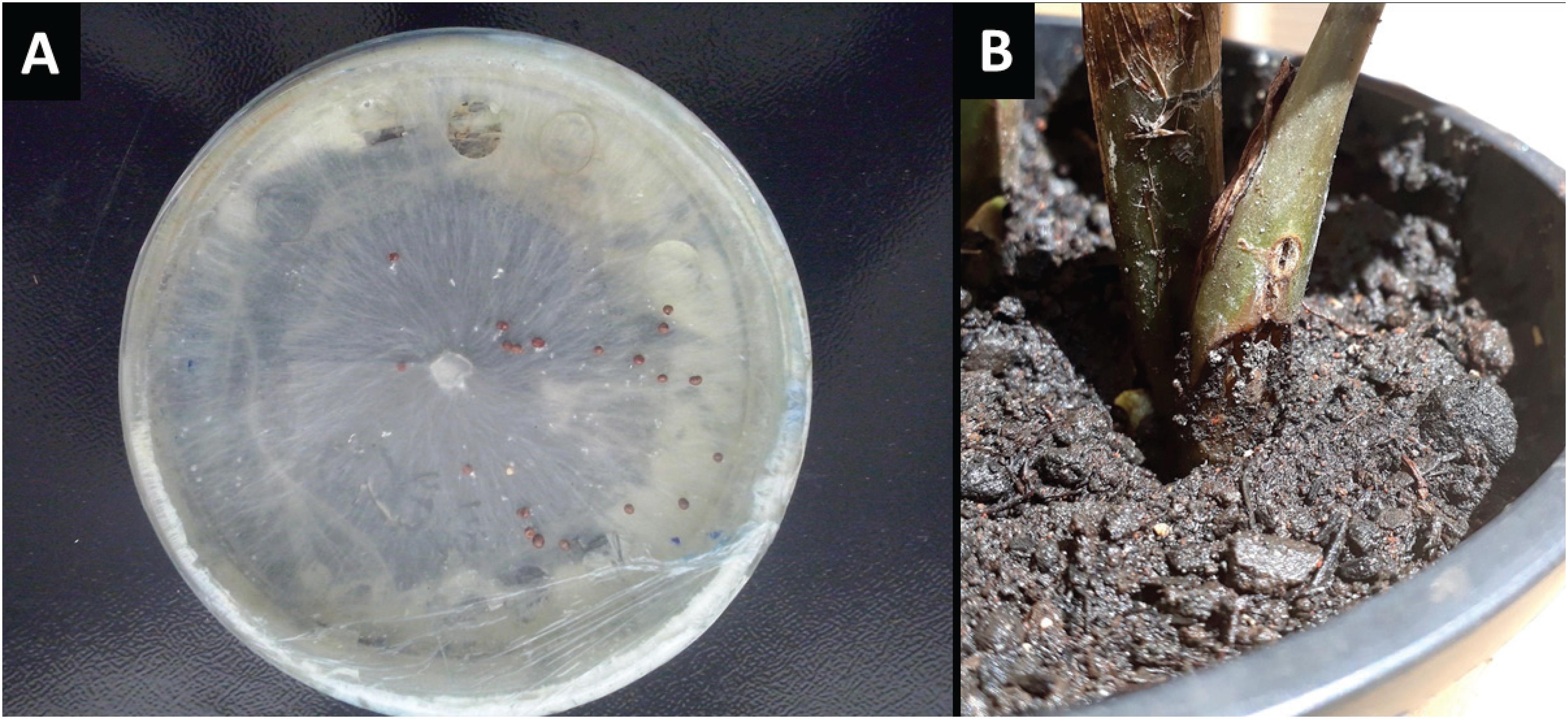
). Based on these morphological characteristics, the SR-001 isolate was identified as Sclerotium rolfsii Sacc (Teleomorph: Atelia rolfsii) (Punja e Damiani, 1996PUNJA, Z.K.; DAMIANI, A. Comparative growth, morphology and physiology of three Sclerotium species. Mycologia, v.88, n.5, p.694-706, 1996.) (Figure 1A).
A) Sclerotium rolfsii in PDA medium; B) Symptom of rot on the stem of a Zamioculcas zamiifolia seedling 15 days after inoculation of S. rolfsii by sclerotia soil infestation.
Amplification and sequencing of the ITS1/ITS4 region of the SR-001 isolate confirmed S. rolfsii as the phytopathogenic agent. The sequence obtained showed 96% similarity with the SrGhMKS2 isolate (GenBank access no. KP412272.1) from Athelia rolfsii, which is the sexual phase of S. rolfsii. The sequence that represents the SR-001 fungal isolate identified as S. rolfsii was deposited in GenBank (access no. MG694322).
Pathogenecity test
With respect to the pathogenicity test, after 15 days’ inoculation, the same rot symptoms exhibited by the samples from which the fungus was isolated were observed in both the leaves and stems of the plants inoculated with the SR-001 isolate. In mycelium disk inoculation, only uninjured leaf tissues displayed no symptoms. This is likely due to the fungus’ low efficiency in directly colonizing aerial tissues, given that its natural infection is more associated with root and stem rot, an environment characterized by climatic conditions favorable to plant cell infection by the fungus. Results of inoculation by soil infestation with sclerotia show that the pathogen's capacity to infect the plant develops under natural soil moisture and temperature conditions, producing symptoms on the plant stem (Figure 1B).
In symptomatic tissues, fragments were once again used to isolate the pathogen, confirming it to be Sclerotium rolfsii, a sine qua non requirement to complete Koch's postulates. The fungus S. rolfsii, considered an aggressive, difficult-to-control phytopathogen, has been reported in more than 500 plant species, including dicotyledons and monocotyledons (Mahadevakumar et al., 2015MAHADEVAKUMAR, S.; TEJASWINI, G.S.; SHILPA, N.; JANARDHANA, G.R.; DHARANENDRASWAMY, S.; YADAV, V. First report of Sclerotium rolfsii associated with boll rot of cotton in India. Plant Disease, v.100, n.1, p. 229, 2015. DOI: 10.1094/PDIS-06-15-0689-PDN
https://doi.org/10.1094/PDIS-06-15-0689-...
; Mahadevakumar et al., 2018MAHADEVAKUMAR, S.; CHANDANA, C.; DEEPIKA, Y.S.; SUMASHRI, K.S.; YADAV, V.; JANARDHANA, G.R. Pathological studies on the southern blight of China aster (Callistephus chinensis) caused by Sclerotium rolfsii. European Journal of Plant Pathology, v.151, n.2, p.1081-1087, 2018. DOI: https://doi.org/10.1007/s10658-017-1415-2
https://doi.org/10.1007/s10658-017-1415-...
). It causes the disease known as sclerotium rot or sclerotium wilt, widely distributed in tropical and subtropical regions (Punja and Damiani, 1996PUNJA, Z.K.; DAMIANI, A. Comparative growth, morphology and physiology of three Sclerotium species. Mycologia, v.88, n.5, p.694-706, 1996.; Mahadevakumar et al., 2016MAHADEVAKUMAR, S.; YADAV, V.; TEJASWINI, G.S.; JANARDHANA, G.R. Morphological and molecular characterization of Sclerotium rolfsii associated with fruit rot of Cucurbita maxima. European Journal of Plant Pathology, v.145, n.1, p.215-219, 2016. DOI: https://doi.org/10.1007/s10658-015-0818-1
https://doi.org/10.1007/s10658-015-0818-...
). S rolfsii is known to infect several economically important crops in various stages of their growth and development, in addition to producing survival structures (Mahadevakumar et al., 2016MAHADEVAKUMAR, S.; YADAV, V.; TEJASWINI, G.S.; JANARDHANA, G.R. Morphological and molecular characterization of Sclerotium rolfsii associated with fruit rot of Cucurbita maxima. European Journal of Plant Pathology, v.145, n.1, p.215-219, 2016. DOI: https://doi.org/10.1007/s10658-015-0818-1
https://doi.org/10.1007/s10658-015-0818-...
; Paul et al., 2017PAUL, N.C.; HWANG, E.J.; NAM, S.S.; LEE, H.U.; LEE, J.S.; YU, G.D.; KANG, Y.G.; LEE, K.B.; GO, S.; YANG, J.W. Phylogenetic placement and morphological characterization of Sclerotium rolfsii (Teleomorph: Athelia rolfsii) associated with blight disease of Ipomeae batatas in Korea. Mycobiology, v.45, n.3, p.129-138, 2017. DOI: 10.5941/MYCO.2017.45.3.129
https://doi.org/10.5941/MYCO.2017.45.3.1...
; Shrestha et al., 2018SHRESTHA, U.; DEE, M.E.; OWNLEY, B.H.; BUTLER, D.M. Anaerobic soil disinfestation reduces germination and affects colonization of Sclerotium rolfsii sclerotia. Phytopathology, v.108, n.3, p.343-351, 2018. DOI: 10.1094/PHYTO-04-17-0152-R
https://doi.org/10.1094/PHYTO-04-17-0152...
). Studies conducted in Sri Lanka (Jegathambiga et al., 2010JEGATHAMBIGA, V.; WILSON, W.R.S.; WIJESUNDERA, R.L.C. Effect of Trichoderma sp. on Sclerotium rolfsii, the causative agent of collar rot on Zamioculcas zamiifolia and an on farm method to mass produce Trichoderma species. Plant Pathology Journal, v.9, n.2, p.47-55, 2010. DOI: 10.3923/ppj.2010.47.55
https://doi.org/10.3923/ppj.2010.47.55...
) reported the occurrence of rot symptoms in Zamioculcas sp. plants caused by S. rolfsii as the primary cause of the decline in plant exports in the country.
Conclusion
The fungus Sclerotium rolfsii is associated with diseases in Z. zamiifolia. As such, this is the first report of soft stem rot caused by S. rolfsii occurring naturally in the cultivated areas of this ornamental species in Brazil.
Acknowledgments
The authors would like to thank the Conselho Nacional de Desenvolvimento Científico e Tecnológico (CNPq) and Coordenação de Aperfeiçoamento de Pessoal de Nível Superior (Capes), for financial support. This study would not have been possible without the equipment provided by the Fundação de Amparo à Pesquisa do Estado de Goiás (FAPEG) and the Funadação de Apoio a Pesquisa (FUNAPE).
References
- ALEXANDRE, M.A.V.; DUARTE, L.M.L.; RIVAS, E.B.; KITAJIMA, E.W.; HARAKAVA, R. First Report of Konjac mosaic virus in Zamioculcas zamiifolia Plant Disease, v.97, n.11, p.1517, 2013. DOI: http://dx.doi.org/10.1094/PDIS-05-13-0537-PDN
» http://dx.doi.org/10.1094/PDIS-05-13-0537-PDN - BOITEUX, L.S.; FONSECA, M.E.N.; SIMON, P.W. Effects of plant tissue and DNA purification method on randomly amplified polymorphic DNA-based genetic fingerprinting analysis in carrot. Journal of American Society for Horticultural Science, v.124, n.1, p.32-38, 1999.
- JEGATHAMBIGA, V.; WILSON, W.R.S.; WIJESUNDERA, R.L.C. Effect of Trichoderma sp. on Sclerotium rolfsii, the causative agent of collar rot on Zamioculcas zamiifolia and an on farm method to mass produce Trichoderma species. Plant Pathology Journal, v.9, n.2, p.47-55, 2010. DOI: 10.3923/ppj.2010.47.55
» https://doi.org/10.3923/ppj.2010.47.55 - KHAKSAR, G.; TREESUBSUNTORN, C.; THIRAVETYAN, P. Impact of endophytic colonization patterns on Zamioculcas zamiifolia stress response and in regulating ROS, tryptophan and IAA levels under airborne formaldehyde and formaldehyde-contaminated soil conditions. Plant Physiology and Biochemistry, v.114, n.3, p.1-9, 2017. DOI: 10.1016/j.plaphy.2017.02.016.
» https://doi.org/10.1016/j.plaphy.2017.02.016 - MAHADEVAKUMAR, S.; CHANDANA, C.; DEEPIKA, Y.S.; SUMASHRI, K.S.; YADAV, V.; JANARDHANA, G.R. Pathological studies on the southern blight of China aster (Callistephus chinensis) caused by Sclerotium rolfsii. European Journal of Plant Pathology, v.151, n.2, p.1081-1087, 2018. DOI: https://doi.org/10.1007/s10658-017-1415-2
» https://doi.org/10.1007/s10658-017-1415-2 - MAHADEVAKUMAR, S.; TEJASWINI, G.S.; SHILPA, N.; JANARDHANA, G.R.; DHARANENDRASWAMY, S.; YADAV, V. First report of Sclerotium rolfsii associated with boll rot of cotton in India. Plant Disease, v.100, n.1, p. 229, 2015. DOI: 10.1094/PDIS-06-15-0689-PDN
» https://doi.org/10.1094/PDIS-06-15-0689-PDN - MAHADEVAKUMAR, S.; YADAV, V.; TEJASWINI, G.S.; JANARDHANA, G.R. Morphological and molecular characterization of Sclerotium rolfsii associated with fruit rot of Cucurbita maxima European Journal of Plant Pathology, v.145, n.1, p.215-219, 2016. DOI: https://doi.org/10.1007/s10658-015-0818-1
» https://doi.org/10.1007/s10658-015-0818-1 - MOULLEC, A.L.; JUVIK, O.J.; FOSSEN, T. First identification of natural products from the African medicinal plant Zamioculcas zamiifolia - a drought resistant survivor through millions of years. Fitoterapia, v.106, n.4, p.280-285, 2015. DOI: 10.1016/j.fitote.2015.09.011
» https://doi.org/10.1016/j.fitote.2015.09.011 - NANDI, S.; HEMBARAM, S.; ADHIKARI, A.; TIWARI, B.K.; DUTTA, S. Host infection beyond the traditional range of Sclerotium (Athelia) rolfsii with Physalis minima Bioinformation, v.13, n.10, p.333-338, 2017. DOI: 10.6026/97320630013333
» https://doi.org/10.6026/97320630013333 - PARK, M.S., KIM, Y.G., LEE, S.W.; PARK, C.G.; KIM, Y.I.;LEE, E.S.; CHANG, J.K.; AN, T.J. Sclerotium rolfsii causes stem rot on Ixeridium dentatum in Korea. Australasian Plant Disease Notes, v.13, n.10, p.2-3, 2018. DOI: https://doi.org/10.1007/s13314-018-0294-5
» https://doi.org/10.1007/s13314-018-0294-5 - PAUL, N.C.; HWANG, E.J.; NAM, S.S.; LEE, H.U.; LEE, J.S.; YU, G.D.; KANG, Y.G.; LEE, K.B.; GO, S.; YANG, J.W. Phylogenetic placement and morphological characterization of Sclerotium rolfsii (Teleomorph: Athelia rolfsii) associated with blight disease of Ipomeae batatas in Korea. Mycobiology, v.45, n.3, p.129-138, 2017. DOI: 10.5941/MYCO.2017.45.3.129
» https://doi.org/10.5941/MYCO.2017.45.3.129 - PUNJA, Z.K.; DAMIANI, A. Comparative growth, morphology and physiology of three Sclerotium species. Mycologia, v.88, n.5, p.694-706, 1996.
- SANAHUJA, G.; LOPEZ, P.; PALMATEER, A.J. First report of Phytophthora nicotianae causing foliar blight on Zamioculcas zamiifolia in Florida. Plant Disease, v.100, n.4, p.864, 2016. DOI: 10.1094/PDIS-09-15-1034-PDN
» https://doi.org/10.1094/PDIS-09-15-1034-PDN - SHRESTHA, U.; DEE, M.E.; OWNLEY, B.H.; BUTLER, D.M. Anaerobic soil disinfestation reduces germination and affects colonization of Sclerotium rolfsii sclerotia. Phytopathology, v.108, n.3, p.343-351, 2018. DOI: 10.1094/PHYTO-04-17-0152-R
» https://doi.org/10.1094/PHYTO-04-17-0152-R - WHITE, T.J.; BRUNS, T.; LEE, S.; TAYLOR, J. Amplification and direct sequencing of fungal ribosomal RNA genes for phylogenetics. In: INNIS, M.A.; GELFAND, D.H.; SHINSKY, J.J.; WHITE, T.J. Protocols: a guide to methods and applications New York: Academic, 1990. p.315-322.
- ZHOU, Z.; LI, Y.L. First Report of Colletotrichum cliviae Causing Anthracnose on Zamioculcas zamiifolia in Henan Province, China. Plant Disease, v.101, n.5, p.838, 2017. DOI: https://doi.org/10.1094/PDIS-10-16-1466-PDN
» https://doi.org/10.1094/PDIS-10-16-1466-PDN
Publication Dates
-
Publication in this collection
24 Jan 2020 -
Date of issue
Oct-Dec 2019
History
-
Received
19 Feb 2019 -
Accepted
22 Oct 2019