
ABSTRACT
BACKGROUND AND OBJECTIVES:
Temporomandibular disorders are the problems involving the masticatory muscles and/or the temporomandibular joint and, among them, the bone and joint disc degenerative processes stand out. However, an effective treatment for these cases has not yet been identified in the literature. Thus, the primary objective of this study was to evaluate the reparative potential of mesenchymal stem cells on degenerative changes in structures associated with the temporomandibular joint in humans and animal models.
CONTENTS:
This narrative review included intervention trials in humans and animals that presented as an outcome variable the repair of joint discs and/or temporomandibular joint. The following databases were used: Pubmed, LILACS, Scielo and Google Scholar. Titles and abstracts were analyzed for the pre-selection of articles potentially eligible for inclusion in this review. The information collected from each article was included in a specific spreadsheet for this purpose containing the year of publication, article title, author’s name, study location, type of study, methodology, results, and conclusions. Two human studies and four animal studies were selected to compose the narrative review. In all studies presented, the presence of stem cells was able to improve the clinical, histological, and morphological parameters of the temporomandibular joint.
CONCLUSION:
The use of stem cells seems to be effective in treating degenerative changes in temporomandibular joint associated structures in both animal and human models. However, due to the small number of studies and their heterogeneity, the results presented should be evaluated sparingly.
Keywords:
Stem cells; Temporomandibular joint disc; Temporomandibular joint disorders
RESUMO
JUSTIFICATIVA E OBJETIVOS:
As desordens temporomandibulares constituem-se em um termo coletivo de problemas que envolvem os músculos mastigatórios e/ou a articulação temporomandibular. Dentre esses, destacam-se os processos degenerativos ósseos e do disco articular, contudo, ainda não foi identificado na literatura um tratamento eficaz para esses casos. Dessa forma, o objetivo desse estudo foi avaliar o potencial reparador das células-tronco mesenquimais sobre as alterações degenerativas das estruturas associadas à articulação temporomandibular em humanos e em modelos animais.
CONTEÚDO:
Foram incluídos ensaios de intervenção em humanos e em animais que apresentassem como variável desfecho o reparo dos discos articulares e/ou da articulação temporomandibular. Foram realizadas buscas nas seguintes bases de dados: Pubmed, LILACS, Scielo e Google Acadêmico. Os títulos e resumos foram analisados para a pré-seleção dos artigos potencialmente elegíveis para sua inclusão. As informações coletadas de cada artigo foram incluídas em planilha específica para essa finalidade contendo o ano de publicação, título do artigo, nome do autor, local do estudo, tipo de estudo, metodologia, resultado e conclusões. Foram selecionados 2 estudos em humanos e 4 estudos em animais para compor este estudo. Em todas essas pesquisas apresentadas, a presença de células-tronco foi capaz de melhorar parâmetros clínicos, histológicos e morfológicos da articulação temporomandibular.
CONCLUSÃO:
O uso de células-tronco parece ser eficaz no tratamento das alterações degenerativas das estruturas associadas à articulação temporomandibular. Todavia, devido ao reduzido número de estudos e sua heterogeneidade, os resultados apresentados devem ser avaliados com parcimônia.
Descritores:
Células-tronco; Disco da articulação temporomandibular; Transtornos da articulação temporomandibular
INTRODUCTION
Temporomandibular disorders (TMD) are the problems involving the masticatory muscles and/or the temporomandibular joint (TMJ). Pain, clicking and jaw limitation form the classic triad of TMD symptoms, which may be related to the masticatory muscles, the pre-auricular area or both11 Okeson JP, de Leeuw R. Differential diagnosis of temporomandibular disorders and other orofacial pain disorders. Dent Clin North Am. 2011;55(1):105-20.,22 Ohrbach R, Dworkin SF. The evolution of TMD diagnosis: past, present, future. J Dent Res. 2016;95(10):1093-101.. The most common subtypes include muscular and joint pain, mainly the joint disc displacements and the degenerative joint diseases22 Ohrbach R, Dworkin SF. The evolution of TMD diagnosis: past, present, future. J Dent Res. 2016;95(10):1093-101.. Epidemiological studies have shown that 20-50% of the population presented at least one symptom associated to TMD22 Ohrbach R, Dworkin SF. The evolution of TMD diagnosis: past, present, future. J Dent Res. 2016;95(10):1093-101..
When the conservative alternatives for the treatment of joint alterations are not effective, the disease may progress, resulting in the modification of the bone structures and the joint disc. The mechanism of the pain phenomenon is not yet completely clear, but it’s known that when the synovial membrane of the TMJ is damaged, lots of inflammatory cytokines are produced and secreted into the synovial liquid, promoting the degenerative and painful process, which can largely vary for each patient33 Kellesarian SV, Al-Kheraif AA, Vohra F, Ghanem A, Malmstrom H, Romanos GE, et al. Cytokine profile in the synovial fluid of patients with temporomandibular joint disorders: a systematic review. Cytokine. 2016;77:98-106..
The joint surfaces of the TMJ are composed of the temporal bone, the mandibular fossa, the joint tubercle, and the mandibular condyle (MC). The scaly part of the temporal bone is part of the formation of the zygomatic arch and the TMJ, including in its extension the mandibular fossa and the joint tubercle, which acts as a bulkhead for the mandibular condyle during movement44 Grossmann E. Algias Craniofaciais: Diagnóstico e Tratamento. São Paulo: Editora dos Editores; 2019..
From the anatomical point of view, the MC is mediolaterally longer than in the anteroposterior direction, forming an ellipse in the transversal plane. The fibrous connective tissue extends to the periphery of the disc, fixating the joint disc to the mandibular condyle below and to the temporal bone above. Anteriorly and posteriorly, the MC connects to the TMJ disc through the capsular ligaments, while mediolaterally, it connects to the disc via collateral ligaments. This arrangement ensures close contact between the disc and the MC during joint movement44 Grossmann E. Algias Craniofaciais: Diagnóstico e Tratamento. São Paulo: Editora dos Editores; 2019..
The TMJ disc is featured as a fibrocartilage and is located between the MC and the temporal bone, providing the mandibular rotation and traverse movements55 Willard VP, Kalpakci KN, Reimer AJ, Athanasiou KA. The regional contribution of glycosaminoglycans to temporomandibular joint disc compressive properties. J Biomech Eng. 2012;134(1):1-8.. The extracellular matrix of the joint disc is composed by a mesh of type 1 collagen, which represents 80-90% of its dry weight55 Willard VP, Kalpakci KN, Reimer AJ, Athanasiou KA. The regional contribution of glycosaminoglycans to temporomandibular joint disc compressive properties. J Biomech Eng. 2012;134(1):1-8., and glycosaminoglycans (GAGs), which represent up to 10% of its weight66 Fazaeli S, Ghazanfari S, Everts V, Smit TH, Koolstra JH. The contribution of collagen fibers to the mechanical compressive properties of the temporomandibular joint disc. Osteoarthritis Cartilage. 2016;24(7):1292-301.. Approximately two thirds of its cells are fibroblasts, while one third have a morphology similar to chondroblasts77 Mäenpää K, Ellä V, Mauno J, Kellomäki M, Suuronen R, Ylikomi T, et al. Use of adipose stem cells and polylactide discs for tissue engineering of the temporomandibular joint disc. J R Soc Interface. 2010;7(42):177-88..
Although the functions and mechanical properties of the TMJ joint disc are already well described, its biomechanical characteristics in inflammatory and degenerative processes are still unclear33 Kellesarian SV, Al-Kheraif AA, Vohra F, Ghanem A, Malmstrom H, Romanos GE, et al. Cytokine profile in the synovial fluid of patients with temporomandibular joint disorders: a systematic review. Cytokine. 2016;77:98-106.,55 Willard VP, Kalpakci KN, Reimer AJ, Athanasiou KA. The regional contribution of glycosaminoglycans to temporomandibular joint disc compressive properties. J Biomech Eng. 2012;134(1):1-8.,88 Colombo V, Palla S, Gallo LM. Temporomandibular joint loading patterns related to joint morphology: a theoretical study. Cells Tissues Organs. 2008;187(4):295-306.. In this context, the study99 Wang XD, Cui SJ, Liu Y, Luo Q, Du RJ, Kou XX, et al. Deterioration of mechanical properties of discs in chronically inflamed TMJ. J Dent Res. 2014;93(11):1170-6. highlights that both the joint disc and synovial membrane can undergo degenerative processes after chronic inflammatory processes.
Chondrocyte death caused by apoptosis or necrosis is considered a central feature in clinical or experimental osteoarthritic pathology. Several therapies were applied with the intention of recovering the joint injury presented in animal models, but they were not able to stimulate local chondrocyte proliferation1010 Wang XD, Kou XX, Mao JJ, Gan YH, Zhou YH. Sustained inflammation induces degeneration of the temporomandibular joint. J Dent Res. 2012;91(5):499-505..
Mesenchymal stem cells (MSC) are multipotent cells present in a wide variety of tissues. They are a source of tissue originating from the mesoderm, such as bone, cartilaginous and adipose tissue. These cells are capable of adopting a fibroblast morphology and, under special conditions, differentiate into adipocytes, chondrocytes and osteocytes1111 Farrar WB. Characteristics of the condylar path in internal derangements of the TMJ. J Prosthet Dent. 1978;39(3):319-23.
12 Saleh R, Reza HM. Short review on human umbilical cord lining epithelial cells and their potential clinical applications. Stem Cell Res Ther. 2017;8(1):222.-1313 Huh Y, Ji RR, Chen G. Neuroinflammation, bone marrow stem cells, and chronic pain. Front Immunol. 2017;8:1014..
Evidence also suggests the existence of MSCs associated with synovial fluid obtained from patients undergoing TMJ arthrocentesis1414 de Souza Tesch R, Takamori ER, Menezes K, Carias RBV, Dutra CLM, de Freitas Aguiar M, et al. Temporomandibular joint regeneration: proposal of a novel treatment for condylar resorption after orthognathic surgery using transplantation of autologous nasal septum chondrocytes, and the first human case report. Stem Cell Res Ther. 2018;9(1):94.. Moreover, recent studies suggest that the use of stem cells in animal models of inflammatory pain, neuropathic pain and pain associated with cancer produce powerful analgesic effects1313 Huh Y, Ji RR, Chen G. Neuroinflammation, bone marrow stem cells, and chronic pain. Front Immunol. 2017;8:1014..
From the perspective that an effective treatment for cases of degeneration of TMJ-associated structures and the tissue forming and analgesic potential of MSCs has not yet been identified in the literature, the primary objective of this study was to assess the repair potential of MSCs on degenerative changes in structures associated with TMJ in human and animal models.
CONTENTS
A literature review using the Pubmed, LILACS, Scielo and Google (gray literature) databases was performed. The search strategy was divided to make the selection of articles more sensitive. For the search in humans, the following descriptors were used: ((“temporomandibular joint” [MeSH Terms] OR (“temporomandibular”[All Fields] AND “joint”[All Fields]) OR “temporomandibular joint” [All Fields]) AND (“stem cells”[MeSH Terms] OR (“stem”[All Fields] AND “cells”[All Fields]) OR “stem cells” [All Fields])) AND “humans” [MeSH Terms]. For the search on animals, the following were used: ((“temporomandibular joint”[MeSH Terms] OR (“temporomandibular”[All Fields] AND “joint” [All Fields]) OR “temporomandibular joint” [All Fields]) AND (“stem cells” [MeSH Terms] OR (“stem” [All Fields] AND “cells” [All Fields]) OR “stem cells” [All Fields])) AND “animals” [MeSH Terms:noexp].
Manual search strategies were performed in the reference list of publications included in the study. Titles and abstracts were analyzed for pre-selection of articles that were potentially eligible for inclusion in the study.
The articles which were included performed animal or human intervention trials and presented as an outcome variable the repair of TMJ discs and/or bone structures using MSCs. Literature review studies, in vitro studies, subcutaneous studies and those that did not present the outcome variable were excluded. No restrictions regarding the period of publication of the manuscript were included, except in the case of Google Scholar, where the search was conducted in the last 3 years in order to identify the most recent studies. The information collected for the qualitative analysis were authors, year, place of publication, methodology, results and conclusions.
527 articles using the stipulated descriptors to evaluate the effectiveness of MSC use in the regeneration of ATM-related structures in humans were found. Of these, 52 were found through Pubmed and 475 through Google Scholar. No articles were found in LILACS and Scielo databases. After reading the titles and abstracts, 3 articles were selected for full reading, as shown in figure 1. Of these, only 2 were included for qualitative analysis, totaling 41 patients (28 women and 13 men), with ages ranging from 23 to 47 years old. The studies originated from Brazil and Italy.

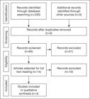
For animal studies, 330 articles were found, 65 in Pubmed and 265 in Google Scholar. No articles were found in LILACS and Scielo databases. After reading the titles and abstracts, 14 articles were selected for full reading, as shown in figure 2. Of those, 10 were excluded, leaving 4 articles for full reading. The studies were conducted in Finland, Egypt, China, United States, and Italy.
The data was tabulated and presented in table 1 (human studies) and table 2 (animal studies).
DISCUSSION
After the critical reading of the studies, it was possible to identify the promising results for the MSCs in humans and animal models on the clinical, histological, and morphological perspectives.
The tissue engineering has been searching for substances that could substitute the removed discs, including autogenous substances as the temporal muscle dermis and fascia, as well as the synthetic materials produced from silicone and polytetrafluorethane. Over the last decades, the autologous chondrocytes implantation (ACI) has developed rapidly. However, the limited speed of cell proliferation and chondrocyte differentiation during in vitro cultures has restricted the use of ACI 2020 Nejadnik H, Hui JH, Feng Choong EP, Tai BC, Lee EH. Autologous bone marrow-derived mesenchymal stem cells versus autologous chondrocyte implantation: an observational cohort study. Am J Sports Med. 2010;38(6):1110-6..
On the other hand, MSCs have become the object of increasing study in this area due to their high proliferative capacity, lower cost, and lower morbidity to the donor site2020 Nejadnik H, Hui JH, Feng Choong EP, Tai BC, Lee EH. Autologous bone marrow-derived mesenchymal stem cells versus autologous chondrocyte implantation: an observational cohort study. Am J Sports Med. 2010;38(6):1110-6.. Its attainment has been associated with the tooth pulp, adipose tissue, umbilical cord and, more recently, the nasal septum1414 de Souza Tesch R, Takamori ER, Menezes K, Carias RBV, Dutra CLM, de Freitas Aguiar M, et al. Temporomandibular joint regeneration: proposal of a novel treatment for condylar resorption after orthognathic surgery using transplantation of autologous nasal septum chondrocytes, and the first human case report. Stem Cell Res Ther. 2018;9(1):94.. During the in vitro growth, MSCs form colonies called fibroblastic colony forming units (CFU-F). The phenotypic characterization of these cells reveals the expression of CD44, CD29, CD105, CD73 and CD166 and the absence of hematopoietic lineage markers such as CD45 and CD342121 Kanafi MM, Ramesh A, Gupta PK, Bhonde RR. Dental pulp stem cells immobilized in alginate microspheres for applications in bone tissue engineering. Int Endod J. 2014;47(7):687-97..
Several therapeutic alternatives have been proposed for the recuperation of the function and the improvement of the clinical parameters in individuals that presented joint alterations. In this context, the joint disc changes are classified into 4 stages with progressive characteristics: a) Joint disc displacement. Stage 1: joint disc displacement with reduction; Stage 2: joint disc displacement with reduction and intermittent closed locking; Stage 3: joint disc displacement without reduction (closed locking); Stage 4: joint disc displacement without reduction with evidence of joint disc perforation or degenerative joint disease2222 Salash JR, Hossameldin RH, Almarza AJ, Chou JC, McCain JP, Mercuri LG, et al. Potential indications for tissue engineering in temporomandibular joint surgery. J Oral Maxillofac Surg. 2016;74(4):705-11.. Thus, it became clear that therapeutic approaches must be related to the severity of the alteration presented by the patient and its clinical implication, with MSCs joint infiltration being another promising treatment for these diseases2222 Salash JR, Hossameldin RH, Almarza AJ, Chou JC, McCain JP, Mercuri LG, et al. Potential indications for tissue engineering in temporomandibular joint surgery. J Oral Maxillofac Surg. 2016;74(4):705-11..
Regarding the evidences presented in the two human studies, the improvement of clinical parameters could be observed, as well as the presence of bone repair assessed through imaging examinations. However, the number of studies and patients is still quite small and should be expanded in order to provide more robust and concrete results regarding the use of MSCs. In addition, gold standards should be established for the definition of diagnostic criteria in order to standardize the studies and their derived results.
As for the researches performed in animals, characterized as pre-clinical studies, it was possible to observe similar behavior to that in humans regarding the recovery of morphology of structures affected by TMJ osteoarthritis models. However, it’s worth noting that the different studies presented quite heterogeneous models. In this context, the study2323 Helgeland E, Shanbhag S, Pedersen TO, Mustafa K, Rosén A. Scaffold-based temporomandibular joint tissue regeneration in experimental animal models: a systematic review Tissue Eng Part B Rev. 2018;24(4):300-16. reported that studies performed in a single animal species are not able to provide standard experimental results with repeated TMJ changes, and more reliable animal models are still necessary2424 Zhang M, Yang H, Lu L, Wan X, Zhang J, Zhang H, et al. Matrix replenishing by BMSCs is beneficial for osteoarthritic temporomandibular joint cartilage. Osteoarthritis Cartilage. 2017;25(9):1551-62..
The exact same mechanism by which the presence of MSCs is able to improve the degenerative changes present in the TMJ is still little known. However, there is a belief that growth factors such as tumor growth factor β1 (TGF-β1) and the family of bone morphogenetic proteins (BMPs) are directly involved in this process2323 Helgeland E, Shanbhag S, Pedersen TO, Mustafa K, Rosén A. Scaffold-based temporomandibular joint tissue regeneration in experimental animal models: a systematic review Tissue Eng Part B Rev. 2018;24(4):300-16.. Moreover, the authors2525 Zhou Y, Wang T, Hamilton JL, Chen D. Wnt/?-catenin signaling in osteoarthritis and in other forms of arthritis. Curr Rheumatol Rep. 2017;19(9):53. have demonstrated that the Wnt pathway regulation promotes the repair of cartilages mediated by the presence of MSCs.
As for the reduction of pain and inflammatory process, several studies report pain relief with the administration of MSCs in rodent models after systemic or local injection. Percutaneous injection of MSCs also caused long-term relief in a pilot study of lumbar discogenic pain in humans2626 Chen G, Park CK, Xie RG, Ji RR. Intrathecal bone marrow stromal cells inhibit neuropathic pain via TGF-? secretion. J Clin Invest. 2015;125(8):3226-40.. Furthermore, another study2727 Di Cesare Mannelli L, Tenci B, Micheli L, Vona A, Corti F, Zanardelli M, et al. Adipose-derived stem cells decrease pain in a rat model of oxaliplatin-induced neuropathy: role of VEGF-A modulation. Neuropharmacology. 2018;131:166-75. demonstrated that the use of MSCs may be a new approach to the treatment of neuropathic pain, due to its significant reduction of pain. These results further contribute to the understanding of pain relief mediated using MSCs.
Regarding the animal studies, only one presented sample calculation for the performance of the experiments. In addition, the necessity for laboratory apparatus for the differentiation of the MSCs still hinders the approach of the technique within the dentistry practice, since the establishment of more simple protocols that could be used in the dentistry routine are necessary.
CONCLUSION
The use of MSCs seems to be effective in treating degenerative changes in TMJ-associated structures in both animal and human models. Due to the small number of studies and their heterogeneity, the results presented should be evaluated sparingly. More research is still necessary to produce more robust evidences for the implementation of this new therapy.
REFERENCES
-
1Okeson JP, de Leeuw R. Differential diagnosis of temporomandibular disorders and other orofacial pain disorders. Dent Clin North Am. 2011;55(1):105-20.
-
2Ohrbach R, Dworkin SF. The evolution of TMD diagnosis: past, present, future. J Dent Res. 2016;95(10):1093-101.
-
3Kellesarian SV, Al-Kheraif AA, Vohra F, Ghanem A, Malmstrom H, Romanos GE, et al. Cytokine profile in the synovial fluid of patients with temporomandibular joint disorders: a systematic review. Cytokine. 2016;77:98-106.
-
4Grossmann E. Algias Craniofaciais: Diagnóstico e Tratamento. São Paulo: Editora dos Editores; 2019.
-
5Willard VP, Kalpakci KN, Reimer AJ, Athanasiou KA. The regional contribution of glycosaminoglycans to temporomandibular joint disc compressive properties. J Biomech Eng. 2012;134(1):1-8.
-
6Fazaeli S, Ghazanfari S, Everts V, Smit TH, Koolstra JH. The contribution of collagen fibers to the mechanical compressive properties of the temporomandibular joint disc. Osteoarthritis Cartilage. 2016;24(7):1292-301.
-
7Mäenpää K, Ellä V, Mauno J, Kellomäki M, Suuronen R, Ylikomi T, et al. Use of adipose stem cells and polylactide discs for tissue engineering of the temporomandibular joint disc. J R Soc Interface. 2010;7(42):177-88.
-
8Colombo V, Palla S, Gallo LM. Temporomandibular joint loading patterns related to joint morphology: a theoretical study. Cells Tissues Organs. 2008;187(4):295-306.
-
9Wang XD, Cui SJ, Liu Y, Luo Q, Du RJ, Kou XX, et al. Deterioration of mechanical properties of discs in chronically inflamed TMJ. J Dent Res. 2014;93(11):1170-6.
-
10Wang XD, Kou XX, Mao JJ, Gan YH, Zhou YH. Sustained inflammation induces degeneration of the temporomandibular joint. J Dent Res. 2012;91(5):499-505.
-
11Farrar WB. Characteristics of the condylar path in internal derangements of the TMJ. J Prosthet Dent. 1978;39(3):319-23.
-
12Saleh R, Reza HM. Short review on human umbilical cord lining epithelial cells and their potential clinical applications. Stem Cell Res Ther. 2017;8(1):222.
-
13Huh Y, Ji RR, Chen G. Neuroinflammation, bone marrow stem cells, and chronic pain. Front Immunol. 2017;8:1014.
-
14de Souza Tesch R, Takamori ER, Menezes K, Carias RBV, Dutra CLM, de Freitas Aguiar M, et al. Temporomandibular joint regeneration: proposal of a novel treatment for condylar resorption after orthognathic surgery using transplantation of autologous nasal septum chondrocytes, and the first human case report. Stem Cell Res Ther. 2018;9(1):94.
-
15Carboni A, Amodeo G, Perugini M, Arangio P, Orsini R, Scopelliti D. Temporomandibular disorders clinical and anatomical outcomes after fat-derived stem cells injection. J Craniofac Surg. 2019;30(3):793-7.
-
16Ahtiainen K, Mauno J, Ellä V, Hagström J, Lindqvist C, Miettinen S, et al. Autologous adipose stem cells and polylactide discs in the replacement of the rabbit temporomandibular joint disc. J R Soc Interface. 2013;10(85):20130287.
-
17Ciocca L, Donati D, Ragazzini S, Dozza B, Rossi F, Fantini M, et al. Mesenchymal stem cells and platelet gel improve bone deposition within CAD-CAM custom-made ceramic HA scaffolds for condyle substitution. Biomed Res Int. 2013;2013:549762.
-
18Zhang M, Yang H, Lu L, Wan X, Zhang J, Zhang H, et al. Matrix replenishing by BMSCs is beneficial for osteoarthritic temporomandibular joint cartilage. Osteoarthritis Cartilage. 2017;25(9):1551-62.
-
19Zaki AA, Zaghloul M, Helal ME, Mansour NA, Grawish ME. Impact of autologous bone marrow-derived stem cells on degenerative changes of articulating surfaces associated with the arthritic temporomandibular joint: an experimental study in Rabbits. J Oral Maxillofac Surg. 2017;75(12):2529-39.
-
20Nejadnik H, Hui JH, Feng Choong EP, Tai BC, Lee EH. Autologous bone marrow-derived mesenchymal stem cells versus autologous chondrocyte implantation: an observational cohort study. Am J Sports Med. 2010;38(6):1110-6.
-
21Kanafi MM, Ramesh A, Gupta PK, Bhonde RR. Dental pulp stem cells immobilized in alginate microspheres for applications in bone tissue engineering. Int Endod J. 2014;47(7):687-97.
-
22Salash JR, Hossameldin RH, Almarza AJ, Chou JC, McCain JP, Mercuri LG, et al. Potential indications for tissue engineering in temporomandibular joint surgery. J Oral Maxillofac Surg. 2016;74(4):705-11.
-
23Helgeland E, Shanbhag S, Pedersen TO, Mustafa K, Rosén A. Scaffold-based temporomandibular joint tissue regeneration in experimental animal models: a systematic review Tissue Eng Part B Rev. 2018;24(4):300-16.
-
24Zhang M, Yang H, Lu L, Wan X, Zhang J, Zhang H, et al. Matrix replenishing by BMSCs is beneficial for osteoarthritic temporomandibular joint cartilage. Osteoarthritis Cartilage. 2017;25(9):1551-62.
-
25Zhou Y, Wang T, Hamilton JL, Chen D. Wnt/?-catenin signaling in osteoarthritis and in other forms of arthritis. Curr Rheumatol Rep. 2017;19(9):53.
-
26Chen G, Park CK, Xie RG, Ji RR. Intrathecal bone marrow stromal cells inhibit neuropathic pain via TGF-? secretion. J Clin Invest. 2015;125(8):3226-40.
-
27Di Cesare Mannelli L, Tenci B, Micheli L, Vona A, Corti F, Zanardelli M, et al. Adipose-derived stem cells decrease pain in a rat model of oxaliplatin-induced neuropathy: role of VEGF-A modulation. Neuropharmacology. 2018;131:166-75.
Publication Dates
-
Publication in this collection
27 July 2020 -
Date of issue
Jul-Sep 2020
History
-
Received
12 Apr 2020 -
Accepted
01 June 2020


