Evaluation of bioactive compounds from Libidibia ferrea (Mart. Ex Tul) LP Queiroz: Antioxidant and antifungal activities, acetylcholinesterase inhibition, and implications in Alzheimer’s disease
Abstract
Fungal infections caused by Candida albicans represent a global health concern, with increasing antifungal resistance and potential links to neurodegenerative diseases such as Alzheimer’s. In this context, this study evaluated the antifungal, antioxidant and acetylcholinesterase inhibitory activities of the hydroethanolic leaf extract of Libidibia ferrea, a species used in traditional medicine. The antioxidant potential was determined using DPPH and ABTS assays, while acetylcholinesterase inhibition was assessed by an in vitro enzymatic assay. Antifungal activity was assessed against Candida albicans strains by broth microdilution and molecular docking was employed to predict the interactions of the main phenolic compounds with acetylcholinesterase and sterol 14-α-demethylase. The extract showed potent antioxidant activity (CI₅₀ = 9.68 ± 0.51 μg/mL for DPPH) and high inhibition of acetylcholinesterase (CI₅₀ = 15.02 ± 0.16 μg/mL). It also showed fungistatic and fungicidal effects, with MIC values ranging from 0.118 to 0.469 mg/mL. In silico analyses highlighted rutin, catechin and ellagic acid as compounds with strong binding affinities to both enzymes, suggesting dual antifungal and neuroprotective effects. These results corroborate Libidibia ferrea as a promising source of bioactive compounds with potential therapeutic applications for fungal infections and neurodegenerative diseases, such as Alzheimer’s.
Key words
Alzheimer; Antioxidant; Candida; Libidibia ferrea
INTRODUCTION
Fungal infections represent a growing challenge to global public health, especially in immunocompromised patients, due to the limited therapeutic options and the emergence of strains resistant to conventional antifungal agents (Vitiello et al. 2023). Among the most relevant etiological agents, Candida albicans stands out for its broad pathogenic capacity, causing infections ranging from superficial to potentially fatal systemic conditions. Amphotericin B remains one of the most effective therapeutic options, although its use is limited due to its high toxicological profile (Fairuz et al. 2022).
In addition to the local and systemic impacts, infections caused by C. albicans have been associated with neuropathological alterations characteristic of Alzheimer’s disease (AD). Evidence shows that this yeast is capable of crossing the blood-brain barrier, inducing the formation of amyloid-beta-like peptides, triggering neuroinflammation and causing cognitive deficits in animal models. Furthermore, a reduction in acetylcholinesterase (AChE) activity has been observed in affected brain regions, which exacerbates the cholinergic dysfunction typical of AD (Wu et al. 2023, Phuna & Madhavan 2022). These findings reinforce that fungal infections may contribute to neurodegenerative processes, highlighting the importance of antifungal therapies not only for infection control but also for mitigating factors associated with AD (Alonso et al. 2017, Yashkin et al. 2022).
In this context, the search for new therapeutic agents, especially of natural origin, has gained increasing relevance. The bioprospecting of plant secondary metabolites emerges as a promising strategy for identifying bioactive molecules with lower toxicity and relevant therapeutic effects (Costa-Lotufo et al. 2009). Libidibia ferrea, popularly known as jucá, is widely used in Brazilian traditional medicine for treating various ailments, including infections. Previous studies have already demonstrated its pharmacological potential, with antioxidant (Prazeres et al. 2019), anti-inflammatory (Almeida et al. 2021b) and antimicrobial activities (Venancio et al. 2014).
Among its constituents, phenolic compounds such as flavonoids and hydrolyzable tannins stand out, recognized for their antioxidant capacity and their role in combating oxidative stress, a central factor in the development of neurodegenerative diseases, including AD (Frota et al. 2025). In parallel, acetylcholinesterase (AChE) remains one of the main therapeutic targets in the treatment of AD, as its inhibition contributes to increased acetylcholine availability in the synaptic cleft, improving cognitive symptoms (Chen et al. 2022, Marucci et al. 2021).
Computational biology tools, such as molecular docking, have been widely employed to predict the interactions between natural compounds and molecular targets of interest (Bartocci & Lió 2016), allowing for a rational approach in the search for new drug candidates. In this study, two enzymes of high pharmacological relevance were selected: sterol 14-α-demethylase, involved in the biosynthesis of ergosterol in fungi (Lepesheva & Waterman 2007), and human recombinant acetylcholinesterase, associated with cholinergic neurotransmission and the pathophysiology of AD.
Therefore, this study aimed to evaluate the antifungal activity of the crude leaf extract of Libidibia ferrea against both standard and clinical strains of Candida albicans, and to investigate, through molecular docking, the interactions of its major phenolic constituents with sterol 14-α-demethylase and human recombinant acetylcholinesterase. By integrating experimental and computational approaches and addressing the lack of studies simultaneously exploring antifungal, antioxidant, and anticholinesterase activities of L. ferrea this work contributes to elucidating the multifunctional therapeutic potential of this species and provides a foundation for future efforts focused on compound isolation, structural optimization, and the development of novel multitarget drug candidates.
MATERIALS AND METHODS
Chemicals and reagents
To carry out the tests here, solvents from Neon - São Paulo, BR; Exodo Cientifica - São Paulo, BR; J.T. BAKER – Radnor, EUA. The reagents are from Sigma-Alcrich – St. Luis, EUA; Chemical - St. Louis, USA; Carvalhaes – Alvorada, RS. The equipment used is by Eyela - Bukit Merah, SIN; Thermo Fisher Scientific - Massachusetts, EUA; Biotek - Vermont, USA; Shimadzu - Kyoto, JP.
Material Collection
The leaves of jucá were collected in March 2023 at the State University of Ceará (UECE). A voucher specimen is deposited in the Prisco Bezerra Herbarium (EAC) at the Federal University of Ceará (UFC), identified as Libidibia ferrea (Mart. Ex Tul.) L.P. Queiroz, under number 66363 and authenticated by botanist Hugo Pereira de Nascimento in March 2023. SisGen registration nº AFB0E62.
Preparation of Libidibia ferrea Leaf Extract
Leaves of Libidibia ferrea (100 g) were collected, oven-dried at 80 °C, ground, and subjected to a single static maceration process, carried out continuously using 70% ethanol, at a solvent-to-plant material ratio of 1:1, at room temperature (25 °C). The plant material remained continuously immersed in the same volume of solvent for seven consecutive days, without interruption, reuse, or replacement of the solvent, in order to obtain the crude extract. After this period, the extract was filtered through a Büchner funnel to remove solid residues and subsequently concentrated using a rotary evaporator at 50 °C. The resulting concentrate was lyophilized, yielding the hydroethanolic extract of L. ferrea leaves (LJ).
Evaluation of Antioxidant Activities by DPPH and ABTS+ Free Radical Inhibition Methods
The antioxidant activity was evaluated using the DPPH method (2,2-diphenyl-1-picrylhydrazyl) following the methodology described by Becker et al. (2019) with modifications and the ABTS+ method (2,2’-azino-bis(3-ethylbenzothiazoline-6-sulfonic acid)) as described by Rhee et al. (2001) Both tests were conducted in a 96-well flat-bottom microplate using a BioTek Elisa reader, model ELX 800. The mean inhibitory concentration (IC50; mg/mL) was obtained using calibration curves, collected by plotting the different absorptions relative to the concentrations and subsequently analyzed by linear regression. All samples were analyzed in triplicate.
In vitro Evaluation of Acetylcholinesterase Inhibition
The inhibitory activity of the enzyme recombinant acetylcholinesterase (AChE) was measured in 96-well flat-bottom plates using a BioTek ELISA reader, model ELX 800, with the “Gen5 V2.04.11” software, based on the methodology described by Re et al. (1999) and Trevisan et al. (2003). The mean inhibitory concentration (IC50; mg/mL) was obtained using calibration curves, collected by plotting the different absorptions relative to the concentrations and subsequently analyzed by linear regression. All solutions were used as negative standards, except the sample. All samples were analyzed in triplicate.
Antifungal Assay
Yeast Strains
The antifungal activity was evaluated against four Candida albicans strains: one standard strain (ATCC90028) obtained from the American Type Culture Collection and three clinical isolates (LABMIC 0102, LABMIC 0104, and LABMIC 0105) provided by Santa Casa de Misericórdia de Sobral and Hospital Regional Norte (Ceará, Brazil). The strains were identified phenotypically and molecularly through CHROMagar™ Candida (CHROMagar, France), Vitek 2 system (BioMérieux Vitek, Hazelwood, France).
Inoculum Preparation
The inoculum was prepared from cultures grown on Sabouraud Dextrose Agar (SDA; Difco, Detroit, MI) for 24 h at 35 ± 2 °C. Yeast colonies were suspended in sterile PBS to achieve a turbidity equivalent to 0.5 on the McFarland scale (1x10⁶ CFU/mL). These suspensions were then diluted 1:2000 in RPMI-1640 medium (Sigma-Aldrich, USA) supplemented with L-glutamine, resulting in a final concentration of 2x10² CFU/mL, following the CLSI M27-A3 guidelines (CLSI 2008).
Minimum Inhibitory Concentration (MIC) and Minimum Fungicidal Concentration (MFC)
The Minimum Inhibitory Concentration (MIC) and Minimum Fungicidal Concentration (MFC) of the hydroethanolic leaf extract of Libidibia ferrea (LJ) were determined using the broth microdilution method following the Clinical and Laboratory Standards Institute M27-A3 protocol (CLSI 2008). The extract stock solution was prepared by solubilizing 15 mg of LJ in 50 μL of 5% dimethyl sulfoxide (DMSO) and 950 μL of Tween 80%, yielding a final concentration of 15 mg/mL.
Assays were conducted in 96-well microplates. Initially, 50 μL of RPMI-1640 medium was dispensed into each well. Then, 50 μL of the LJ stock solution was added to the first column, followed by twofold serial dilutions across the plate. After adding 50 μL of the standardized inoculum to each well (final volume: 100 μL), the resulting extract concentrations ranged from 7.50 to 0.058 mg/mL, covering the working interval in which the MIC (0.118–0.469 mg/mL) and MFC (0.234–0.938 mg/mL) values were obtained. Amphotericin B (AMB) was used as the positive control (0.016 to 1.5×10⁻⁵ mg/mL).
The plates were incubated at 37 °C for 48 h, and the MIC was defined as the lowest concentration that completely inhibited visible fungal growth. For MFC determination, 100 μL aliquots from wells showing no visible growth were subcultured onto Potato Dextrose Agar (PDA) and incubated at 28 °C for 48 h. The MFC corresponded to the lowest concentration producing no colony growth. All assays were performed in triplicate.
Molecular Modeling
Protein Preparation
The receptor structures used were sterol 14-alpha-demethylase (CYP51) (PDB: 5TZ1) (Hargrove et al. 2017), rystallized in complex with the antifungal compound VT1161 at a resolution of 2.00 Å, and human recombinant acetylcholinesterase (PDB: 4EY6) (Cheung et al. 2012), crystallized with galantamine at a resolution of 2.40 Å. Both structures were obtained from the RCSB Protein Data Bank (Berman 2000). Water molecules and co-crystallized ligands were removed. Polar hydrogen atoms and Kollman charges were added.
Ligand Preparation
The two-dimensional structures of the compounds amphotericin B, galantamine, caffeic acid, catechin, chlorogenic acid, ellagic acid, gallic acid and rutin were drawn using ChemSketch (free version). Their three-dimensional structures were generated using the 3D Viewer (free version).
Molecular Docking
Molecular docking was performed to predict the binding orientation and estimate the binding energies of the ligands (amphotericin B, galantamine, caffeic acid, catechin, chlorogenic acid, ellagic acid, gallic acid and rutin) with the receptors sterol 14-alpha-demethylase and human recombinant acetylcholinesterase (Zhang et al. 2022). Amphotericin B was used as a reference ligand for sterol 14-alpha-demethylase and galantamine was used as the reference for human recombinant acetylcholinesterase.
The calculations accounted for hydrogen bonding, attractive van der Waals interactions, electrostatic interactions, and hydrophobic interactions (Goodsell et al. 2021). AutoDock 4.2 software (Morris et al. 2009) was employed for automated docking to explore possible binding modes between receptors and ligands.
During the calculations, the receptor structures were kept rigid, while the ligands were considered flexible. The Lamarckian genetic algorithm was chosen for the search parameters. Each docking run was set to 50 runs, allowing each ligand to adopt 50 different conformations within the grid box. The grid box was centered according to the original crystallographic binding site, with dimensions set to 60 × 60 × 60 Å and a grid spacing of 0.375 Å.
Docking results were analyzed using AutoDock Tools and visualized with Chimera software (Pettersen et al. 2004).
RESULTS AND DISCUSSION
The leaf extract of Libidibia ferrea exhibited high antioxidant capacity, with an average DPPH radical inhibitory concentration (IC₅₀) of 9.68 ± 0.51 μg/mL, being statistically more active than the standard Trolox (IC₅₀ 12.35 ± 0.05 μg/mL) (q = 12.24, p < 0.0001; LJ vs. Trolox) [(F7, 16) = 181.4] (Table I). The strong antioxidant activity of the extract can be attributed to its high total phenolic content (554.94 ± 1.24 mg GAE/g), mainly characterized by hydrolyzable tannins, with the presence of caffeic, chlorogenic, ellagic and gallic acids, as well as catechin, being identified (Frota et al. 2025). These phenolic compounds were evaluated for their antioxidant, anticholinesterase activities and demonstrated excellent free radical scavenging capacity when compared to the standard. Among them, gallic acid showed the lowest IC₅₀ (2.33 ± 0.10 μg/mL), followed by the other phenolic compounds, all statistically equivalent (p > 0.05) in DPPH radical inhibition.
Antioxidant and acetylcholinesterase inhibitory activities of the hydroethanolic extract from Libidibia ferrea leaves and its constituents.
For ABTS⁺ radical inhibition (Table I), caffeic acid, ellagic acid and rutin stood out, showing statistically similar IC₅₀ values (6.78 ± 0.28, 6.38 ± 0.71 and 7.62 ± 0.10 μg/mL, respectively) and were significantly more active than the Trolox standard (12.13 ± 0.07 µg/mL) [(F7, 16) = 72.87]. Regarding AChE inhibitory activity (Table I), catechin and chlorogenic acid exhibited IC₅₀ values statistically similar to the reference drug galantamine (7.52 ± 0.96, 5.90 ± 0.99 and 5.82 ± 0.02 μg/mL, respectively) [(F7, 16) = 82.81]. It is worth mentioning that all tested samples demonstrated high AChE inhibitory activity (IC₅₀ < 20 μg/mL) (dos Santos et al. 2018).
Chlorogenic acid exhibits various therapeutic activities, including neuroprotective effects (Singh et al. 2020). Studies have shown that chlorogenic acid improves scopolamine-induced memory deficits in animal models and inhibits acetylcholinesterase activity in the hippocampus and frontal cortex, both in vitro (IC₅₀ = 98.17 μg/mL) and ex vivo. It also demonstrates potent antioxidant activity (DPPH IC₅₀ = 3.09 μg/mL) (Kwon et al. 2010). Its mechanisms of action involve acetylcholinesterase inhibition, malondialdehyde reduction, modulation of neuroreceptors, ion channels, activation of the Nrf2 and AMPK pathways, promoting anti-inflammatory, antioxidant effects and restoring metabolic homeostasis. In addition to neuroprotection, chlorogenic acid provides benefits for cardiovascular, inflammatory, metabolic diseases and cancer (Nguyen et al. 2024). However, further studies are needed to improve its bioavailability and confirm its clinical efficacy.
Catechin, caffeine and theobromine are three bioactive compounds present in plant-based foods and are major constituents of tea, coffee and cocoa, respectively. Although not structurally related, catechin, caffeine and theobromine have been reported to exhibit psychostimulant properties. These compounds demonstrated antioxidant activity and showed inhibitory effects on AChE and BChE enzymes. Catechin displayed the strongest antioxidant activity, while theobromine exhibited the highest enzyme inhibition effect. These findings provide new insights into the effects of these bioactive compounds commonly found in foods, particularly regarding their antioxidant and neuroprotective properties (Ademola et al. 2024).
Ellagic acid is a phenolic phytoconstituent found in grains and fruits, with notable antioxidant effects and the ability to modulate several endogenous molecular pathways beneficially in humans. Ellagic acid is considered a green multitarget compound capable of reducing oxidative stress and inflammation, thereby preventing and improving the condition of Alzheimer’s disease (Alfei & Zuccari 2025).
Extracts of L. ferrea in in vivo models do not show toxicity in their use (Almeida et al. 2021a). LJ showed better inhibitory activity (MIC), as well as better fungicidal activity (MFC) against the standard strain ATCC 90028 (0.118 and 0.234 mg/mL, respectively) and the clinical isolate LABMIC 0102 (0.118 and 0.234 mg/mL, respectively), indicating greater sensitivity to the extract. On the other hand, the clinical isolates LABMIC 0104 and LABMIC 0105 showed higher MIC and MFC values (0.234 and 0.469 mg/mL; 0.469 and 0.938 mg/mL, respectively), suggesting lower susceptibility compared to the standard strain. In comparison with the positive control, amphotericin B exhibited significantly lower MIC values (0.0005 mg/mL for all strains), which reinforces its well-established profile of high antifungal potency (Table II). Despite its potential toxicity, amphotericin B remains a valuable therapeutic option for the treatment of invasive fungal infections due to its broad spectrum of activity, low resistance rate and proven clinical and pharmacological efficacy (Cavassin et al. 2021).
MIC and MFC of the hydroethanolic extract from Libidibia ferrea leaves against Candida albicans.
However, it is important to emphasize that although LJ exhibited higher MIC values compared to amphotericin B, the levels obtained still indicate relevant activity, especially considering the use of crude natural products without purification processes.
The antifungal activity observed for LJ can be attributed to its well-documented antimicrobial potential described in the literature, mainly associated with the presence of bioactive compounds such as tannins, flavonoids and phenolic acids (Paiva et al. 2015). These compounds act through different mechanisms against microorganisms, including fungi such as Candida albicans, which reinforces the relevance of this species as a promising source of natural antifungal agents, since the minimum inhibitory concentrations (MIC) of the extract ranged from 0.118 to 0.469 mg/mL against C. albicans (Table II). Additionally, hydrolyzable tannins, such as those present in this extract, have shown efficacy in inhibiting biofilm formation and inducing apoptosis in fungal cells, without loss of efficacy when combined with conventional antifungal drugs (Moreira et al. 2024). Condensed tannins, in turn, are effective against both planktonic growth and biofilms of C. albicans, inducing morphological alterations in fungal cells, such as the formation of dumbbell-shaped blastoconidia (Luiz et al. 2015).
Other extracts from jucá were also considered for testing. However, the methanolic seed fraction showed only moderate inhibitory activity, with MICs of 0.938 mg/mL for the ATCC 90028 strain and 0.469 mg/mL for LABMIC 0102, but showed no inhibitory effect against LABMIC 0104 and LABMIC 0105, nor fungicidal activity (MFC not determined, indicating that it did not kill fungal cells at the tested concentrations). The hydroethanolic extract of the bark did not exhibit antifungal activity against any of the tested strains and was considered inactive. The flower decoction showed inhibitory activity only against the ATCC 90028 strain (MIC of 0.938 mg/mL) and LABMIC 0102 (MIC of 0.938 mg/mL), being inactive against LABMIC 0104, LABMIC 0105 and with no fungicidal effect (MFC not determined).
Therefore, among all the extracts evaluated, only LJ exhibited both fungistatic and fungicidal activity against C. albicans strains. This antifungal activity observed exclusively in the leaf extract, despite the presence of similar phenolic compounds in the other extracts (Frota et al. 2025), suggests that the bioactivity is not directly related solely to the isolated presence of these metabolites, but possibly to a synergistic effect among them, a greater phytochemical diversity, or even the absence of antagonistic compounds present in the other extracts. This result highlights the importance of considering not only the qualitative composition but also the chemical interactions within the extract matrix. Therefore, future studies focused on evaluating the complete metabolic profile, testing of fractions, as well as combinatorial analysis approaches and mechanism of action studies will be essential to confirm whether the activity results from synergism, antagonism, or the predominance of specific compounds in LJ.
In silico evaluation
Molecular docking is a crucial tool to explore the interactions between the target protein, called the receptor and a small molecule, called the ligand, which correspond to the main compounds found in LJ. Molecular docking performed between the seven ligands and the two evaluated receptors, the sterol 14-α-demethylase protein (PDB 5TZ1), associated with antifungal activity and acetylcholinesterase (PDB 4EY6), related to cholinesterase activity, resulted in binding energy values presented in Table III.
Binding energy values calculated using AutoDock and the amino acid fragments present in the active site that are interacting with the ligands.
Amphotericin B, the reference ligand, showed a binding energy of -9.94 kcal/mol, indicating high affinity with the receptor’s active site. The compounds rutin (-8.48 kcal/mol) and chlorogenic acid (-8.04 kcal/mol) exhibited similar values, suggesting potential antifungal activity.
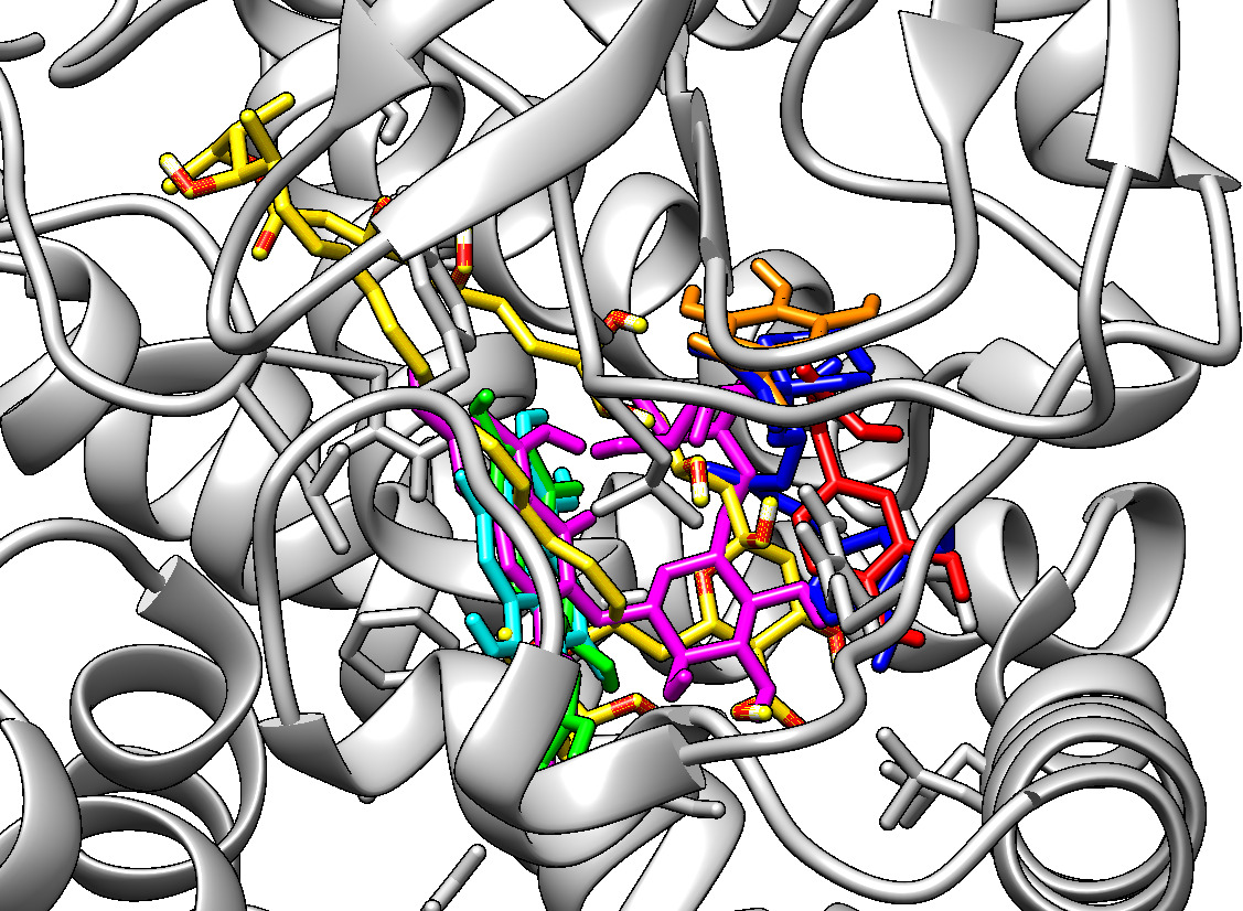
Analysis of the binding site (Figure 1) revealed that all compounds share the same interaction site with the enzyme. Due to its size, amphotericin B occupies most of the site and forms hydrophobic interactions (TYR75, LEU80, ALA284, LEU385) as well as hydrogen bonds (ASP12, LEU80, GLN186, SER228, GLY387). LEU80, LEU385 and ALA284 are responsible for forming a hydrophobic pocket that accommodates the ligand. GLY387, located in the C-terminal region and GLN186 may participate in a water-mediated hydrogen bonding network with the ligand, assisting in stabilizing the reactive intermediate and the reaction complex transition (Strushkevich et al. 2010).
Position of amphotericin B (yellow), caffeic acid (red), catechin (green), chlorogenic acid (blue), ellagic acid (cyan), gallic acid (orange), and rutin (magenta) in the active site of sterol 14-α-demethylase, calculated by molecular docking.
Among the phenolics, chlorogenic acid established the greatest number of favorable interactions, including hydrogen bonds with ARG64, HIS96, LEU285, TRP343 and hydrophobic interactions with LEU99, PHE310 and ILE246. Caffeic acid and gallic acid, although interacting with similar residues, formed fewer interactions, resulting in less favorable energies.
On the opposite side of the active site, catechin and ellagic acid showed relevant interactions. Catechin formed hydrophobic interactions with LEU80, PHE231 and ALA232, as well as hydrogen bonds with GLN186, SER228 and ASN386. Ellagic acid stood out due to its π-π interaction with PHE231, in addition to hydrogen bonds with LEU80 and ASN386.
Rutin, due to its larger size, interacts with regions on both sides of the active site, establishing hydrophobic interactions (LEU80, PHE231, PHE310, LEU385) and hydrogen bonds (LEU80, GLN186, SER228, LEU285, ASN386). Residues such as SER228, GLY387 and GLN186 may form hydrogen bonds with the ligand and reactive intermediates, aiding in the transition of the reaction complex (Strushkevich et al. 2010).
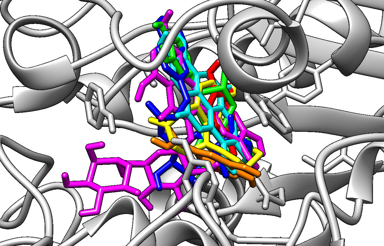
In silico evaluation of the activity of phenolic compounds present in L. ferrea leaf extract on acetylcholinesterase Galantamine, a known AChE inhibitor, presented a binding energy of -7.84 kcal/mol. However, catechin (-9.13 kcal/mol) and ellagic acid (-8.23 kcal/mol) showed higher values, suggesting greater affinity for the enzyme’s catalytic site.
All compounds were located within the AChE active center (Figure 2), potentially blocking acetylcholine hydrolysis (Dvir et al. 2010). Catechin, with the best binding energy, hydrophobically interacted with TRP86 and formed hydrogen bonds with catalytically essential residues such as SER203 and HIS447, as well as ASN87, GLU202 and TYR133. SER203, for example, is a key residue in AChE’s catalytic process, participating in acetylcholine hydrolysis. Interaction with the serine hydroxyl group through hydrogen bonds stabilizes the inhibitor’s binding to the enzyme, blocking the active site. HIS447 also assists in stabilizing the reaction intermediate during acetylcholine hydrolysis. It acts as a base accepting a proton from SER203, allowing the cleavage of the bond between acetylcholine’s acetyl group and the rest of the molecule (De Boer et al. 2021).
Position of galantamine (yellow), caffeic acid (red), catechin (green), chlorogenic acid (blue), ellagic acid (cyan), gallic acid (orange), and rutin (magenta) in the active site of human recombinant acetylcholinesterase, calculated by molecular docking.
Ellagic acid showed a similar interaction profile, including interactions with TRP86 (hydrophobic and π-π), and hydrogen bonds with GLN71, ASP74, ASN87, GLU202 and TYR337. Although TRP86 does not directly participate in the catalytic hydrolysis mechanism like SER203 and HIS447, it significantly contributes to the structural stability of the active site (Sussman et al. 1991). This may explain why gallic acid exhibited the poorest binding energy with the enzyme (-4.55 kcal/mol), as results indicate that TRP86 does not interact with this ligand.
Despite its bulky structure, rutin showed a binding energy of -7.62 kcal/mol, close to that of galantamine. Its interactions involve important residues for substrate recognition and blockage, reinforcing its potential as a multitarget compound.
These results reinforce the potential of the evaluated phenolic compounds as inhibitors of pharmacologically relevant target enzymes and provide a basis for future in vitro and in vivo studies aimed at validating their biological activities.
CONCLUSIONS
The hydroethanolic leaf extract of Libidibia ferrea (LJ) demonstrated strong antioxidant and anticholinesterase activities, along with relevant antifungal effects against Candida albicans strains. Molecular docking supported these findings, identifying rutin, catechin and ellagic acid as promising multitarget compounds with potential antifungal and neuroprotective actions.
Together, the in vitro and in silico results indicate that L. ferrea is a valuable source of bioactive metabolites with multifunctional therapeutic potential. Future studies should focus on isolating the active constituents, clarifying their mechanisms of action, improving their pharmacological properties, and exploring synergistic combinations with established antifungal and neuroprotective agents to support the development of innovative and less toxic therapeutic strategies
Acknowledgements
We thank the Ceará Foundation for Scientific and Technological Development (FUNCAP) for the postdoctoral fellowship granted to Lucas Soares Frota (FPD-00213-00262.02.00/23). We would like to thank the National Council for Scientific and Technological Development (CNPq) for the funding granted through the Productivity Project (312597/2023-1), and the Ceará Foundation for Support of Scientific and Technological Development (FUNCAP) for the financial support through the Technological Innovation Project (31032.003403/2024-42), linked to researcher Selene Maia de Morais. Thanks to the support of the members of the Research Group of the Natural Products Chemistry Laboratory (LQPN) at the State University of Ceará (UECE).
References
- ADEMOLA IO, OBOH G & ADEMOSUN AO. 2024. Comparative Antioxidant and Anti-cholinesterase Properties of Catechin, Caffeine and Theobromine. Niger J Biochem Mol Biol 39: 91-98.
- ALFEI S & ZUCCARI G. 2025. Ellagic Acid: A Green Multi-Target Weapon That Reduces Oxidative Stress and Inflammation to Prevent and Improve the Condition of Alzheimer’s Disease. Int J Mol Sci 26: 844.
- ALMEIDA NCOS, FURTADO SC & BARCELLOS JFM. 2021a. A Narrative Review of Libidibia ferrea: Botanical Aspects, Ethnopharmacological Properties, Phytochemical Characteristics, Toxicity, and Experimental Tests. European J Med Plants 32(12): 16-30.
- ALMEIDA NCOS, SILVA FRP, CARNEIRO ALB, LIMA ES, BARCELLOS JFM & FURTADO SC. 2021b. Libidibia ferrea (jucá) anti-inflammatory action: A systematic review of in vivo and in vitro studies. In: Dalmarco EM (Ed), PLoS ONE 16: e0259545.
- ALONSO R, PISA D, AGUADO B & CARRASCO L. 2017. Identification of Fungal Species in Brain Tissue from Alzheimer’s Disease by Next-Generation Sequencing. J Alzheimer’s Dis 58: 55-67.
- BARTOCCI E & LIÓ P. 2016. Computational Modeling, Formal Analysis, and Tools for Systems Biology. In: Kwiatkowska MZ (Ed), PLoS Comput Biol 12: e1004591.
- BECKER M, NUNES G, RIBEIRO D, SILVA F, CATANANTE G & MARTY J. 2019. Determination of the Antioxidant Capacity of Red Fruits by Miniaturized Spectrophotometry Assays. J Braz Chem Soc 3: 223-227.
- BERMAN HM. 2000. The Protein Data Bank. Nucleic Acids Res 28: 235-242.
- CAVASSIN FB, BAÚ-CARNEIRO JL, VILAS-BOAS RR & QUEIROZ-TELLES F. 2021. Sixty years of Amphotericin B: An Overview of the Main Antifungal Agent Used to Treat Invasive Fungal Infections. Infect Dis Ther 10: 115-147.
- CHEN Z-R, HUANG J-B, YANG S-L & HONG F-F. 2022. Role of Cholinergic Signaling in Alzheimer’s Disease. Molecules 27: 1816.
- CHEUNG J, RUDOLPH MJ, BURSHTEYN F, CASSIDY MS, GARY EN, LOVE J, FRANKLIN MC & HEIGHT JJ. 2012. Structures of human acetylcholinesterase in complex with pharmacologically important ligands. J Med Chem 55: 10282-10286.
- CLSI. 2008. Reference Method for Broth Dilution Antifungal Susceptibility Testing of Yeasts, Third Edit ed., Pennsylvania: Clinical and Laboratory Standards Institute, 40 p.
- COSTA-LOTUFO LV, WILKE DV, JIMENEZ PC & EPIFANIO RA. 2009. Organismos marinhos como fonte de novos fármacos: histórico & perspectivas. Quim Nova 32: 703-716.
- DE BOER D, NGUYEN N, MAO J, MOORE J & SORIN EJ. 2021. A Comprehensive Review of Cholinesterase Modeling and Simulation. Biomolecules 11: 580.
- DOS SANTOS TC, GOMES TM, PINTO BAS, CAMARA AL & PAES AMA. 2018. Naturally Occurring Acetylcholinesterase Inhibitors and Their Potential Use for Alzheimer’s Disease Therapy. Front Pharmacol 9: 1192.
- DVIR H, SILMAN I, HAREL M, ROSENBERRY TL & SUSSMAN JL. 2010. Acetylcholinesterase: From 3D structure to function. Chem Biol Interact 187: 10-22.
- FAIRUZ S, NAIR RS & BILLA N. 2022. Orally Administered Amphotericin B Nanoformulations: Physical Properties of Nanoparticle Carriers on Bioavailability and Clinical Relevance. Pharmaceutics 14: 1823.
- FROTA LS, BARBOSA SICG, FEITOSA CC, LOPES FFS, DE OLIVEIRA DP, FREITAS LS, MATTOS ALA, DA SILVA WMB, ISHIKI HM & DE MORAIS SM. 2025. Chemical Characterization, Antioxidant, and Anticholinesterase Activities of Libidibia ferrea (Mart. Ex Tul.) L.P.Queiroz and In Silico Studies with the Acetylcholinesterase Enzyme. Chem Biodivers 22(10): e00550.
- GOODSELL DS, SANNER MF, OLSON AJ & FORLI S. 2021. The AutoDock suite at 30. Protein Sci 30: 31-43.
- HARGROVE TY, FRIGGERI L, WAWRZAK Z, QI A, HOEKSTRA WJ, SCHOTZINGER RJ, YORK JD, GUENGERICH FP & LEPESHEVA GI. 2017. Structural analyses of Candida albicans sterol 14α-demethylase complexed with azole drugs address the molecular basis of azole-mediated inhibition of fungal sterol biosynthesis. J Biol Chem 292: 6728-6743.
- KWON S-H ET AL. 2010. Neuroprotective effects of chlorogenic acid on scopolamine-induced amnesia via anti-acetylcholinesterase and anti-oxidative activities in mice. Eur J Pharmacol 649: 210-217.
- LEPESHEVA GI & WATERMAN MR. 2007. Sterol 14alpha-demethylase cytochrome P450 (CYP51), a P450 in all biological kingdoms. Biochim Biophys Acta 1770: 467-477.
- LUIZ RLF, VILA TVM, DE MELLO JCP, NAKAMURA CV, ROZENTAL S & ISHIDA K. 2015. Proanthocyanidins polymeric tannin from Stryphnodendron adstringens are active against Candida albicans biofilms. BMC Complement Altern Med 15: 68.
- MARUCCI G, BUCCIONI M, BEN DD, LAMBERTUCCI C, VOLPINI R & AMENTA F. 2021. Efficacy of acetylcholinesterase inhibitors in Alzheimer’s disease. Neuropharmacology 190: 108352.
- MOREIRA LEA ET AL. 2024. Antifungal activity of tannic acid against Candida spp. and its mechanism of action. Brazilian J Microbiol 55: 3679-3690.
- MORRIS GM, RUTH H, LINDSTROM W, SANNER MF, BELEW RK, GOODSELL DS & OLSON AJ. 2009. Software news and updates AutoDock4 and AutoDockTools4: Automated docking with selective receptor flexibility. J Comput Chem 30: 2785-2791.
- NGUYEN V, TAINE EG, MENG D, CUI T & TAN W. 2024. Chlorogenic Acid: A Systematic Review on the Biological Functions, Mechanistic Actions, and Therapeutic Potentials. Nutrients 16: 924.
- PAIVA WDS, DE SOUZA NETO FE, LIMA BANDEIRA MG, ABRANTES MR, DE LIMA BATISTA AC & DA SILVA JBA. 2015. ATIVIDADE ANTIBACTERIANA DA CASCA DO JUCÁ (Libidibia ferrea (Mart. ex Tul.) L. P. Queiroz), FRENTE A Staphylococcus spp. ISOLADOS DO LEITE DE CABRAS COM MASTITE. Arch Vet Sci 20(2): 141-146.
- PETTERSEN EF, GODDARD TD, HUANG CC, COUCH GS, GREENBLATT DM, MENG EC & FERRIN TE. 2004. UCSF Chimera - A visualization system for exploratory research and analysis. J Comput Chem 25: 1605-1612.
- PHUNA ZX & MADHAVAN P. 2022. A closer look at the mycobiome in Alzheimer’s disease: Fungal species, pathogenesis and transmission. Eur J Neurosci 55: 1291-1321.
- PRAZERES LDKT ET AL. 2019. Antioxidant and Antiulcerogenic Activity of the Dry Extract of Pods of Libidibia ferrea Mart. ex Tul. (Fabaceae). Oxid Med Cell Longev 2019: 1-23.
- RE R, PELLEGRINI N, PROTEGGENTE A, PANNALA A, YANG M & RICE-EVANS C. 1999. Antioxidant activity applying an improved ABTS radical cation decolorization assay. Free Radic Biol Med 26: 1231-1237.
- RHEE IK, MEENT M, INGKANINAN K & VERPOORTE R. 2001. Screening for acetylcholinesterase inhibitors from Amaryllidaceae using silica gel thin-layer chromatography in combination with bioactivity staining. J Chromatogr A 915: 217-223.
- SINGH SS, RAI SN, BIRLA H, ZAHRA W, RATHORE AS, DILNASHIN H, SINGH R & SINGH SP. 2020. Neuroprotective Effect of Chlorogenic Acid on Mitochondrial Dysfunction-Mediated Apoptotic Death of DA Neurons in a Parkinsonian Mouse Model. Oxid Med Cell Longev 2020: 1-14.
- STRUSHKEVICH N, USANOV SA & PARK H-W. 2010. Structural Basis of Human CYP51 Inhibition by Antifungal Azoles. J Mol Biol 397: 1067-1078.
- SUSSMAN JL, HAREL M, FROLOW F, OEFNER C, GOLDMAN A, TOKER L & SILMAN I. 1991. Atomic Structure of Acetylcholinesterase from Torpedo californica : A Prototypic Acetylcholine-Binding Protein. Science 253: 872-879.
- TREVISAN MTS, MACEDO FVV, VAN DE MEENT M, RHEE IK & VERPOORTE R. 2003. Seleção de plantas com atividade anticolinasterase para tratamento da doença de Alzheimer. Quim Nova 26: 301-304.
- VENANCIO GN ET AL. 2014. Evaluation of the stability and antimicrobial activity of an ethanolic extract of Libidibia ferrea. Clin Cosmet Investig Dent 9.
- VITIELLO A, FERRARA F, BOCCELLINO M, PONZO A, CIMMINO C, COMBERIATI E, ZOVI A, CLEMENTE S & SABBATUCCI M. 2023. Antifungal Drug Resistance: An Emergent Health Threat. Biomedicines 11: 1063.
- WU Y ET AL. 2023. Toll-like receptor 4 and CD11b expressed on microglia coordinate eradication of Candida albicans cerebral mycosis. Cell Rep 42: 113240.
- YASHKIN A, AKUSHEVICH I, YASHIN A, GORBUNOVA G & UKRAINTSEVA S. 2022. FUNGAL INFECTIONS, USE OF ANTIFUNGAL AGENTS, AND THE RISK OF ALZHEIMER’S DISEASE. Innov Aging 6: 1-2.
- ZHANG B, LI H, YU K & JIN Z. 2022. Molecular docking-based computational platform for high-throughput virtual screening. CCF Trans High Perform Comput 4: 63-74.
Edited by
-
Handling editor
Marilia Goulart
The authors declare that no supplementary material is available for this study. Any additional data or information supporting the findings of this work may be obtained from the corresponding author upon reasonable request.