Abstract
The objectives of this study are to prepare a 5 wt% lidocaine/aspirin ionic liquid drug-loaded gelatin/polyvinyl alcohol composite film using a freeze-thaw procedure and to evaluate their physicochemical characteristics, in vitro drug release, and stability. Lidocaine/aspirin ionic liquid drugs can be prepared by an ion-pair reaction between the hydrochloride salts of lidocaine and the sodium salts of aspirin, which showed a significant change in their thermal properties when compared to those pure drugs. The results showed that a transdermal patch could feasibly be used in pharmaceutical transdermal patches with good physicochemical properties. A chemical interaction between the drug and polymer base was not found. Decomposition of the lidocaine/aspirin ionic liquid drug was found in the patch; however, the properties of the patch were not changed after drug loading. The patch controlled the drug release and showed good stability during the studied period of three months when kept at 4°C more than at ambient temperature and 45°C.
Key words
Ionic liquid drug; lidocaine; aspirin; transdermal patch; characterization
INTRODUCTION
Transdermal polymeric patches are low-cost systems used to treat numerous non-communicable diseases (e.g., non-infectious and non-transmissible pathologies). For example, nicotine and anticholinergic transdermal patches are now on the World Health Organization’s (WHO) model list as necessary medicines for a basic care health system (Kataria et al. 2014KATARIA K, GUPTA A, RATH G, MATHUR RB & DHAKATE SR. 2014. In vivo wound healing performance of drug loaded electrospun composite nanofibers transdermal patch. Int J Pharm 1: 102-110., Pichayakorn et al. 2012PICHAYAKORN W, SUKSAEREE J, BOONME P, AMNUAIKIT T, TAWEEPREDA W & RITTHIDEJ GC. 2012. Nicotine transdermal patches using polymeric natural rubber as the matrix controlling system: Effect of polymer and plasticizer blends. J Membr Sci, p. 81-90., Suksaeree et al. 2013SUKSAEREE J, CHAROENCHAI L, MONTON C, CHUSUT T, SAKUNPAK A, PICHAYAKORN W & BOONME P. 2013. Preparation of a pseudolatex-membrane for ketoprofen transdermal drug delivery systems. Ind Eng Chem Res 45: 15847-15854.). Today, second- and third-generation transdermal systems, such as for the delivery of macromolecules, vaccines, and drugs avoiding the gastrointestinal tract and for high dose administration, are being studied using novel approaches (Chen et al. 2012CHEN MC, LING MH, LAI KY & PRAMUDITYO E. 2012. Chitosan microneedle patches for sustained transdermal delivery of macromolecules. Biomacromolecules 12: 4022-4031., Zhang et al. 2018ZHANG Y, WANG D, GAO M, XU B, ZHU J, YU W, LIU D & JIANG G. 2018. Separable microneedles for near-infrared light-triggered transdermal delivery of metformin in diabetic rats. ACS Biomater Sci Eng 8: 2879-2888.). In recent years, ionic liquids have been used in pharmaceutical applications due to the scope of both their physical and chemical properties (MacFarlane & Seddon 2007MACFARLANE DR & SEDDON KR. 2007. Ionic liquids progress on the fundamental issues. Aust J Chem 1: 3-5., Ferraz et al. 2011FERRAZ R, BRANCO LC, PRUDÊNCIO C, NORONHA JP & PETROVSKI Ž. 2011. Ionic liquids as active pharmaceutical ingredients. ChemMedChem 6: 975-985., Torimoto et al. 2010TORIMOTO T, TSUDA T, OKAZAKI K-I & KUWABATA S. 2010. New frontiers in materials science opened by ionic liquids. Adv Mater 11: 1196-1221.) .
Ionic liquids can form as an organic salt between a large asymmetric organic cations and inorganic or organic anions. Ionic liquids reach liquid form at room temperature and have a melting point below 100° C (MacFarlane et al. 2006MACFARLANE DR, PRINGLE JM, JOHANSSON KM, FORSYTH SA & FORSYTH M. 2006. Lewis base ionic liquids. Chem Commun 18: 1905-1917., Sowmiah et al. 2009SOWMIAH S, SRINIVASADESIKAN V, TSENG MC & CHU YH. 2009. On the chemical stabilities of ionic liquids. Molecules 14(9): 3780-3813., Torimoto et al. 2010TORIMOTO T, TSUDA T, OKAZAKI K-I & KUWABATA S. 2010. New frontiers in materials science opened by ionic liquids. Adv Mater 11: 1196-1221.). They have good peculiar properties such as high chemical and thermal stability, low volatility, lack of inflammability, and negligible vapor pressure (and, therefore, the ability to dissolve organic, inorganic, and polymeric materials). Therefore, interest has been shown in developing their use in medical and pharmaceutical applications (Dobler et al. 2013DOBLER D, SCHMIDTS T, KLINGENHÖFER I & RUNKEL F. 2013. Ionic liquids as ingredients in topical drug delivery systems. Int J Pharm 1: 620-627., Huddleston et al. 2001HUDDLESTON JG, VISSER AE, REICHERT WM, WILLAUER HD, BROKER GA & ROGERS RD. 2001. Characterization and comparison of hydrophilic and hydrophobic room temperature ionic liquids incorporating the imidazolium cation. Green Chem 4: 156-164., Tokuda et al. 2005TOKUDA H, HAYAMIZU K, ISHII K, SUSAN MABH & WATANABE M. 2005. Physicochemical properties and structures of room temperature ionic liquids. 2. Variation of alkyl chain length in imidazolium cation. J Phys Chem B 13: 6103-6110., 2006), such as for acyclovir (Moniruzzaman et al. 2010MONIRUZZAMAN M, TAMURA M, TAHARA Y, KAMIYA N & GOTO M. 2010. Ionic liquid-in-oil microemulsion as a potential carrier of sparingly soluble drug: Characterization and cytotoxicity evaluation. Int J Pharm 1: 243-250.), cefadroxil (Zakrewsky et al. 2014ZAKREWSKY M ET AL. 2014. Ionic liquids as a class of materials for transdermal delivery and pathogen neutralization. Natl Acad Sci 37: 13313-13318.), imidazolium (Zhang et al. 2017ZHANG D, WANG HJ, CUI XM & WANG CX. 2017. Evaluations of imidazolium ionic liquids as novel skin permeation enhancers for drug transdermal delivery. Pharm Dev Technol 4: 511-520.), albendazole, danazol, acetaminophen, and caffeine (Mizuuchi et al. 2008MIZUUCHI H, JAITELY V, MURDAN S & FLORENCE AT. 2008. Room temperature ionic liquids and their mixtures: Potential pharmaceutical solvents. Eur J Pharm Sci 4: 326-331.), as well as for penicillin V potassium, dexamethasone, progesterone, and dehydroepiandrosterone (Jaitely et al. 2008JAITELY V, KARATAS A & FLORENCE AT. 2008. Water-immiscible room temperature ionic liquids (RTILs) as drug reservoirs for controlled release. Int J Pharm 1: 168-173.).
Ionic liquids can also be used in synthesis as active pharmaceutical ingredients with modified solubility and increased thermal stability, bringing significant enhancement to the efficacy of topical analgesia compared to the liquids’ starting materials, as is the case with an acetylsalicylic acid-salicylic acid ionic liquid drug (Bica et al. 2010BICA K, RIJKSEN C, NIEUWENHUYZEN M & ROGERS RD. 2010. In search of pure liquid salt forms of aspirin: ionic liquid approaches with acetylsalicylic acid and salicylic acid. Phys Chem Chem Phys 8: 2011-2017.), a lidocaine-etodolac ionic liquid drug (Miwa et al. 2016MIWA Y, HAMAMOTO H & ISHIDA T. 2016. Lidocaine self-sacrificially improves the skin permeation of the acidic and poorly water-soluble drug etodolac via its transformation into an ionic liquid. Eur J Pharm Biopharm, p. 92-100.), a lidocaine-ibuprofen ionic liquid drug (Berton et al. 2017BERTON P, DI BONA KR, YANCEY D, RIZVI SAA, GRAY M, GURAU G, SHAMSHINA JL, RASCO JF & ROGERS RD. 2017. Transdermal bioavailability in rats of lidocaine in the forms of ionic liquids, salts, and deep eutectic. ACS Med Chem Lett 5: 498-503., Park & Prausnitz 2015PARK HJ & PRAUSNITZ MR. 2015. Lidocaine-ibuprofen ionic liquid for dermal anesthesia. AIChE J 9: 2732-2738.), and a lidocaine docusate-lidocaine hydrochloride ionic liquid drug (Hough et al. 2007HOUGH WL ET AL. 2007. The third evolution of ionic liquids: active pharmaceutical ingredients. New J Chem 8: 1429-1436.). In addition, ionic liquids exhibit antimicrobial activity, which can make them useful as active pharmaceutical ingredients or formulation preservatives (Pernak et al. 2003PERNAK J, SOBASZKIEWICZ K & MIRSKA I. 2003. Anti-microbial activities of ionic liquids. Green Chem 1: 52-56., Dobler et al. 2013DOBLER D, SCHMIDTS T, KLINGENHÖFER I & RUNKEL F. 2013. Ionic liquids as ingredients in topical drug delivery systems. Int J Pharm 1: 620-627.).
Polyvinyl alcohol (PVA) is a synthetic polymer approved by the FDA for the development of biomedical devices such as eye lenses, artificial tears, and soft tissue replacement (Campoccia et al. 2013CAMPOCCIA D, MONTANARO L & ARCIOLA CR. 2013. A review of the biomaterials technologies for infection-resistant surfaces. Biomaterials 34: 8533-8554., Gonzalez & Alvarez 2014GONZALEZ JS & ALVAREZ VA. 2014. Mechanical properties of polyvinylalcohol/hydroxyapatite cryogel as potential artificial cartilage. J Mech Behav Biomed Mater, p. 47-56.). A physical method of gelation and solidification of PVA has been examined to avoid component leaching associated with traditional chemical cross-linking techniques. This method involves the casting of a polymeric and aqueous solutions of PVA, which are then frozen at -20° C, then thawed back to room temperature over several cycles (Hassan & Peppas 2000aHASSAN CM & PEPPAS NA. 2000a. Structure and applications of poly(vinyl alcohol) hydrogels produced by conventional crosslinking or by freezing/thawing methods. In: Chang JY et al. (Eds). Biopolymers-PVA hydrogels, anionic polymerisation nanocomposites, Berlin Heidelberg: Springer-Verlag, New York, USA, p. 37-65.). This preparation is nontoxic in nature and a stable hydrogel film, which has been demonstrated to enhance mechanical properties such as high mechanical strength and high elasticity, particularly for medical and pharmaceutical applications.
A porous matrix in their structure is obtained where molecules can be allocated and also diffused along the film (Hassan & Peppas 2000aHASSAN CM & PEPPAS NA. 2000a. Structure and applications of poly(vinyl alcohol) hydrogels produced by conventional crosslinking or by freezing/thawing methods. In: Chang JY et al. (Eds). Biopolymers-PVA hydrogels, anionic polymerisation nanocomposites, Berlin Heidelberg: Springer-Verlag, New York, USA, p. 37-65.). In a previous work, the structure and morphology of PVA were prepared by a freeze-thawed method. The freeze-thawed PVA hydrogel showed a long-term morphological change in water at 37° C for six months (Hassan & Peppas 2000bHASSAN CM & PEPPAS NA. 2000b. Structure and morphology of freeze/thawed PVA hydrogels. Macromolecules 7: 2472-2479.). Moreover, PVA is prepared by mixing it with pectin to deliver keratinase and enrofloxacin, acting as a patch in the place of the injury with or without infection, to treat eschars caused by burns and ulcers (Martínez et al. 2013MARTÍNEZ YN, CAVELLO I, HOURS R, CAVALITTO S & CASTRO GR. 2013. Immobilized keratinase and enrofloxacin loaded on pectin PVA cryogel patches for antimicrobial treatment. Bioresour Technol, p. 280-284., Gonzalez et al. 2016GONZALEZ JS, MARTÍNEZ YN, CASTRO GR & ALVAREZ VA. 2016. Preparation and characterization of polyvinyl alcohol–pectin cryogels containing enrofloxacin and keratinase as potential transdermal delivery device. Adv Mater Lett 8: 640-645.). The sustained release of keratinase and enrofloxacin from the PVA-pectin matrix was acquired. Thus, the preparation of the PVA film by the freeze-thawed method is interesting for the transdermal patch development of liquid ionic drugs.
The present work aims to prepare and characterize the lidocaine/aspirin ionic liquid patch. The transdermal patch made from PVA and gelatin blends are prepared by freezing for 8 h at -20° C and thawing for 4 h at 25° C for ten cycles. Characterizations were determined using dynamic scanning calorimetry (DSC), thermogravimetric analysis (TGA), and an x-ray diffractometer (XRD). The in vitro study and stability of the lidocaine/aspirin ionic liquid patch were then studied.
MATERIALS AND METHODS
Materials
Lidocaine hydrochloride, aspirin, gelatin type B, PVA (Mw 360,000), and glycerin were purchased from Sigma-Aldrich, USA. All chemicals were of analytical grade and were obtained from Merck KGaA, Germany.
Preparation of lidocaine/aspirin ionic liquid drug
The aspirin was neutralized with sodium hydroxide in a 1:1 mole ratio to produce aqueous aspirin sodium salt. Ten mmol aspirin and 10 mmol sodium hydroxide were dissolved in 100 mL of distilled water, stirring for 1 h. Subsequently, lidocaine hydrochloride was added at 10 mmol and continuously stirred and heated at 80° C for 2 h. The product was extracted with 100 mL dichloromethane. The dichloromethane phase was then washed with distilled water to remove inorganic salt. The solvent was removed by keeping it in a chemical hood for one day and, subsequently, in a low-pressure chamber (<400 mm Hg) for 10 min to remove residual solvent. The HPLC method, residual solvent, and chloride ions were checked, as reported in our previous publication (Maneewattanapinyo & Suksaeree 2019MANEEWATTANAPINYO P & SUKSAEREE J. 2019. Development and validation of HPLC method for characterization of the acidic and basic drugs prepared into ionic liquid. Lat Am J Pharm 5: 961-967.).
Characterization of the lidocaine/aspirin ionic liquid drug
Thermal behaviors of lidocaine/aspirin ionic liquid drug were determined by the DSC and TGA methods. The DSC method used the DSC-7 instrument (Perkin Elmer, USA) to determine. A sample of about 5 mg was weighed into the DSC pan and then hermetically sealed. The heating rate was 10° C/min from 25° C to 300° C in a liquid nitrogen atmosphere. The DSC thermogram was reported, and the endothermic transition was observed. The TGA method also used the Perkin Elmer TGA-7 instrument to determine. The sample was prepared and placed into the TGA pan. It was analyzed at a heating rate of 10° C/min from 50° C to 500° C under nitrogen flushing with 100 mL/min of gas flow. The TGA thermogram was reported, and the derivative thermogravimetry (DTG) was calculated.
Preparation of blank and lidocaine/aspirin ionic liquid-loaded patches
Aqueous gelatin and PVA solutions were prepared by dissolving 10 wt% gelatin and 10 wt% PVA in deionized water for 6 h at 90°C, and the glycerin, used as a plasticizer, was added to the polymer mixture after cooling it to room temperature. In some cases, the prepared lidocaine/aspirin ionic liquid drug was mixed at 5 wt%. The prepared aqueous solution was cast in a Petri dish with surface areas of 70.88 cm2. The sample was then exposed to ten cycles of freezing for 8 h at -20 °C and thawing for 4 h at 25 °C.
Characterization of the lidocaine/aspirin ionic liquid-loaded patch
Thermal behaviors of blank and lidocaine/aspirin ionic liquid-loaded patches were determined by the DSC and TGA methods. The DSC method used the DSC-7 instrument (Perkin Elmer, USA) to determine. A sample of about 5 mg was weighed into the DSC pan and then hermetically sealed. The heating rate was 10°C/min from 25°C to 400°C under a liquid nitrogen atmosphere. The DSC thermogram was reported, and the endothermic transition was observed. TGA method used the Perkin Elmer TGA-7 instrument (Perkin Elmer, USA) to determine. The sample was prepared and placed into the TGA pan. It was analyzed at the heating rate of 10 °C/min from 50 °C to 500 °C under nitrogen flushing with 100 mL/min of gas flow. The TGA thermogram was reported, and the DTG was calculated.
The crystallinity (the degree of structural order in a solid) of the blank and lidocaine/aspirin ionic liquid-loaded patches was determined by the XRD method; the instrument X’Pert MPD XRD (Philips, the Netherlands) was also used. The generator’s operating voltage and the x-ray source’s current were 40 kV and 45mA, respectively, with an angular of 5 - 40° (2θ) and a stepped angle of 0.02° (2θ)/s. The cross-section morphology of blank and lidocaine/aspirin ionic liquid-loaded patches was photographed under a scanning electron microscope (SEM; JSM-5800LV; JEOL, Japan) at an appropriate magnification. The blank and lidocaine/aspirin ionic liquid-loaded patches were coated with gold in a sputter coater.
Determination of entrapped drug in the lidocaine/aspirin ionic liquid-loaded patch
The lidocaine/aspirin ionic liquid drug was extracted by distilled water; the HPLC instrument was used for drug analysis. The lidocaine/aspirin ionic liquid-loaded patch was cut into 1 cm × 1 cm squares and sonicated for 90 min. The solution was diluted with distilled water and filtered using a 0.45 μm cellulose acetate membrane. The lidocaine and aspirin content in the sample was determined by comparison to the calibration curve.
In vitro release study of the lidocaine/aspirin ionic liquid-loaded patch
The in vitro release of the drug used the modified Franz-type diffusion cell with an effective diffusion area of 1.77 cm2 to study. The isotonic phosphate buffer solution pH 7.4 was used as a receptor medium with 12 mL, thermoregulated with a water jacket at 37 ± 0.5°C, and stirred constantly at 100 rpm with a magnetic stirrer. The lidocaine/aspirin ionic liquid-loaded patch was cut into 2 cm × 2 cm pieces and placed on the cellulose membrane (MWCO 3,500) between the donor and receptor compartments. At an appropriate time, 1 mL of the receptor medium was withdrawn and fresh receptor medium was immediately replaced at an equal volume. The content of the drug at various times was analyzed by the HPLC instrument. The in vitro experiment was performed in triplicate.
In vitro skin permeation study of the lidocaine/aspirin ionic liquid-loaded patch
A modified Franz-type diffusion cell (Hanson® 57-6M, Hanson Research Corporation, USA) with an effective diffusion area of 1.77 cm2 was used to study the in vitro skin permeation of the drug from the patch for 12 h. The isotonic phosphate buffer solution pH 7.4 was used as a receptor medium with 12 mL, thermoregulated with a water jacket at 37±0.5°C and stirred constantly at 600 rpm with a magnetic stirrer. The newborn pigs of 1.4 to 1.8 kg weight that had died by natural causes shortly after birth were freshly purchased from a local pig farm in Thailand. The full thickness of flank pig skin was excised, epidermal hairs were surgically removed, and subcutaneous fat and other extraneous tissues were trimmed with a scalpel, cleaned, dried by blotting, wrapped with aluminum foil and stored frozen. Before the skin permeation experiments, the isolated pig skin, which varied from 30 to 140 µm in thickness, was soaked overnight in receptor medium and mounted on the modified Franz-type diffusion cell with the epidermis facing upward on the donor compartment. The transdermal patch was cut into 2 cm × 2 cm squares and subsequently placed onto the pig skin. One mL of the receptor medium was withdrawn and fresh receptor medium was immediately replaced at an equal volume. The drug content in the sample was analyzed by the HPLC method. The experiment was performed in triplicate.
Stability study of the lidocaine/aspirin ionic liquid-loaded patch
Lidocaine/aspirin ionic liquid-loaded patch was kept for three months at various temperatures (i.e., 4±1° C, ambient temperature (≈28±4° C), and 45±1° C). They were then evaluated for the possibility of change in appearance, drug content, and drug release profiles by the previously-described methods.
Analytical method
The drugs were analyzed with the HPLC method, which was performed by the Agilent 1260 Infinity System (Agilent Technologies, USA), which consisted of a quaternary pump system (1260 VL), an autosampler (1260 TCC), and a UV-Vis with a diode array detector (1260 DAD VL). The ionic liquid drugs were separated with a reverse-phase ACE Generix 5 C18 (4.6 mm × 150 mm; 5 µm particle size; DV12-7219, USA.). The HPLC system was operated with a gradient elution mode at a flow rate of 1 mL/min., using acetonitrile and 0.01% of 85% orthophosphoric acid (pH=2.87) as a mobile phase of 25% to 70% of acetonitrile for 0-10 min, 70% to 25% of acetonitrile for 10-11 min, and 25% of acetonitrile for 11-14 min. The injection volume was 10 µL, and the UV detection was set at 220 nm. The calibration curve was constructed by running drug standard solutions for every series of samples.
Validation of the method was performed to ensure that the calibration curve between 10 and 100 μg/mL of lidocaine and aspirin solutions and peak areas was in the linearity range (r2 > 0.999) and that coefficients of variation were less than 2% for both intraday and interday. The validation method was described and reported by our research group (Maneewattanapinyo & Suksaeree 2019MANEEWATTANAPINYO P & SUKSAEREE J. 2019. Development and validation of HPLC method for characterization of the acidic and basic drugs prepared into ionic liquid. Lat Am J Pharm 5: 961-967.); validation results are shown in Table I. In addition, the blank patch was analyzed by the same method to check the specificity and selectivity of the drug and to ensure that no interference from the patch’s components affected the assay results.
RESULTS AND DISCUSSION
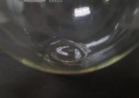
The prepared lidocaine/aspirin ionic liquid was formed by hydrochloride salts of lidocaine and sodium salt aspirins as anionic counterions by an ion-pair reaction. The dichloromethane was used as an organic solvent to extract the final product of a lidocaine/aspirin ionic liquid drug. However, it was completely removed by retaining the liquid in a chemical hood for one day and, subsequently, in a low-pressure chamber for 10 min to remove residual solvent. As a result, dichloromethane was not found in the ionic liquid drug when checked by gas chromatography (Maneewattanapinyo & Suksaeree 2019MANEEWATTANAPINYO P & SUKSAEREE J. 2019. Development and validation of HPLC method for characterization of the acidic and basic drugs prepared into ionic liquid. Lat Am J Pharm 5: 961-967.). These combinations produced an ionic liquid drug at room temperature and were observed as a viscous liquid, believed to be the lidocaine-aspirin ionic liquid drug, after evaporating off the dichloromethane (Figure 1).
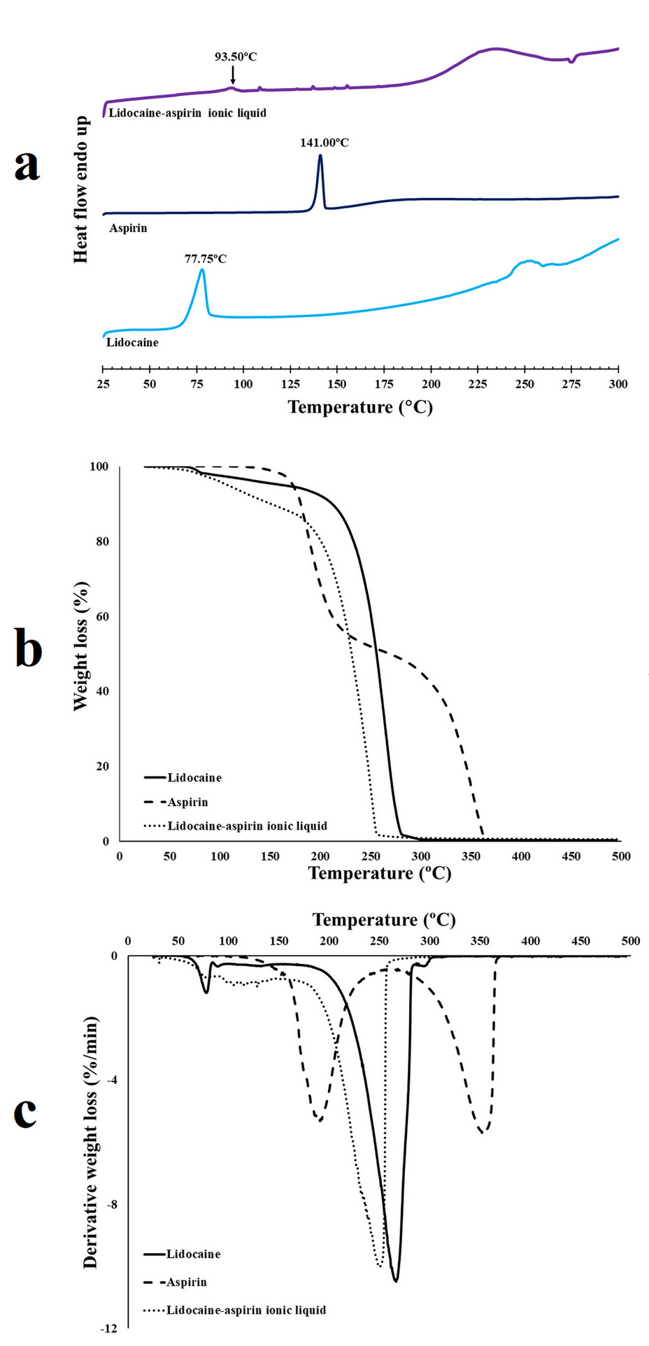
The pure powder of lidocaine and aspirin showed a melting point at 77.75° C and 141° C, respectively (Figure 2a). The lidocaine-aspirin ionic liquid drug showed a single melting point at 93.50° C. The thermal degradation of the pure powders of lidocaine and aspirin, and of the lidocaine-aspirin ionic liquid drug, is shown in Figures 2b and 2c. It was found that the pure powder of aspirin presented two stages of decomposition. The first stage of decomposition, the elimination of the acetic acid and the formation of the salicylic acid, was in the 150-240° C temperature range with Tmax = 166.7°C. The second stage, the elimination of CO2 and the formation of phenol, took place in the 300-360° C temperature range with Tmax= 325.5°C. The pure powder of lidocaine showed one stage of decomposition in the 116-288° C temperature range with Tmax= 240.2°C. The lidocaine-aspirin ionic liquid drug showed one stage of decomposition in the 105-265° C temperature range with Tmax= 220.4°C. Therefore, the prepared lidocaine-aspirin ionic liquid drug showed different thermal behavior from those pure drugs, and it might form an ionic liquid.
Characterization of the lidocaine/aspirin ionic liquid: (a) DSC thermogram, (b) TGA thermogram, and (c) DTG thermogram.
The prepared lidocaine-aspirin ionic liquid drug was mixed in the polymeric solution between PVA and gelatin with glycerin as a plasticizer. The transdermal patch was produced by a freeze-thawed procedure by exposure to ten cycles of freezing for 8 h at -20° C and thawing for 4 h at 25° C. The thickness and weight of the patches were measured at five different positions on the samples. The average thickness and weight variation of blank patches were 253.64±25.36 µm and 122.33±18.41 mg, respectively while the average thickness and weight variation of lidocaine/aspirin ionic liquid-loaded patches were 262±20.96 µm and 126±15.80 mg, respectively.
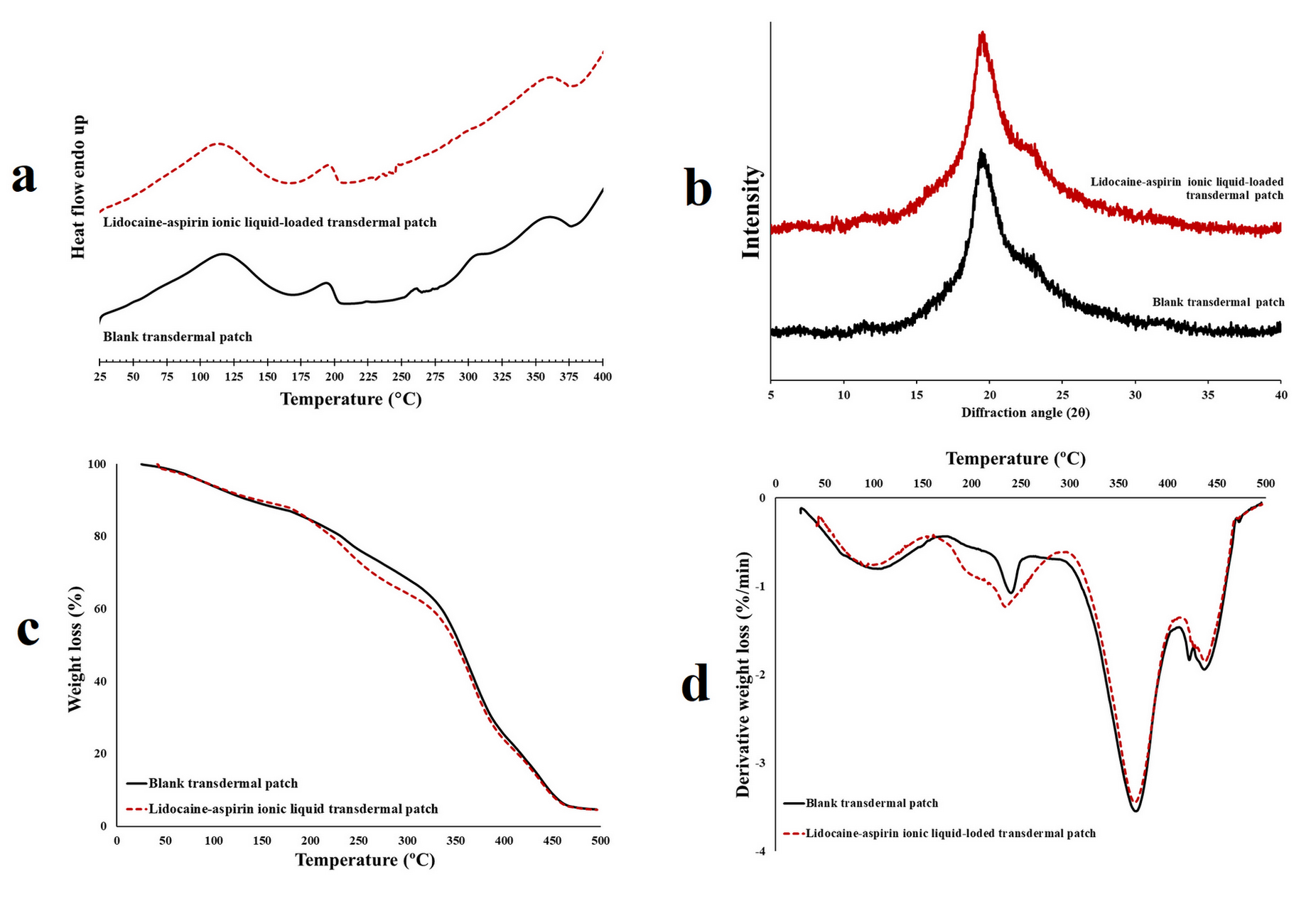
The characterization and cross-section morphology of blank and lidocaine/aspirin ionic liquid-loaded patches is shown in Figures 3 and 4. Figure 3a reveals the DSC thermogram of the blank patch, where the endothermic peak at approximately 115.7° C can be attributed to the evaporation of bound water, representing the energy required to vaporize bound water present in the films. Following this, another endothermic peak observed at about 190° C relates to the melting point of the blank patch (Aluigi et al. 2008ALUIGI A, VINEIS C, CERIA A & TONIN C. 2008. Composite biomaterials from fibre wastes: Characterization of wool–cellulose acetate blends. Compos A 1: 126-132., Nakano et al. 2007NAKANO Y, BIN Y, BANDO M, NAKASHIMA T, OKUNO T, KUROSU H & MATSUO M. 2007. Structure and mechanical properties of chitosan/poly (vinyl alcohol) blend films. Macromol Symp 1: 63-81., Dou et al. 2015DOU Y, ZHANG B, HE M, YIN G, CUI Y & SAVINA IN. 2015. Keratin/polyvinyl alcohol blend films cross-linked by dialdehyde starch and their potential application for drug release. Polymers 3: 580-591.).
Characterization of the lidocaine/aspirin ionic liquid-loaded transdermal patch: (a) DSC thermogram, (b) XRD patterns, (c) TGA thermogram, and (d) DTG thermogram.
SEM images of (a) a blank patch and (b) a lidocaine/aspirin ionic liquid-loaded transdermal patch.
Following previous studies, the XRD profile of pure PVA showed two characteristic peaks centered semi-crystalline at 19.69° and 22° (2θ) (Suksaeree et al. 2017SUKSAEREE J, NAWATHONG N, ANAKKAWEE R & PICHAYAKORN W. 2017. Formulation of polyherbal patches based on polyvinyl alcohol and hydroxypropylmethyl cellulose: characterization and in vitro evaluation. AAPS PharmSciTech 7: 2427-2436., Gonzalez et al. 2016GONZALEZ JS, MARTÍNEZ YN, CASTRO GR & ALVAREZ VA. 2016. Preparation and characterization of polyvinyl alcohol–pectin cryogels containing enrofloxacin and keratinase as potential transdermal delivery device. Adv Mater Lett 8: 640-645.). In this preparation, PVA was mixed with gelatin, using glycerin as a plasticizer and producing film formation by the freeze-thaw procedure. The blank patch showed an amorphous form (Figure 3b). This was due to the gelatin, which may decrease the semi-crystalline state of the PVA by intermolecular hydrogen bonding interaction among PVA chains. As the intensity of the intermolecular H-bonding interaction decreased, it is possible that new hydrogen bridges were established between the PVA and gelatin.
The thermal degradation of the blank patch is shown in Figures 3c and 3d. It was found that the blank patch showed four stages of decomposition. The first stage was the decomposition in the 50-150° C temperature range with Tmax= 100.3°C, manifesting as the elimination of the water or moisture in the patch. The second through fourth stages are the decomposition of all ingredients in the patch, which took place, respectively, in the 220-250° C temperature range with Tmax= 245.5° C; in the 300-400° C temperature range with Tmax= 360.3° C; and in the 410-450° C temperature range with Tmax= 440.2° C.
While the decomposition of the lidocaine-aspirin ionic liquid drug showed in the 105-265° C temperature range with Tmax= 220.4°C (Figures 2b and c), the decomposition of the lidocaine/aspirin ionic liquid-loaded patch was found to broadly peak in the 220-250° C temperature range with Tmax= 245.5°C (Figures 3c and 3d). Thus, the lidocaine-aspirin ionic liquid drug was found in the transdermal patch. Overall, the DSC thermogram, XRD pattern, TGA thermogram, and DTG thermogram of the lidocaine/aspirin ionic liquid-loaded patch were similar to that patch; therefore, the properties of the patch were not changed after loading the prepared lidocaine/aspirin ionic liquid drug. The SEM images showed the physical structure of the cross-section of the blank and lidocaine/aspirin ionic liquid-loaded patches. The formation of some porous, interconnecting networks and a rugged and dense continuous patch (Figure 4) were found.
The lidocaine/aspirin ionic liquid-loaded patch was cut and extracted in distilled water, and the drug was analyzed by an HPLC instrument. It was found that the contents of the drug in the patch were 2.54±0.51 mg/cm2 and 1.90±0.33 mg/cm2 for lidocaine and aspirin, respectively (Figure 5a) which could be calculated as the percentages of drug loading were 101.46±20.53% and 95.15±16.66% for lidocaine and aspirin, respectively. Because the drug contents obtained from five different points on the patch were not significantly different (p>0.05), the results exhibited that the drug could be thoroughly distributed in the matrix of the transdermal patch. Hence, this preparation method was suitable to obtain a transdermal patch with uniformity of the loaded drug.
(a) Drug contents of lidocaine/aspirin ionic liquid-loaded transdermal patches kept at various conditions for three months. In vitro release profiles of (b) lidocaine and (c) aspirin from the lidocaine/aspirin ionic liquid-loaded transdermal patch (n=3). (d) In vitro skin permeation profiles of lidocaine and aspirin from the lidocaine/aspirin ionic liquid-loaded transdermal patch (n=3).
The solubility of the pure drug was determined by equilibrium method (shake-flask technique), known amounts of drug were added in isotonic phosphate buffer solution pH 7.4 (receptor medium) until to reach saturation and the mixture was subjected to agitation for 72 h at 37 °C. It was found that the values of solubility as 43.8±3.56 mM and 76.4±10.93 mM for lidocaine and aspirin, respectively.
The in vitro releases of both lidocaine and aspirin are shown in Figures 5b and 5c. The drug could diffuse from the matrix of the patch and be released into the receptor medium. The cumulative drug release of both lidocaine and aspirin also had high values. The in vitro skin permeation of the drug is shown in Figure 5d. These results are related to the in vitro study. In other words, it was possible to use the gelatin blended with the PVA, using glycerin as a plasticizer, as the base of a transdermal patch prepared by the freeze-thaw procedure because it provides a controlled drug release, implying treatment efficacy.
The appearances of the lidocaine/aspirin ionic liquid-loaded patches after being kept at 4°C at ambient temperature for three months were quite similar compared to the initial preparation. However, the patch stored for three months at 45° C had darkened in color. Figure 5a shows the drug contents of the lidocaine/aspirin ionic liquid-loaded patch after being stored at various conditions; they were not significantly different from the initial condition. The patch kept at 4° C seemed to change slowest and least compared with those stored at ambient temperatures and at 45° C.
The release profiles of the drug from the lidocaine/aspirin ionic liquid-loaded patch after being kept at various temperatures for three months are presented in Figures 5b and 5c. No significant change in the drug’s release profiles was found when the lidocaine/aspirin ionic liquid-loaded patch was kept at 4° C and at ambient temperature; however, an additional decrease in the amount of drug released was observed when the lidocaine/aspirin ionic liquid-loaded patch was kept at higher temperature conditions. The matrix structures of the transdermal patches could be tighter at higher storage temperatures for long durations due to rearrangement of the molecular chain, leading to difficulty in drug extraction and release.
CONCLUSIONS
A gelatin/polyvinyl alcohol composite film-loaded lidocaine/aspirin ionic liquid drug can be prepared by a freeze-thawed procedure with a high feasibility to be used in pharmaceutical transdermal patches. In this study, a lidocaine/aspirin ionic liquid drug was prepared by an ion-pair reaction between hydrochloride salts of lidocaine and sodium salt aspirins as anionic counterions, which exhibited a significant change in their thermal properties when compared to the pure drugs. The properties of the obtained lidocaine/aspirin ionic liquid-loaded patch were similar to the blank patch; thus, the lidocaine/aspirin ionic liquid drug that was loaded did not change the patch’s properties. In addition, the decomposition of the lidocaine/aspirin ionic liquid drug was obviously found in the patch. The patch could control the release behavior of the drug; however, it should be kept at 4° C and ambient temperature to protect it from a decrease in the amount of drug released.
ACKNOWLEGMENTS
This work was supported by the Research Institute of Rangsit University for financial support (Grant No.6/2561).
REFERENCES
- ALUIGI A, VINEIS C, CERIA A & TONIN C. 2008. Composite biomaterials from fibre wastes: Characterization of wool–cellulose acetate blends. Compos A 1: 126-132.
- BERTON P, DI BONA KR, YANCEY D, RIZVI SAA, GRAY M, GURAU G, SHAMSHINA JL, RASCO JF & ROGERS RD. 2017. Transdermal bioavailability in rats of lidocaine in the forms of ionic liquids, salts, and deep eutectic. ACS Med Chem Lett 5: 498-503.
- BICA K, RIJKSEN C, NIEUWENHUYZEN M & ROGERS RD. 2010. In search of pure liquid salt forms of aspirin: ionic liquid approaches with acetylsalicylic acid and salicylic acid. Phys Chem Chem Phys 8: 2011-2017.
- CAMPOCCIA D, MONTANARO L & ARCIOLA CR. 2013. A review of the biomaterials technologies for infection-resistant surfaces. Biomaterials 34: 8533-8554.
- CHEN MC, LING MH, LAI KY & PRAMUDITYO E. 2012. Chitosan microneedle patches for sustained transdermal delivery of macromolecules. Biomacromolecules 12: 4022-4031.
- DOBLER D, SCHMIDTS T, KLINGENHÖFER I & RUNKEL F. 2013. Ionic liquids as ingredients in topical drug delivery systems. Int J Pharm 1: 620-627.
- DOU Y, ZHANG B, HE M, YIN G, CUI Y & SAVINA IN. 2015. Keratin/polyvinyl alcohol blend films cross-linked by dialdehyde starch and their potential application for drug release. Polymers 3: 580-591.
- FERRAZ R, BRANCO LC, PRUDÊNCIO C, NORONHA JP & PETROVSKI Ž. 2011. Ionic liquids as active pharmaceutical ingredients. ChemMedChem 6: 975-985.
- GONZALEZ JS & ALVAREZ VA. 2014. Mechanical properties of polyvinylalcohol/hydroxyapatite cryogel as potential artificial cartilage. J Mech Behav Biomed Mater, p. 47-56.
- GONZALEZ JS, MARTÍNEZ YN, CASTRO GR & ALVAREZ VA. 2016. Preparation and characterization of polyvinyl alcohol–pectin cryogels containing enrofloxacin and keratinase as potential transdermal delivery device. Adv Mater Lett 8: 640-645.
- HASSAN CM & PEPPAS NA. 2000a. Structure and applications of poly(vinyl alcohol) hydrogels produced by conventional crosslinking or by freezing/thawing methods. In: Chang JY et al. (Eds). Biopolymers-PVA hydrogels, anionic polymerisation nanocomposites, Berlin Heidelberg: Springer-Verlag, New York, USA, p. 37-65.
- HASSAN CM & PEPPAS NA. 2000b. Structure and morphology of freeze/thawed PVA hydrogels. Macromolecules 7: 2472-2479.
- HOUGH WL ET AL. 2007. The third evolution of ionic liquids: active pharmaceutical ingredients. New J Chem 8: 1429-1436.
- HUDDLESTON JG, VISSER AE, REICHERT WM, WILLAUER HD, BROKER GA & ROGERS RD. 2001. Characterization and comparison of hydrophilic and hydrophobic room temperature ionic liquids incorporating the imidazolium cation. Green Chem 4: 156-164.
- JAITELY V, KARATAS A & FLORENCE AT. 2008. Water-immiscible room temperature ionic liquids (RTILs) as drug reservoirs for controlled release. Int J Pharm 1: 168-173.
- KATARIA K, GUPTA A, RATH G, MATHUR RB & DHAKATE SR. 2014. In vivo wound healing performance of drug loaded electrospun composite nanofibers transdermal patch. Int J Pharm 1: 102-110.
- MACFARLANE DR, PRINGLE JM, JOHANSSON KM, FORSYTH SA & FORSYTH M. 2006. Lewis base ionic liquids. Chem Commun 18: 1905-1917.
- MACFARLANE DR & SEDDON KR. 2007. Ionic liquids progress on the fundamental issues. Aust J Chem 1: 3-5.
- MANEEWATTANAPINYO P & SUKSAEREE J. 2019. Development and validation of HPLC method for characterization of the acidic and basic drugs prepared into ionic liquid. Lat Am J Pharm 5: 961-967.
- MARTÍNEZ YN, CAVELLO I, HOURS R, CAVALITTO S & CASTRO GR. 2013. Immobilized keratinase and enrofloxacin loaded on pectin PVA cryogel patches for antimicrobial treatment. Bioresour Technol, p. 280-284.
- MIWA Y, HAMAMOTO H & ISHIDA T. 2016. Lidocaine self-sacrificially improves the skin permeation of the acidic and poorly water-soluble drug etodolac via its transformation into an ionic liquid. Eur J Pharm Biopharm, p. 92-100.
- MIZUUCHI H, JAITELY V, MURDAN S & FLORENCE AT. 2008. Room temperature ionic liquids and their mixtures: Potential pharmaceutical solvents. Eur J Pharm Sci 4: 326-331.
- MONIRUZZAMAN M, TAMURA M, TAHARA Y, KAMIYA N & GOTO M. 2010. Ionic liquid-in-oil microemulsion as a potential carrier of sparingly soluble drug: Characterization and cytotoxicity evaluation. Int J Pharm 1: 243-250.
- NAKANO Y, BIN Y, BANDO M, NAKASHIMA T, OKUNO T, KUROSU H & MATSUO M. 2007. Structure and mechanical properties of chitosan/poly (vinyl alcohol) blend films. Macromol Symp 1: 63-81.
- PARK HJ & PRAUSNITZ MR. 2015. Lidocaine-ibuprofen ionic liquid for dermal anesthesia. AIChE J 9: 2732-2738.
- PERNAK J, SOBASZKIEWICZ K & MIRSKA I. 2003. Anti-microbial activities of ionic liquids. Green Chem 1: 52-56.
- PICHAYAKORN W, SUKSAEREE J, BOONME P, AMNUAIKIT T, TAWEEPREDA W & RITTHIDEJ GC. 2012. Nicotine transdermal patches using polymeric natural rubber as the matrix controlling system: Effect of polymer and plasticizer blends. J Membr Sci, p. 81-90.
- SOWMIAH S, SRINIVASADESIKAN V, TSENG MC & CHU YH. 2009. On the chemical stabilities of ionic liquids. Molecules 14(9): 3780-3813.
- SUKSAEREE J, CHAROENCHAI L, MONTON C, CHUSUT T, SAKUNPAK A, PICHAYAKORN W & BOONME P. 2013. Preparation of a pseudolatex-membrane for ketoprofen transdermal drug delivery systems. Ind Eng Chem Res 45: 15847-15854.
- SUKSAEREE J, NAWATHONG N, ANAKKAWEE R & PICHAYAKORN W. 2017. Formulation of polyherbal patches based on polyvinyl alcohol and hydroxypropylmethyl cellulose: characterization and in vitro evaluation. AAPS PharmSciTech 7: 2427-2436.
- TOKUDA H, HAYAMIZU K, ISHII K, SUSAN MABH & WATANABE M. 2005. Physicochemical properties and structures of room temperature ionic liquids. 2. Variation of alkyl chain length in imidazolium cation. J Phys Chem B 13: 6103-6110.
- TOKUDA H, ISHII K, SUSAN MABH, TSUZUKI S, HAYAMIZU K & WATANABE M. 2006. Physicochemical properties and structures of room-temperature ionic liquids. 3. Variation of cationic structures. J Phys Chem B 6: 2833-2839.
- TORIMOTO T, TSUDA T, OKAZAKI K-I & KUWABATA S. 2010. New frontiers in materials science opened by ionic liquids. Adv Mater 11: 1196-1221.
- ZAKREWSKY M ET AL. 2014. Ionic liquids as a class of materials for transdermal delivery and pathogen neutralization. Natl Acad Sci 37: 13313-13318.
- ZHANG D, WANG HJ, CUI XM & WANG CX. 2017. Evaluations of imidazolium ionic liquids as novel skin permeation enhancers for drug transdermal delivery. Pharm Dev Technol 4: 511-520.
- ZHANG Y, WANG D, GAO M, XU B, ZHU J, YU W, LIU D & JIANG G. 2018. Separable microneedles for near-infrared light-triggered transdermal delivery of metformin in diabetic rats. ACS Biomater Sci Eng 8: 2879-2888.
Publication Dates
-
Publication in this collection
20 July 2020 -
Date of issue
2020
History
-
Received
9 Sept 2019 -
Accepted
22 Nov 2019


