Testing the occlusal relations between isolated upper and lower molars of an Eocene metatherian (Mammalia): The case of Xenocynus crypticus
 Abstract
Xenocynus crypticus is a metatherian from the Itaboraí Basin (early Eocene) in the state of Rio de Janeiro, Brazil. While this taxon was initially described based on a single upper molar and three isolated lower molars, the occlusal relations between these teeth have never been previously analyzed. In this study, the upper and lower molars assigned to this taxon, along with specimen AMNH 49805, were included in a scatterplot and two principal component analyses (PCA), assessing several morphometric parameters that were diagnostic of this taxon. We inferred the occlusal relationships between the assigned molars to assess their morpho-occlusal compatibility. Our analyses have demonstrated that the Xenocynus crypticus molars are morphometrically distinct from other sympatric taxa of similar size, with m1s having proportionally shorter talonids. Furthermore, the upper and lower molars of X. crypticus are occlusally compatible, exhibiting similar inferred proportions in the length and width of the talonid. These findings support the taxonomic validity of X. crypticus and an association of the lower molars with this species. Additionally, our results indicate that PCAs can reliably assess morphometric similarity between isolated upper and lower molars in metatherians, offering valuable insights for taxonomic studies on fragmentary dental-based taxa.
Key words
Didelphoidea; Itaboraian SALMA; PCA; Sternbergiidae; teeth
INTRODUCTION
The Itaboraí Basin, located in the municipality of Itaboraí, in the state of Rio de Janeiro, in Southeastern Brazil (22º 50’ 20” S; 42º 52’ 30” W), preserves one of the most diverse Cenozoic metatherian faunas in South America. Dated to the early Eocene (Woodburne et al. 2014), this fauna includes a minimum of 29 genera and 44 species (Carneiro et al. 2024a), despite originating from one of the smallest sedimentary basins in Brazil (Bergqvist et al. 2009). Metatherian specimens from this locality are primarily represented by isolated teeth, incomplete jaws, maxillae (Paula Couto 1949, 1952a, b, 1961, 1962, 1970, Marshall 1987, Carneiro & Oliveira 2023), isolated petrosals (Ladevèze 2004, 2007, Ladevèze & Muizon 2010), postcranial elements (Szalay 1994, Szalay & Sargis 2001), and skulls (Simpson 1947, Paula Couto 1952a, b).
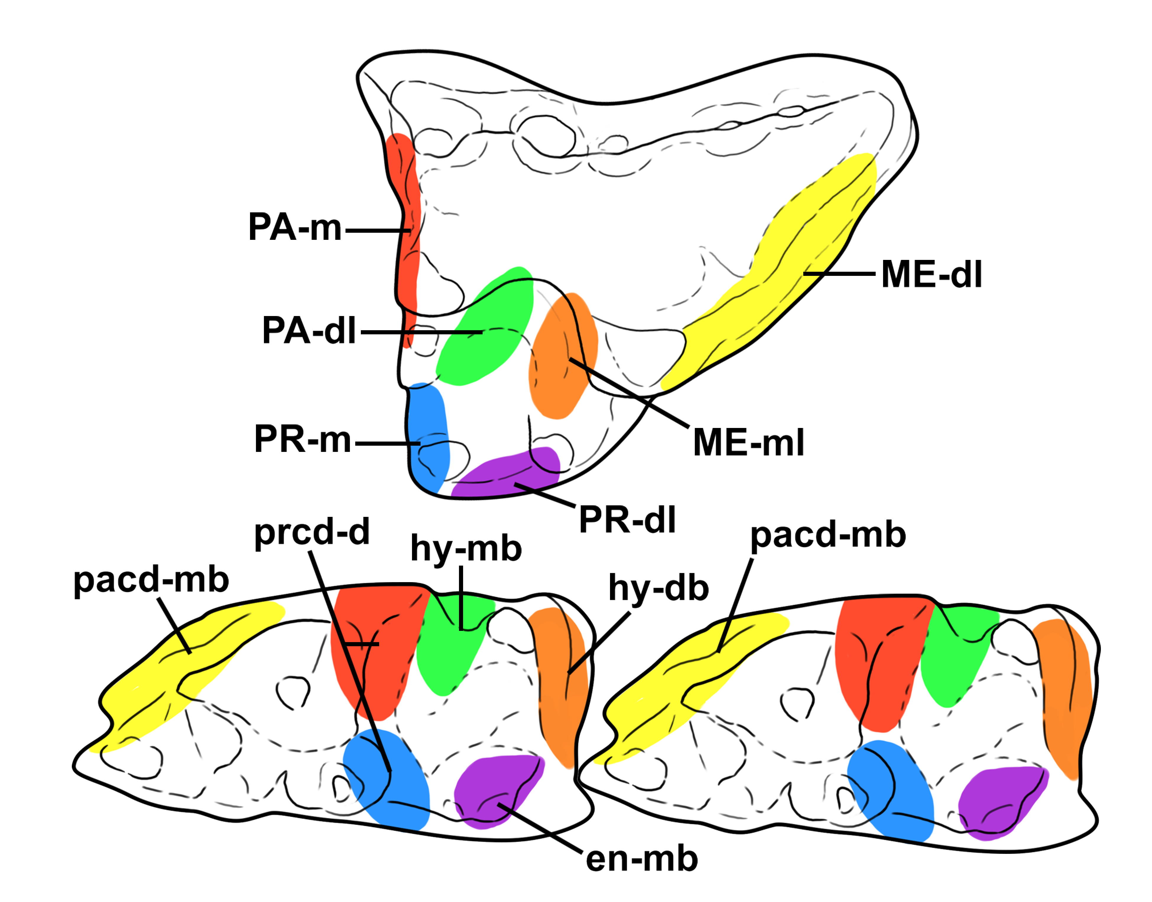
Mammalian fossils in Itaboraí have mostly been recovered from fissure fills within the basin (Bergqvist et al. 2009, 2011), often rendering them fragmentary, disarticulated, and incomplete. The taphonomy of these specimens led Paula Couto (1949) and Bergqvist et al. (2011) to conclude that most specimens in this locality were subaerially exposed and hydrologically transported before their final burial. This taphonomic bias complicates associations between isolated specimens, especially in efforts to assign teeth, isolated petrosals, and postcranial elements to a single taxon (see Lorente 2016 for a discussion on the associations of tarsal elements to Litopterna presented by Cifelli 1983). However, isolated molars can be more readily associated due to their complementary occlusal relationship (Marshall 1987, Oliveira & Goin 2011). During occlusion in taxa with basically ortal (“vertical”) masticatory movements, the protocone of the upper molar occludes between the distal wall of the metaconid and the mesiolabial wall of the entoconid of the antagonistic lower molar (Crompton & Hiiemae 1970, Davis 2011, N. Zimicz, unpublished data, Williamson et al. 2014, Carneiro et al. 2024c) (Figure 1). The hypoconid apex occludes against the centrocrista between the paracone and metacone (see Crompton & Hiiemae 1970; Figure 1), and the postmetacrista occludes with the paracristid of the posterior molar (i.e., the postmetacrista of M1 occludes with the paracristid of m2; see Crompton & Hiiemae 1970; Figure 1). Consequently, the metrics between antagonistic molars (i.e., upper and lower) should be similar. Thus, the length of the protocone and the talonid basin should be similar, and the distance between the apex of the protocone and the centrocrista should be similar to the width of the talonid (Figure 1).

Among the metatherians from the Itaboraí Basin, Xenocynus crypticus Carneiro et al. 2024a is a relatively large taxon (between 1 kg and 3.3 kg sensu Carneiro et al. 2024a) with an inferred generalized carnivorous diet (insectivory-carnivory sensu Rangel et al. 2023a; see Carneiro et al. 2024a) and uncertain taxonomic affinities (possibly a sparassodont). This taxon was described based on an incomplete maxilla with an M2 (MCT.M.2832, holotype) from the Museu de Ciências da Terra (MCTer) in Rio de Janeiro, Brazil and three isolated lower molars (MCN-PV 1793, MCN-PV 1813, two m1s, and MCN-PV 1788, an m2) from the Museu de Ciências Naturais (MCN-PV), in Porto Alegre, Brazil (see Carneiro et al. 2024a). Carneiro et al. (2024a) were aware of another possible specimen of X. crypticus (AMNH 49805 [former MN 1334-V]), an isolated M3. However, Carneiro et al. (2024a) did not examine the occlusal relations between the M2 of the holotype of this species and the referred isolated lower molars (MCN-PV 1793, MCN-PV 1813, and MCN-PV 1788). Instead, they based their conclusions on these specimens’ similar size and compatible morphology.
In this study, we assess whether the lower molars assigned to Xenocynus crypticus by Carneiro et al. (2024a) can be distinguished from other lower molars from the Itaboraí Basin with similar dimensions and if these lower molars are occlusally/morphometrically compatible with the upper molars of X. crypticus. We also examine whether the putative new specimen of Xenocynus crypticus, AMNH 49805 (former MN 1334-V), exhibits a morphology consistent with referral to this species.
Anatomical abbreviations. cent, centrocrista; hyp, hypoconid; M/m, upper/lower molars, with numbers indicating their locus (M/m1 to 4); met, metacone; metcr, metacristid (i.e., premetacristid + postprotocristid sensu Carneiro et al. 2024a); par, paracone; pas, parastyle; PM, postmetacrista; PPA, preparacrista; pro, protocone; prob, protoconal basin; prof, protofossa (i.e., the space between the paracone and the metacone); parcr, paracristid; tal, talonid; tri, trigonid.
Abbreviations for dental measurements. A, area (L x W); L, length (mesiodistal axis); ra, relative area (=Atal/Atri); rl, relative length (=Ltal/Ltri); W, width (labiolingual axis).
Institutional abbreviations. AMNH, American Museum of Natural History, New York, NY, USA; MCN-PV, Coleção de Paleovertebrados do Museu de Ciências Naturais da Secretaria de Meio Ambiente (SEMA; former Fundação Zoobotânica do Rio Grande do Sul), Porto Alegre, RS, Brazil; MCT, Museu de Ciências da Terra, Serviço Geológico do Brasil (MCTer-SGB), Rio de Janeiro, RJ, Brazil; MN, Museu Nacional (MN/UFRJ), Universidade Federal do Rio de Janeiro, Rio de Janeiro, RJ, Brazil; UFRJ-DG, Coleção de Macrofósseis do Departamento de Geologia da Universidade Federal do Rio de Janeiro, Rio de Janeiro, RJ, Brazil.
Other abbreviations. SALMA, South American Land Mammal Age; SEM, scanning electronic microscope.
MATERIALS AND METHODS
All the studied specimens are from the Itaboraí Basin, in the municipality of Itaboraí, Rio de Janeiro, Southeastern Brazil, at coordinates 22° 45’ 9.9144” S, 42° 51’ 53.5536” W (Figure 2a). These materials are housed in the collections of the AMNH, MCN-PV, MCTer (former DGM), MN/UFRJ, and UFRJ-DG. The fauna from this locality has been considered early Eocene (sensu Woodburne et al. 2014) or late Paleocene to early Eocene (Bergqvist et al. 2009) in age (Figure 2b). For further information on the geology and fauna of the Itaboraí Basin, see Bergqvist et al. (2009), and Rangel et al. ( 2023a).

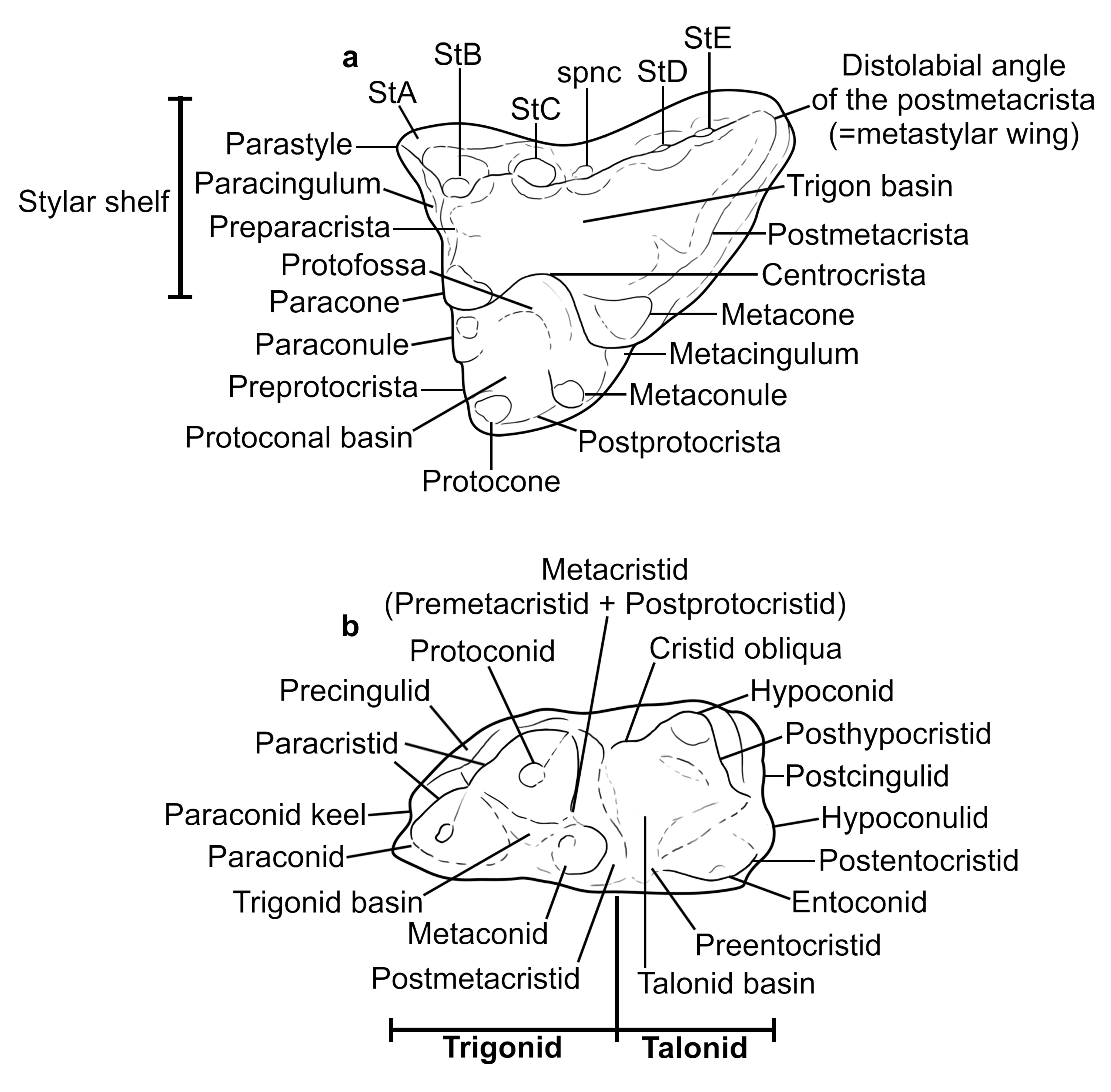
The nomenclature of the considered dental structures (cusps, crests) is based on Carneiro et al. (2024a), and it is represented below in Figure 3.

Occlusal relations between antagonistic upper and lower molars of a metatherian. Wear facets nomenclature is based on Schultz et al. (2020). Similar colors in upper and lower molars indicate occlusally compatible facets/structures. d, distal; db, distobuccal; dl, distolingual; en, entoconid; hy, hypoconid; m, lower molar; mb, mesiobuccal; ME, metacone; ml, mesiolingual; PA, paracone; pacd, paracristid; PR, protocone; prcd, protocristid (=metacristid).
A scatterplot and two principal component analyses (PCAs) were conducted to distinguish the molar morphology (based on specific morphometric parameters) of Xenocynus crypticus and other similarly sized metatherians from the Itaboraí basin, including Didelphopsis cabrerai Paula Couto 1952a, Didelphopsis sp. (sensu Carneiro & Oliveira 2023), Itaboraidelphys camposi Marshall & Muizon 1984; and Patene simpsoni Paula Couto 1952b. The included specimens were identified by their molar locus (M/m1, M/m2, M/m3, and/or m4).
The scatterplot was constructed considering two variables from the lower molars: lower molar length (Lm) and the relative size of the talonid (ra = Atal/Atri). Carneiro et al. (2024a) noted that the proportionally smaller Xenocynus crypticus talonids were distinctive compared to the similar-sized metatherians Itaboraidelphys camposi and Didelphopsis cabrerai.
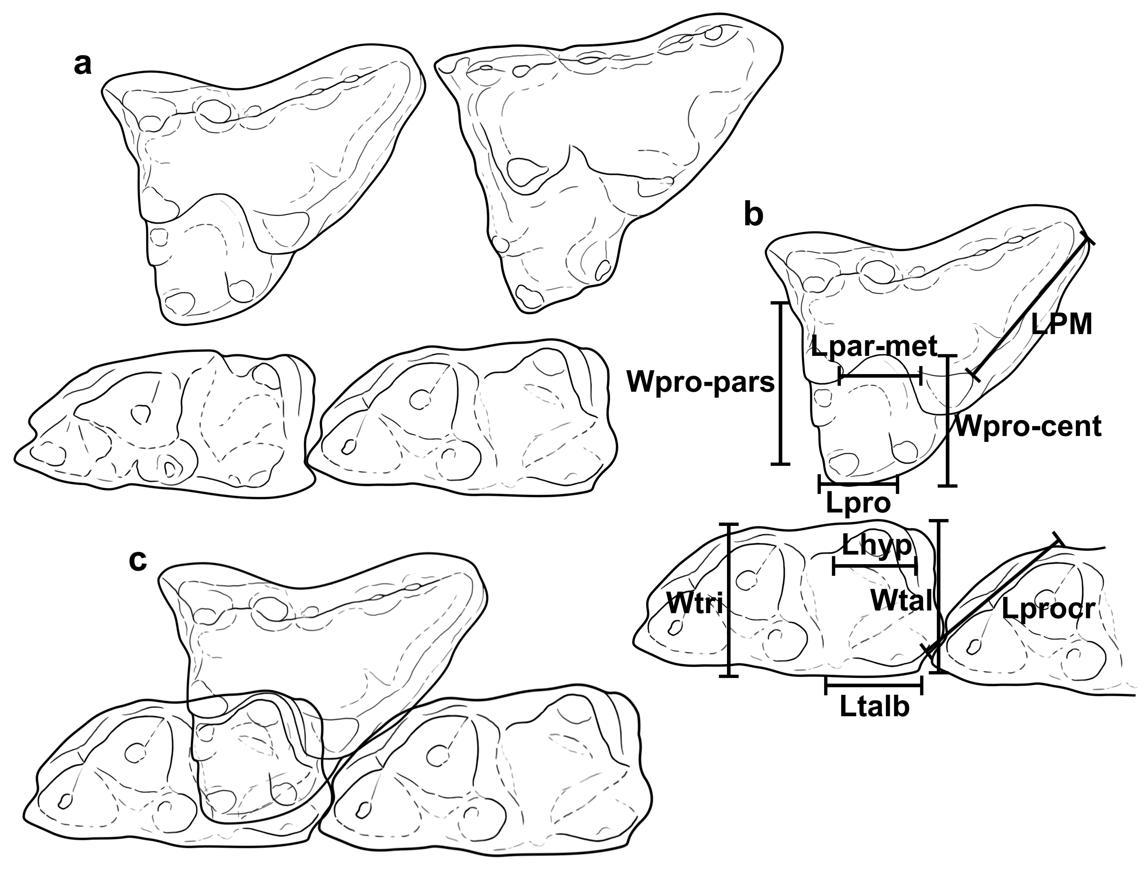
The first PCA considered four variables (PPA/PM = relative length between the preparacrista and the postmetacrista; rlprof = relative distance between the apices of the paracone and the metacone [Lpar-met/LM]; rlpro = relative length of the protocone [Lpro/LM]; and rlprob = relative width between the protocone-centrocrista to the protocone-parastyle [Wpro-cent/Wpro-pas]; see Figure 4). These dental variables are crucial because they provide occlusal complementary parameters to those of the lower molars (e.g., the talonid length, relative width between the talonid to the trigonid, relative length, and angulation of the paracristid). These dental variables help support morphometric patterns of occlusal compatibility between the upper and lower molars assigned to Xenocynus crypticus.

The Itaboraí fauna: locality and biochron. a) Map showing the region and the coordinates of the Itaboraí Basin, municipality of Itaboraí, Rio de Janeiro, Brazil (22° 45’ 9’’ S; 42° 51’ 53’’ W). b) The stratigraphic relationships of the Itaboraian SALMA with other Paleogene SALMAs are indicated. A plausible age range for the Itaboraian SALMA is highlighted in grey (sensu Carneiro et al. 2024a). PETM, Paleocene-Eocene Thermal Maximum; SALMA, South American Land Mammal Age. The scale bar represents 1000 Km.
The second PCA considered three correspondent variables for the upper and lower molars: rlhyp (=rlprof), relative length of the hypoconid (Lhyp/Lm); RLtal/Lm (=rlpro), relative length of the talonid to the length of the lower molar; RtalW/L (=rlprob), relative talonid width to length (see Figure 4). These complementary variables enable the inclusion of both the upper and lower molars in the same analysis, allowing the examination of similar morphometric parameters between antagonistic molars of a locus.
The database and R script are presented as supplementary material (Tables SI-VI, Figures S1-S2) and Appendix. All analyses were performed in R software (R Core Team 2013).
RESULTS
Biplot analysis (Lower molars)
Considering the included taxa, the lower molars of Xenocynus crypticus are similar to the m1-2s of Didelphopsis cabrerai and Itaboraidelphys camposi in length (Lm). However, they differ by having smaller talonids (ra). The lower molars of X. crypticus reveal similar ra values to the m4s of Itaboraidelphys camposi (Figure 5).

Upper (a) and lower (b) molar structures (cusps and crests) nomenclature considered by Carneiro et al. (2024a) and adhered to in this study. St, stylar cusp, with the letters (a, b, c, d, e) indicating its placement from the parastyle to the distolabial angle of the postmetacrista/metastylar wing.
First PCA (Upper molars)
PCA1 explains 47.5% of the total variation, whereas PCA2 explains 32.1% (Figure 6), with the rlprof, rlprob, and rlpro the variables that better explain the data variation of PC1 (about 41%, 28%, and 26%, respectively) and PPA/PM and rlprob variables better explaining the data variation of the PC2 (about 67% and 25%; see Supplementary Material - Figure S1). The M3s have longer preparacristae (PPA/PM) than the M1-2, as indicated by the higher scores of the M3s compared to the M1-2s. The M1s exhibit less variation in width between the protocone-centrocrista and the protocone-parastyle (rlprob), resulting in a lower placement than the M2-3s. These results indicate that the mesial lobe of the stylar shelf increases in width from M1 to M3, resulting in a longer preparacrista and a greater distance between the protocone and the parastyle on M3 than M1.

Occlusal and metric relations between antagonistic upper and lower molars of Xenocynus crypticus Carneiro et al. 2024a. a) schematic drawings of known upper and lower molars loci of X. crypticus (MCT.M.2382 [holotype], showing the left M2; AMNH 49805 [former MN 1334-V], a right M2 [reversed]; MCN-PV 1813, a right m1; MCN-PV 1788, a right m1); b) specimens MCT.M.2382 and MCN-PV 1788, with similar numbers indicating occlusally compatible structures between antagonistic molars; c) occlusal relation between M/m in X. crypticus. cent, centrocrista; L, length; hyp, hypoconid; met, metacone; par, paracone; pars, parastyle; PM, postmetacrista; pro, protocone; parcr, paracristid; tal, talonid; talb, talonid basin; tri, trigonid; W, width.
In the first PCA, Patene simpsoni (blue convex hull) and Xenocynus crypticus (pink convex hull) differ from all other examined metatherians by displaying closer paracones and metacones (low values of rlprof), shorter protocones (low values of rlpro), and a greater width between the protocone and the parastyle compared to the width between the protocone-centrocrista (low values of rlprob). In contrast, sternbergiids (Didelphopsis spp. [red and yellow convex hulls] and Itaboraidelphys camposi [green convex hull]), especially Didelphopsis sp., display a more separated paracone and metacone (higher values of rlprof), longer protocones (rlpro), and a broader protocone-centrocrista compared to that between the protocone and the parastyle. Furthermore, the morphometric parameters displayed by the upper molars of Xenocynus crypticus (MCT.M.2832 [holotype] and AMNH 49805 [former MN 1334-V]) are more similar to those of the M2 of the holotype of Patene simpsoni (MN 1331-V).
Second PCA (Upper and lower molars)
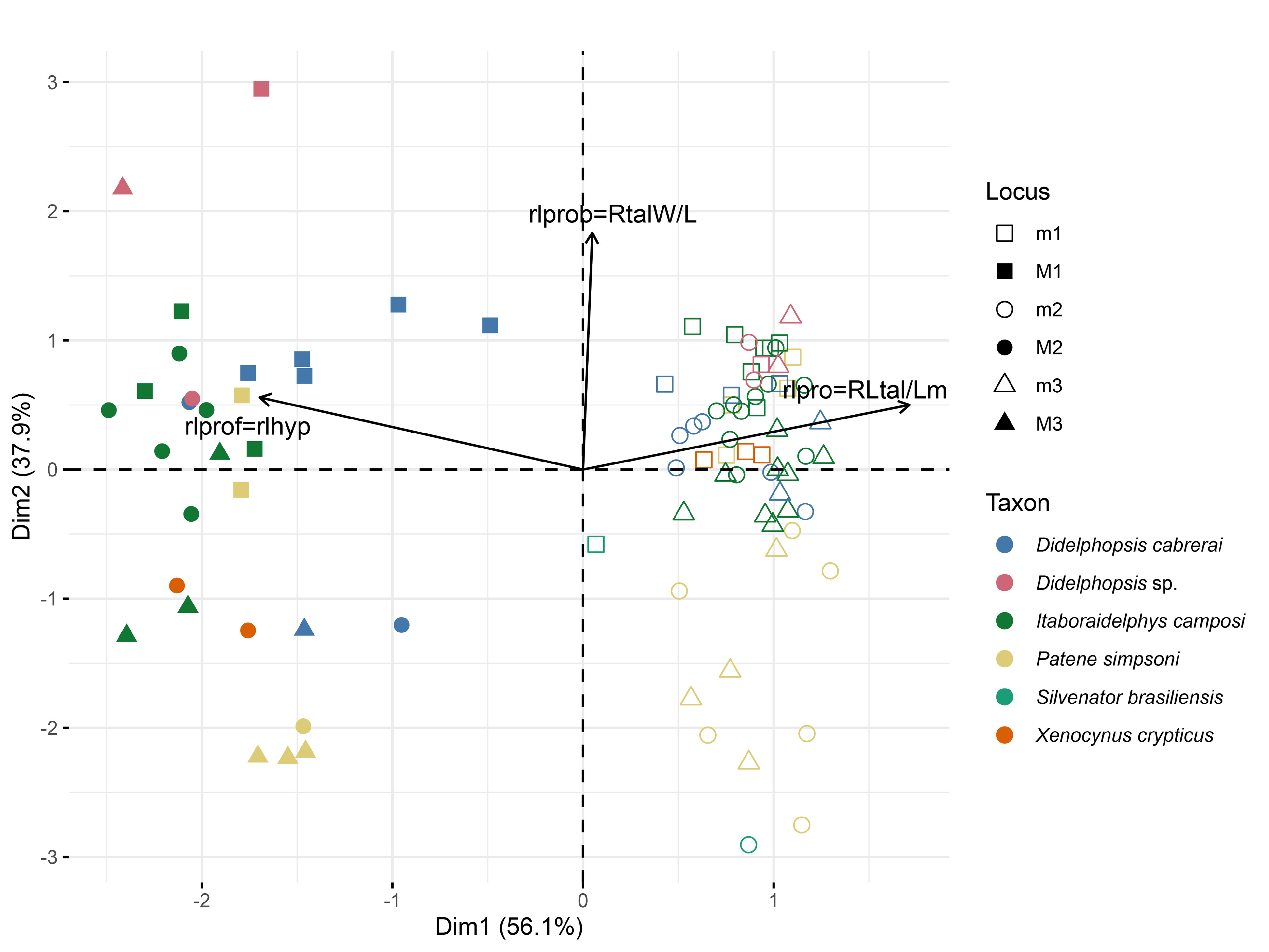
PCA1 explains 56.1% of total variation, whereas PC2 explains 37.9% (Figure 7), with the rlpro=RLtal/Lm and rlprof=rlhyp the variables that better explain the data variation of PC1 (about 50% and 49%, respectively) and rlprob=rltalW/L the variable better explaining the data variation of the PC2 (about 86%; Figure S2). This analysis positions the upper and lower molars in a “mirrored” arrangement, in which the lower molars are located on the right side of the morphospace and the upper molars on the left side of the morphospace. The distances between the paracone and metacone of the upper molars are proportionally longer (occlusal complementary to the length of the hypoconid; rlprof=rlhyp). The greater distance between the paracone and metacone, compared to the length of the hypoconid, may be interpreted as an adaptation of tribosphenic mammals, facilitating the movement of the hypoconid in the protofossa (i.e., the space between the paracone and the metacone, labially limited by the centrocrista) during masticatory movements (see Crompton & Hiiemae 1970).

Scatterplot of two morphometric and morphofunctional indices based on lower molar measurements for Didelphopsis cabrerai Paula Couto 1952a, Didelphopsis sp. (see Carneiro & Oliveira 2023), Itaboraidelphys camposi Marshall & Muizon 1984; Patene simpsoni Paula Couto 1952b; and Xenocynus crypticus Carneiro et al. 2024a.
Similarly, the longer talonid compared to the protocone (rlpro=RLtal/Lm) is a consequence of the masticatory movements of the jaw, as the lower molar structures “slide” between/against the upper molar features during mastication (see Crompton & Hiiemae 1970). Consequently, the morphometric parameters recovered here replicate the expected occlusal relationships between the upper and lower molars of metatherians
In general, the M/m1-2s exhibit higher PCA scores than the M/m3s (except for Didelphopsis sp.) due to the smaller differences in width between the protocone-centrocrista and the protocone-parastyle (complementary to talonids that are subequal to or broader than the trigonids in the lower molars; rlprob=RtalW/L). The M3s display greater distances between the protocone-parastyle (=trigonid width) than the protocone-centrocrista (=talonid width), with antagonistic m3s having broader trigonids than the talonids. This pattern is indicated by their lower scores in the PCA2. The M1-2s display a relatively more separated paracone and metacone, with antagonistic lower molars (m1-2) having longer hypoconids (rlprof=rlhyp) than the M/m3s. This pattern is observed by the former (M/m1-2), which has more negative values in PCA1 than the M/m3s.
As mentioned for other taxa, the lower molars of Xenocynus crypticus exhibit higher scores in PCA2 than the upper molars of this species. The lower molars belong to a different locus than the upper molars since lower molars have broader talonids than trigonids, but inferred proportions of the upper molars suggest narrower talonids than trigonids for their antagonistic lower molars (see discussion below).
DISCUSSION
Comparisons between the lower molars of Xenocynus, Didelphopsis, Itaboraidelphys, and Patene
The lower molars of Xenocynus crypticus (MCN-PV 1788, MCN-PV 1793, and MCN-PV 1813) cannot be assigned to Didelphopsis or Itaboraidelphys due to several distinct features: the paraconid and metaconid on m1-2 are separated and/or not in contact (sensu Muizon & Ladevèze 2020), whereas they are in contact in Didelphopsis and Itaboraidelphys on m1-2; the paraconid is larger than the metaconid on m2 (the paraconid is always smaller than the metaconid on m1-4 in Didelphopsis and Itaboraidelphys); and the metaconid is distal to the protoconid on m2 (m2 with metaconid and protoconid transversally aligned in Didelphopsis and Itaboraidelphys). For additional dental features that distinguish Xenocynus from Didelphopsis and Itaboraidelphys, see Carneiro et al. (2024a).
The lower molars of Xenocynus crypticus (m1s), in terms of the talonid size (ra), are mostly similar to the size of the m4 of Itaboraidelphys camposi (see Figure 5). This finding suggests that the m1s of X. crypticus have smaller talonids than those of the sympatric taxa of comparable size (Didelphopsis cabrerai and Itaboraidelphys camposi). Therefore, these lower molars are outside the natural individual variation in Didelphopsis and Itaboraidelphys and cannot be assigned to these two taxa.
The upper molars of Xenocynus crypticus display shorter protocones (rlpro) than those of Itaboraidelphys camposi and Didelphopsis cabrerai (see Table II and Figures 6-7). This observation would require X. crypticus to reveal proportionally shorter talonids to maintain proper occlusion between the opposing teeth (see occlusal relations in Figures 1 and 4). Consequently, the MCN-PV lower molars cannot be assigned to Didelphopsis or Itaboraidelphys.
Considered metrics for the lower molars of Didelphopsis cabrerai Paula Couto 1952a, Didelphopsis sp. (sensu Carneiro & Oliveira 2023), Itaboraidelphys camposi Marshall & Muizon 1984; Patene simpsoni Paula Couto 1952b; and Xenocynus crypticus Carneiro et al. 2024a from the Itaboraí Basin. Holotypes are in bold. The minimum and maximum values are presented. L, length; m, lower molar, with numbers indicating its locus (M/m1 to 4); ra, relative area (Atal/Atri = [Ltal x Wtal] / [Ltri x Wtri]).
Considered metrics for the upper molars of Didelphopsis cabrerai Paula Couto 1952a, Didelphopsis sp. (sensu Carneiro & Oliveira 2023), Itaboraidelphys camposi Marshall & Muizon 1984; Patene simpsoni Paula Couto 1952b, and Xenocynus crypticus Carneiro et al. 2024a from the Itaboraí Basin. Holotypes are in bold. M, upper molar, with numbers indicating its locus (M/m1 to 3); PM, postmetacrista; PPA, preparacrista; PPA/PM, relative length between the preparacrista by the postmetacrista; pro, protocone; prob, protoconal basin; prof, distance between the paracone and metacone (=protofossa); rlpro = relative length of the protocone (Lpro/LM); rlprob = relative width between the protocone-centrocrista by protocone-parastyle (Wpro-cent/Wpro-pas); rlprof = relative distance between the paracone and metacone (Lpar-met/LM).
The relative size of the protocones and talonids in Xenocynus crypticus (ra; see Table I) and rlpro (see Table II) are more similar to those of the second upper and lower molars of Patene simpsoni. In P. simpsoni, the talonid is subequal to slightly broader than the trigonid only on m1 (see Rangel et al. 2019).
Initially, the specimens of Xenocynus crypticus were assigned as the M/m1s of P. simpsoni (e.g., AMNH and MCN-PV’s specimens), with morphological differences (e.g., smaller talonids and flattened edges of the paracone and metacone) representing the natural morphological variability of the latter. However, the lower molars of Xenocynus crypticus differ from Patene simpsoni due to their smaller talonids (see ra in Table I and Figure 5), which implies proportionally shorter protocones on M1 (smaller talonids on m1) for X. crypticus. This pattern supports the initial observation of Carneiro et al. (2024a), who concluded that X. crypticus has smaller talonids compared to sympatric similarly sized taxa (Didelphopsis spp., Itaboraidelphys camposi, Patene simpsoni, Protodidelphis vanzolinii, and Robertbutleria mastodontoidea). Consequently, specimens MCN-PV 1788, MCN-PV 1793, and MCN-PV 1813 are outside the natural range of the individual variation observed in P. simpsoni.
In short, the lower molars assigned to Xenocynus crypticus by Carneiro et al. (2024a) cannot be assigned to any other currently recognized medium-to-large animalivorous metatherian in the Itaboraí Basin, including Didelphopsis spp., Itaboraidelphys camposi, nor Patene simpsoni, due to their differential morphometric pattern (see Figure 5). Thus, the upper and lower molars assigned to X. crypticus may confidently be identified as belonging to a unique taxon of a large-bodied metatherian from the Itaboraí Basin.
Identifying molar loci of Xenocynus crypticus
Carneiro et al. (2024a) identified specimens MCN-PV 1793 and MCN-PV 1813 as m1s and specimen MCN-PV 1788 as an m2 based on the presence of a paraconid larger than the metaconid and the larger size of the protoconid on the latter than specimens MCN-PV 1793 and MCN-PV 1813. Rangel et al. ( 2019, 2023a, b) observed that the m1s of early sparassodonts have paraconids that are subequal in size to metaconids and proportionally shorter (in height) than protoconids. This conclusion led Carneiro et al. (2024a) to consider MCN-PV 1788 as an m2 due to its larger paraconid in relation to the metaconid and a proportionally larger protoconid than the two other lower molars (MCN-PV 1793 and MCN-PV 1813).
However, the metrics of these three lower molars (MCN-PV 1793, MCN-PV 1813, and MCN-PV 1788) differ very little, suggesting that they represent a single lower molar locus (m1) of Xenocynus crypticus. Moreover, these specimens are similar in length to the M1 alveoli of the holotype (MCT.M.2832) and specimen MCT.M.4590, and the m1 alveoli of the assigned edentulous dentaries (UFRJ-DG 14(a)-M and UFRJ-DG 14(b+c)-M); although they are smaller than the M2 of the holotype, the M2 alveoli of specimen MCT.M.4590, and the m2 alveoli of specimens UFRJ-DG 14(a)-M and UFRJ-DG 14(b+c)-M assigned to this species.
It cannot be ruled out that specimen MCN-PV 1788 could represent an m2 of a small individual of Xenocynus crypticus. Examination of the hypodigms of the Itaboraí metatherians reveals considerable intraspecific size variation, including in Bobbschaefferia fluminensis, Epidolops ameghinoi, Gaylordia macrocynodonta, Guggenheimia crocheti, Itaboraidelphys camposi, and Patene simpsoni (L.M. Carneiro, unpublished data). In large-sized taxa, i.e., between 1 kg and 3.3 kg (sensu Carneiro et al. 2024a), the average size variation of a lower molar locus can reach up to 0.80 mm (see L.M. Carneiro, unpublished data). The smaller size of specimen MCN-PV 1788 (if an m2) could be more parsimoniously explained by natural size variation. However, the M/m2 loci (alveoli and the M2 of the holotype) of the known specimens of X. crypticus range from 3.95 mm to 4.58 mm.
Consequently, the enlarged paraconid on specimen MCN-PV 1788 is the only morphological feature supporting a different molar locus for this specimen than the other two. However, the scatterplot and the PCAs placed specimens MCN-PV 1793, MCN-PV 1813, and MCN-PV 1788 in a similar morphospace (see Figures 5 and 7), suggesting no significant differences. In short, it is more probable that all the lower molars assigned to Xenocynus crypticus represent m1s.
The lower molars of Xenocynus crypticus cannot be assigned to an m4 locus, as they display a slightly broader talonid than the trigonid. In general, the m4s of most therians are characterized by smaller, narrower talonids compared to other lower molar loci (Van Valen 1970). This is true for most metatherians, including Didelphopsis cabrerai and Itaboraidelphys camposi, except for some taxa with inferred omnivorous and frugivorous diets (e.g., Didelphopsis sp., Hondadelphys fieldsi, Protodidelphis vanzolinii, Stylocynus paranensis; Marshall 1976, 1979, Carneiro & Oliveira 2023). Consequently, the lower molars of X. crypticus cannot be m4s, especially not m4s of I. camposi, which are known to be very narrow (Marshall 1987, Figure 20b).
The holotype of Xenocynus crypticus (MCT.M.2832) is an incomplete left maxilla with an M2 and preserved alveoli of M1 and M3-4. The M4 is fully erupted, indicating that the holotype pertains to an adult specimen, ruling out the first alveoli tooth as representing a DP3 locus. Therefore, the preserved tooth in this specimen is an M2.
The holotype and specimen AMNH 49805, a right upper molar, have similar morphometric and morphological features, suggesting that these two teeth may represent the same locus. Engelman & Croft (2022) described several differences between the loci of the upper molars of Sparassodonta. These authors concluded that the mesial lobe of the stylar shelf, the ectoflexus, the preparacrista, and the postmetacrista increase in size (length or width) from M1 to M3. By contrast, the centrocrista, the paracone, and the protocone reduce in size from M1 to M3. These conclusions were also recovered by our PCAs (see results and Figures 6-7). Moreover, sparassodonts M2s are, in general, characterized by a parastylar (=mesial) lobe that is equal in width to the width of the stylar shelf at the centrocrista, whereas in M3, the mesial and distal lobes project labially forming a small ectoflexus (see Engelman & Croft 2022). AMNH 49805 shows a state more similar to the former, suggesting it is an M2. These conclusions were also recovered by our PCAs (see results and Figures 6-7).
Despite the morphometric and morphological similarities, specimen AMNH 49805 is larger (approximately 5.15 mm in mesiodistal length) than the M2 of the holotype (~4.38 mm). The alveoli of M/m2 in all known maxillae and dentary assigned to Xenocynus crypticus are between 3.95-4.58 mm, whereas the alveoli of M3 are longer (between 4.43 and 5.21 mm). In short, specimen AMNH 49805 likely represents an M2 of a larger individual of X. crypticus or an M2 of a second, larger species of Xenocynus in the Itaboraí Basin.
Occlusal relations between the upper and lower molars of Xenocynus crypticus
The upper (MCT.M.2832 [holotype] and AMNH 49805 [former MN 1334-V]) and lower molars (MCN-PV 1788, MCN-PV 1793, and MCN-PV 1813) of Xenocynus crypticus are occlusally compatible based on their morphology. The upper molars display an obliquely/distolabially oriented postmetacrista, which is compatible with lower molars with a more mesially oriented paracristid (i.e., it forms a less acute angle with the postprotocristid). In Itaboraidelphys and Didelphopsis, the lower molar paracristids are more angled, i.e., they form a more acute angle with the postprotocristid and, consequently, a vertically/labially oriented postmetacrista on the upper molars.
The short talonids of the assigned lower molars are compatible with the mesiodistally compressed (=short) protocones of Xenocynus crypticus. Shorter protocones are occlusally compatible with shorter talonids (see occlusal studies presented by Crompton & Hiiemae 1970), resulting in potential associations of specimens MCN-PV 1788, MCN-PV 1793, MCN-PV 1813, and MCT.M.2832 to a single taxon (i.e., Xenocynus crypticus).
The narrower width of the protocone to the centrocrista (Wpro-cent) than the one between the apex of the protocone and the parastyle (Wpro-pas) on the two M2s of Xenocynus crypticus (MCT.M.2382 [holotype] and AMNH 49805 [former MN 1334-V]) indicates that the talonids of the antagonistic m2s are narrower than the trigonids. This morphometric pattern indicates that the three lower molars assigned to X. crypticus should not be considered m2s (contra Carneiro et al. 2024a, who considered specimen MCN-PV 1788 as an m2; see above discussion).
The PCAs (Figures 6-7) indicate that the m1s of Xenocynus crypticus display a similar pattern to the m2-3 of Itaboraidelphys camposi, with smaller talonids (see RW/L and RLtal/Lm; Figure 7) and longer paracristids (see PPA/PM=rlmetcr/prtocr; Figure 7). The upper molars of X. crypticus depart from the morphometric pattern of I. camposi mostly by their closer paracone and metacone on M2 (occlusally compatible with shorter [in length] hypoconids; see rlprof=rlhyp; Figure 7), shorter [in length] preparacrista (occlusally compatible with proportionally longer paracristids; see PPA/PM; see Figure 6), and shorter [in length] protocone (occlusally compatible with short [in length] talonids; see rlpro=RLtal/Lm; Figure 7).
In general, the M/m3s of Itaboraidelphys camposi display shorter protocones/talonids, longer preparacristae/metacristids, and closer paracone and metacone/narrower hypoconids than the M/m2s (see similar conclusions for sparassodonts in Engelman & Croft 2022). Consequently, the molars of Xenocynus crypticus have shorter protocones/talonids, closer paracone and metacone/smaller hypoconids, and longer postmetacristae/paracristids than those of I. camposi. These dental features indicate a more carnivorous diet for X. crypticus than I. camposi, as similarly concluded by Carneiro et al. (2024a).
The parameters displayed by Xenocynus crypticus are more similar, although less evident, to Patene simpsoni. Based on the known upper molar parameters of X. crypticus, this taxon differs from P. simpsoni in their more spaced paracone and metacone on M2 (occlusally compatible with longer hypoconids; i.e., higher values of rlprof=rlhyp; Figure 7). However, other morphometric parameters have similar values, showing some degree of overlapping for these two species. An undergoing revision of P. simpsoni’s specimens may conclude that some should be identified as X. crypticus.
Furthermore, the PCAs support our conclusions that while the upper and lower molars of X. crypticus do not belong to the same locus, they can be assigned to a single taxon due to their compatible morphometrics.
Systematic affinities as evidence in support of the association of isolated upper and lower molars
Xenocynus shares certain dental similarities with sternbergiids (Didelphopsis and Itaboraidelphys), such as flat labial walls on the paracone and metacone and a conical, inflated metaconule. However, Xenocynus differs from Sternbergiidae (Didelphopsis and Itaboraidelphys) in several respects: it has a small to reduced StD on M2 (large in Didelphopsis and Itaboraidelphys), slightly merged bases of the paracone and the metacone (see specimen AMNH 49805 in Figure 4a; separated/not in contact in Didelphopsis and Itaboraidelphys), a crested metaconule (non-crested in Didelphopsis and Itaboraidelphys), a short protocone (longer protocone in Didelphopsis and Itaboraidelphys), a keeled paraconid (a condition which is absent in Didelphopsis and Itaboraidelphys), a paraconid larger than the metaconid (a condition which is absent in Didelphopsis and Itaboraidelphys), a metaconid distal to the protoconid (metaconid and protoconid transversally aligned in Didelphopsis and Itaboraidelphys), a convex labial wall of the protoconid (wider at mid-height sensu Muizon et al. 2018; a condition which is absent in Didelphopsis and Itaboraidelphys), the presence of a lateral/completely labial contact of the cristid obliqua (slightly labial contact in Didelphopsis and Itaboraidelphys), and a small entoconid (moderate to large entoconid in Didelphopsis and Itaboraidelphys). Consequently, the observed dental features and morphometric patterns do not support close affinities between Xenocynus and Sternbergiidae (Didelphopsis and Itaboraidelphys).
Xenocynus crypticus shares more similarities to Patene simpsoni than any other Itaboraí metatherian, suggesting that the m2-3 of these two taxa should be morphological/morphometrically similar. This condition resulted in the initial identification of most X. crypticus specimens (e.g., AMNH 49805, MCN-PV 1788, MCN-PV 1793, and MCN-PV 1813) as P. simpsoni (see Carneiro et al. 2024a). However, the upper molars of X. crypticus differ from P. simpsoni in their smaller dimensions, the presence of slightly merged paracone and metacone on M2, flat labial walls of the paracone and metacone, and a conical, inflated metaconule (see Carneiro et al. 2024a). These similarities could result from close systematic affinities (i.e., Xenocynus is a sparassodont), reflecting a retained ancestral pattern, or similar diets (carnivory; see Carneiro et al. 2024a), representing evolutionary convergence between non-related clades.
Suárez et al. (2023) defined Sparassodonta based on five dental features: (1) preparacrista oriented anterobucally/mesiolabially (i.e., obliquely oriented) to the long axis of the tooth, (2) the absence of StD, (3) StE indistinct to absent, (4) orientation of postprotocristid/metacristid parallel or oblique to the lower jaw, and (5) hypoconid/protoconid height (based on m2-3) ratio between 25-35%. However, the preparacrista of Patene simpsoni is mesiolabially/anterobucally (i.e., obliquely) oriented only on M3 and not on M1-2. The M1 of P. simpsoni has a developed StD, while M2-3 possess several small to reduced stylar cusps on the distal lobe of the stylar shelf, making dental homology reconstruction challenging (likely a reduced StD with associated supernumerary cuspules and/or StE?). Moreover, some m2s (e.g., MCT.M.4844, UFRJ-DG 57(b)-M) display a hypoconid that reaches over 35% of the protoconid height (between 37% and 50%). In contrast, only m3s display shorter hypoconids (less than 35% of the height of the protoconid, except for specimen MCT.M.801 [36%]).
Xenocynus crypticus has a reduced/vestigial StE, featuring a reduced supernumerary cuspule twinned to StD, a similar condition to Patene simpsoni [MN 1331-V, holotype; MCT.M.797] and Procladosictis anomala (see Marshall 1979, Rangel et al. 2019, 2023b), a labially oriented preparacrista on M2, and a metaconid distal to the protoconid on m1 (=orientation of postprotocristid/metacristid parallel or oblique to the lower jaw). However, Xenocynus differs from Patene in the presence of a larger StD on M2 (though small to reduced).
The aforementioned similarities in the dentition of Xenocynus crypticus and Patene simpsoni suggest that most dental features traditionally regarded as synapomorphies for Sparassodonta require reassessment. Xenocynus possesses all the major dental features observed in other sparassodonts (e.g., long postmetacrista, merged paracone and metacone, short protocone, enlarged paraconid, metaconid distal to the protoconid, protoconid wider at mid-height, talonid shorter than trigonid sensu Muizon et al. 2018).
Following the definition of Sparassodonta by Suárez et al. (2023), this clade would be characterized solely by the putative absence of StD on M2. Consequently, Xenocynus should not be a sparassodont only because it has a small StD on M2. However, the presence and absence of stylar cups are highly variable in some insectivorous and carnivorous taxa (see discussions in L.M. Carneiro, unpublished data, Carneiro et al. 2024b) and other sparassodonts like Fredszalaya hunteri, a late Oligocene borhyaenoid from Bolivia, display a developed StD on M2 (see Shockey & Anaya 2008). The variability is evident even across different loci within a single individual (see the holotype of Patene simpsoni [MN 1331-V], which has a developed StD on M1 but lacks it on M2-3; and specimen MCT.M.799, a fragment of a right maxilla with an M3 and a partially preserved M4, which displays a small StC on M3). Therefore, sparassodont affinities for Xenocynus cannot be ruled out based solely on the degree of development and/or presence/absence of StD (or any other stylar cusp).
Muizon et al. (2018) and Muizon & Ladevèze (2020) defined Sparassodonta based on the presence of several features: (1) paracone and metacone bases merged at 50% or more of their height (with reversions in Hondadelphys and Stylocynus), (2) conical labial walls on the paracone and metacone (with reversions in Hondadelphys and Stylocynus), (3) mesiodistally short protocones (with reversions in Hondadelphys and Stylocynus), (4) spaced/separated paraconid and metaconid bases, (5) a paraconid keel (sensu Muizon et al. 2018), (6) a wider protoconid at mid-height (=convex labial wall of the protoconid), and (7) a m3-4 protoconid height greater than 90% of the length of the molar. Nevertheless, as observed by some authors (Forasiepi 2009, Muizon et al. 2018, Engelman et al. 2020), Hondadelphys and Stylocynus lack some of these features (e.g., the paracone and metacone are not in contact, and the protocone is not short) but are still confidently included within Sparassodonta. The absence of these features in hondadelphids is more likely a secondary reversion (Forasiepi 2009, Muizon et al. 2018, Engelman et al. 2020, Rangel et al. 2023a).
Xenocynus crypticus displays most of the features mentioned above, except for the paracone and metacone being merged for 50% or more of their total height and conical labial walls of the paracone and metacone. Specimen AMNH 49805 indeed display merged bases of the paracone and metacone, although this merge does not reach 50% of their heights. The merged condition of the paracone and metacone bases also indicates sparassodont affinities for Xenocynus, as no other metatherian clade from the Eocene of South America displays this feature (Muizon et al. 2018, Carneiro & Oliveira 2023). Consequently, the considered states of this dental character (e.g., paracone and metacone not merged, merged at 50% of their height, almost completely merged; see Muizon et al. 2018) should be reviewed to include a simpler definition (i.e., paracone and metacone bases either not in contact or merged/in contact).
Following Muizon et al. (2018) definition of Sparassodonta, Xenocynus also shares more similarities with this group than any other South American Paleogene clade of Metatheria (except for the merging of the paracone and metacone and the conical edges of the paracone and metacone). The considered dental features in this definition are present in members of Sparassodonta but absent in other South American metatherian clades (e.g., merged bases of the paracone and metacone, paraconid and metaconid not in contact on m1-3, protoconid wider at mid-height), supporting Muizon et al. (2018)’s definition as a more reliable definition of Sparassodonta.
Forasiepi (2009) and Suárez et al. (2023)’s definition of Sparassodonta (i.e., the clade that includes Patene and taxa more closely related to it than to Xenocynus, Allqokirus, and Mayulestes) is pending on reliable evidence to be followed, as considered dental features in support of this definition show evident variability in early members of this clade, and Xenocynus shares with Patene the absence of a StE and presence of a metaconid distally placed to the protoconid. This definition of Sparassodonta does not provide any reliable feature that confidently excludes Xenocynus as a member of this clade. Consequently, the restriction of Sparassodonta to Patene and more closely related taxa represents an arbitrary definition at the current state of knowledge, as there is no reliable line of evidence supporting excluding Xenocynus as a member of this clade.
Furthermore, Xenocynus is more likely a sparassodont from the early Eocene of Brazil, as its dental features and morphometrics are more similar to Patene than any other Itaboraí metatherian. By contrast, Xenocynus cannot represent a sternbergiid as its dental pattern is, in most considered features, opposed to the latter.
CONCLUSIONS
MCN-PV 1788, MCN-PV 1793, and MCN-PV 1813 can securely be assigned to Xenocynus crypticus. This species’ lower molars may be distinguished from other similarly sized taxa from the Itaboraí basin by their comparatively reduced talonids on m1 (low values of ra).
Moreover, specimen AMNH 49805 is more likely to be the M2 of a larger individual of Xenocynus crypticus. The three lower molars assigned to this species cannot be considered m2-4s since their occlusal morphology differs from that of the assigned M2s. These three lower molars should be identified as m1s.
Furthermore, the PCAs based on occlusally compatible morphometric parameters are reliable for identifying the m1 and m2-3 loci of early sparassodonts. This suggests that similar methods could be useful for associating upper and lower molars, particularly in sites where isolated metatherian teeth are common (e.g., Las Flores, Argentina; see Goin et al. 1997). We strongly recommend that future studies emphasize on the morphometric parameters of isolated upper and lower molars to determine their loci and occlusal compatibility.
SUPPLEMENTARY MATERIAL
Acknowledgements
We would like to express our gratitude to the institutions and their collection curators - RC Silva (MCTer-SGB), AM Ribeiro (MCN-PV), L Carvalho (MN/UFRJ), RR Machado (MCTer-SGB) - for allowing us to access specimens under their care; and J Meng (AMNH) and J Galkin (AMNH) for allowing the shearing of the AMNH specimen images. RC Silva for sending images of the Itaboraí specimens from the MCTer to LMC, and RK Engelman, J Meng, and J Galkin for sending images of specimen AMNH 49805 (former MN 1334-V) to LMC. F.A. Elias from PaleoZOOBR (https://www.paleozoobr.com/) for the drawings of the Itaboraí metatherians presented in this study. Bryan Stephen Honeyball for the English revision of the manuscript. We thank the reviewers of this manuscript (RK Engelman and an anonymous reviewer) for their contributions to an earlier version of the manuscript. LMC received support from the Conselho Nacional de Desenvolvimento Científico e Tecnológico (CNPq) [CNPq, 140891/2020-0 - GD], Brazil; HB received support from the Coordenação de Aperfeiçoamento de Pessoal de Nível Superior (CAPES) [CAPES, 88887.660826/2022-00], Brazil; and SL received support from the Coordenação de Aperfeiçoamento de Pessoal de Nível Superior (CAPES) [CAPES, 88887.712796/2022-00], Brazil.
References
- BERGQVIST LP, ARAÚJO–JÚNIOR HI & ALMEIDA EB. 2011. Tafonomia da assembleia fossilífera de mamíferos da “Fenda 1968”, Bacia de São José de Itaboraí, Estado do Rio de Janeiro, Brasil. Rev Bras Paleontol 14(1): 75-86. http://doi:10.4072/rbp.2011.1.08.
- BERGQVIST LP, MANSUR K, RODRIGUES MA, RODRIGUES-FRANCISCO BH, PEREZ R & BELTRÃO MC. 2009. Bacia São José de Itaboraí, RJ. Berço dos mamíferos no Brasil. In: Winge M, Schobbenhaus C, Souza CRG, Fernandes ACS, Berbert-Born M & Queiroz ET (Eds), Sítios Geológicos e Paleontológicos do Brasil. Brasilia: CPRM, Brazil, p. 413-432.
-
CARNEIRO LM, BAMPI H, RANGEL CC, GUIMARÃES BMG, SILVA RC & OLIVEIRA ÉV. 2024a. A new metatherian (Mammalia) from the Itaboraí Basin (Early Eocene), Brazil: implications to trophic niche partitioning between large-sized “didelphoid-like” metatherians. J South Am Earth Sci 139: 104895. https://doi.org/10.1016/j.jsames.2024.104895.
» https://doi.org/10.1016/j.jsames.2024.104895 - CARNEIRO LM, GOIN FJ, BAMPI H, SILVA RC, RANGEL CC, GUIMARÃES BMG & ARÊAS MR. 2024b. A tiny-sized Herpetotheriidae (Mammalia, Metatheria) from the Itaboraí Basin (early Eocene), Brazil: Paleobiogeographic and systematic implications for Herpetotheriidae. J South Am Earth Sci 144: 105016.
- CARNEIRO LM & OLIVEIRA ÉV. 2023. Paleogene metatherians from the Itaboraí Basin: diversity and affinities. In: Cáceres NC & Dickman CR (Eds), American and Australasian Marsupials. Cham: Springer, Switzerland, p. 269-324. https://doi.org/10.1007/978-3-030-88800-8_5-1.
-
CARNEIRO LM, ZANESCO T, BERGQVIST LP, GOIN FJ, GUEDES FR, AGNES A & OLIVEIRA ÉV. 2024c. New material of Carolocoutoia ferigoloi (Mammalia, Marsupialia, Didelphimorphia): systematic affinities and dietary inferences. J South Am Earth Sci 134: 104777. https://doi.org/10.1016/j.jsames.2023.104777.
» https://doi.org/10.1016/j.jsames.2023.104777 - CIFELLI RL. 1983. Eutherian tarsals from the late Paleocene of Brazil. Am Mus Novit 2761: 1-31.
-
CROMPTON AW & HIIEMAE K. 1970. Molar occlusion and mandibular movements during occlusion in the American opossum, Didelphis marsupialis L. Zool J Linn Soc 49: 21-47. https://doi.org/10.1111/j.1096-3642.1970.tb00728.x.
» https://doi.org/10.1111/j.1096-3642.1970.tb00728.x -
DAVIS BM. 2011. Evolution of the Tribosphenic Molar Pattern in Early Mammals, with Comments on the “Dual-Origin” Hypothesis. J Mamm Evol 18: 227-244. https://doi.org/10.1007/s10914-011-9168-8.
» https://doi.org/10.1007/s10914-011-9168-8 -
ENGELMAN RK & CROFT DA. 2022. Identifying tooth position of isolated teeth of sparassodonts (Mammalia: Metatheria) using geometric morphometrics. Palaeont Electr 25(1): a8. https://doi.org/10.26879/1111.
» https://doi.org/10.26879/1111 -
ENGELMAN RK, FLYNN JJ, WYSS AR & CROFT DA. 2020. Eomakhaira molossus, A New Saber-Toothed Sparassodont (Metatheria: Thylacosmilinae) from the Early Oligocene (?Tinguirirican) Cachapoal Locality, Andean Main Range, Chile. Am Mus Novit 3957: 1-75. https://doi.org/10.1206/3957.1.
» https://doi.org/10.1206/3957.1 - FORASIEPI AM. 2009. Osteology of Arctodictis sinclairi (Mammalia, Metatheria, Sparassodonta) and phylogeny of Cenozoic metatherian carnivores from South America. Mon Mus Argent Cienc Nat 6: 1-174.
- GOIN FJ, CANDELA A & FORASIEPI A. 1997. New, middle Paleocene marsupials from central Patagonia. J Vert Paleontol 17(3): 49A.
-
LADEVÈZE S. 2004. Metatherian petrosals from the Late Paleocene of Itaboraí (Brazil), and their phylogenetic implications. J Vertebr Paleontol 24: 202-213. https://doi.org/10.1671/16.
» https://doi.org/10.1671/16 -
LADEVÈZE S. 2007. Petrosal bones of metatherian mammals from the Late Palaeocene of Itaboraí (Brazil), and a cladistic analysis of petrosal features in metatherians. Zoo J Linnean Soc 150: 85-115. https://doi.org/10.1111/j.1096-3642.2007.00282.x.
» https://doi.org/10.1111/j.1096-3642.2007.00282.x -
LADEVÈZE S & MUIZON C. 2010. Evidence of early evolution of Australidelphia (Metatheria, Mammalia) in South America: Phylogenetic relationships of the metatherians from the Late Paleocene of Itaboraí (Brazil) based on teeth and petrosal bones. Zoo J Linnean Soc 159: 746-784. https://doi.org/10.1111/j.1096-3642.2009.00577.x.
» https://doi.org/10.1111/j.1096-3642.2009.00577.x -
LORENTE M. 2016. Isolated Litopterna Postcranial Remains from La Barda Tuff (Early Eocene), Paso Del Sapo, Chubut, Argentina: Proposed Association with Dental Taxa and their Implications. Ameghiniana 53: 26-38. https://doi.org/10.5710/AMGH.16.09.2015.2914.
» https://doi.org/10.5710/AMGH.16.09.2015.2914 - MARSHALL LG. 1976. New didelphine marsupials from the La Venta fauna (Miocene) of Colombia, South America. J Paleontol 50: 402-418.
- MARSHALL LG. 1979. Review of the Prothylacyninae, an extinct subfamily of South American “dog-like” marsupials. Fieldiana Geol 3, p. 1-50.
- MARSHALL LG. 1987. Systematics of Itaboraian Age “opossum-like” Marsupials. In: Archer M (Ed), Possums and Opossums: studies in evolution. Sydney: Surrey Beatty and Sons Pty Limited, Chipping Norton, and the Royal Zoological Society of New South Wales, Australia, p. 91-160.
- MARSHALL LG & MUIZON C. 1984. Un nouveau Marsupial didelphidé (Itaboraidelphys camposi nov. gen. nov. sp.) du Paléocène moyen (Itaboraïen) de São José de Itaboraí (Brésil). C R Acad Sci 299(18): 1297-1300.
-
MUIZON C & LADEVÈZE S. 2020. Cranial anatomy of Andinodelphys cochabambensis, a stem metatherian from the early Paleocene of Bolivia. Geodiversitas 42(30): 597-739. https://doi.org/10.5252/geodiversitas2020v42a30.
» https://doi.org/10.5252/geodiversitas2020v42a30 -
MUIZON C, LADEVÈZE S, SELVA C, VIGNAUD R & GOUSSARD F. 2018. Allqokirus australis (Sparassodonta, Metatheria) from the early Paleocene of Tiupampa (Bolivia) and the rise of the metatherian carnivorous radiation in South America. Geodiversitas 40: 363-459. https://doi.org/10.5252/geodiversitas2018v40a16.
» https://doi.org/10.5252/geodiversitas2018v40a16 -
OLIVEIRA EV & GOIN FJ. 2011. A reassessment of bunodont metatherians from the Paleogene of Itaboraí (Brazil): systematics and age of the Itaboraian SALMA. Rev Bras Paleontol 14: 105-136. https://doi.org/10.4072/rbp.2011.2.01.
» https://doi.org/10.4072/rbp.2011.2.01 - PAULA COUTO C. 1949. Novas observações sobre a paleontologia e geologia do depósito calcáreo de São José de Itaboraí. Notas Prelim Estud Serv Geol Mineral 49: 1-13.
- PAULA COUTO C. 1952a. Fossil Mammals from the beginning of the Cenozoic in Brazil Marsupialia: Didelphidae. Am Mus Novit 1567: 1-26.
- PAULA COUTO C. 1952b. Fossil Mammals from the beginning of the Cenozoic in Brazil Marsupialia: Polydolopidae and Borhyaenidae. Am Mus Novit 1559: 1-27.
- PAULA COUTO C. 1961. Marsupiais Fósseis do Paleoceno do Brasil. An Acad Bras Cienc 33: 321-333.
- PAULA COUTO C. 1962. Didelfideos fosiles del Paleoceno de Brasil. Rev Mus Argent Cienc Nat Cienc Zool 12: 135-166.
- PAULA COUTO C. 1970. News on the fossil marsupials from the Riochican of Brazil. An Acad Bras Cienc 42: 19-34.
-
R CORE TEAM. 2013. R: A language and environment for statistical computing. R Foundation for Statistical Computing, Vienna, Austria. ISBN 3-900051-07-0, URL http://www.R-project.org/
» http://www.R-project.org/ -
RANGEL CC, CARNEIRO LM, BERGQVIST LP, OLIVEIRA ÉV, GOIN FJ & BABOT MJ. 2019. Diversity, affinities, and adaptations of the basal sparassodont Patene Simpson, 1935 (Mammalia, Metatheria). Ameghiniana 56(4): 263-289. https://doi.org/10.5710/AMGH.06.05.2019.3222.
» https://doi.org/10.5710/AMGH.06.05.2019.3222 -
RANGEL CC, CARNEIRO LM & OLIVEIRA ÉV. 2023a. Systematics, dental specialization and paleoecology of Silvenator gen. nov., a small carnivorous metatherian (Mammalia, Sparassodonta) from the Paleogene Itaboraí Basin. J S Am Earth Sci 128: 104461. https://doi.org/10.1016/j.jsames.2023.104461.
» https://doi.org/10.1016/j.jsames.2023.104461 -
RANGEL CC, CARNEIRO LM, TEJEDOR MF, BERGQVIST LP & OLIVEIRA ÉV. 2023b. A reassessment of Nemolestes (Mammalia, Metatheria): Systematics and evolutionary implications for Sparassodonta. J Mamm Evol 30: 535-559. https://doi.org/10.1007/s10914-023-09663-7.
» https://doi.org/10.1007/s10914-023-09663-7 - SCHULTZ JA ET AL. 2020. A new wear facet terminology for mammalian dentition. In: Martin T & Koenigswald WV (Eds), Mammalian teeth: Form and function. München: Verlag Dr. Friedrich Pfeil, Germany, p. 11-24. http://doi.org/10.23788/mammteeth.01.
- SHOCKEY BJ & ANAYA F. 2008. Postcranial osteology of mammals from Salla, Bolivia (late Oligocene): form, function, and phylogenetic implications. In: Sargis EJ & Dagosto M (Eds), Mammalian Evolutionary Morphology: A Tribute to Frederick S. Szalay, Dordrecht: Springer Science + Business Media BV, Netherlands, p. 135-157. http://doi.org/10.1007/978-1-4020-6997-0_7.
- SIMPSON GG. 1947. A new Eocene marsupial from Brazil. Am Mus Novit 1357: 1-7.
-
SUÁREZ C, FORASIEPI AM, BABOT MJ, SHINMURA T, LUQUE J, VANEGAS RD, CADENA EA & GOIN FJ. 2023. A sabre-tooth predator from the Neotropics: Cranial morphology of Anachlysictis gracilis Goin, 1997 (Metatheria, Thylacosmilidae), based on new specimens from La Venta (Middle Miocene, Colombia). Geodiversitas 45(18): 497-572. https://doi.org/10.5252/geodiversitas2023v45a18.
» https://doi.org/10.5252/geodiversitas2023v45a18 - SZALAY FS. 1994. Evolutionary history of the marsupials and an analysis of osteological characters, New York: Cambridge University Press, USA, 481 p.
- SZALAY FS & SARGIS EJ. 2001. Model-based analysis of postcranial osteology of marsupials from the Palaeocene of Itaboraí (Brazil) and the phylogenetics and biogeography of Metatheria. Geodiversitas 23(2): 139-302.
- VAN VALEN L. 1970. An analysis of developmental fields. Dev Biol 23: 456-477.
-
WILLIAMSON TE, BRUSATTE SL & WILSON GP. 2014. The origin and early evolution of metatherian mammals: The Cretaceous record. Zookeys 465: 1-76. https://doi.org/10.3897/zookeys.465.8178.
» https://doi.org/10.3897/zookeys.465.8178 -
WOODBURNE MO, GOIN FJ, RAIGEMBORN MS, HEIZLER M, GELFO JN & OLIVEIRA ÉV. 2014. Revised timing of the South American early Paleogene land mammals ages. J S Am Earth Sci 54: 109-119. https://doi.org/10.1016/j.jsames.2014.05.003.
» https://doi.org/10.1016/j.jsames.2014.05.003
APPENDIX
R Scripts Scatterplot and PCAs #read packages library(dplyr) library(factoextra) library(ggplot2) library(FactoMineR) library(readxl) library(gridExtra) Scatterplot
Load the necessary packageslibrary(readxl)
Load the dataset from the Excel spreadsheetdataset >- read_excel(“/#_Supplementary_Appendix_1_Carneiro_et_al_Xeno_Measurements_PCAs.xlsx”, sheet = “Plot”)
Check the levels of Locus to ensure each has a unique symbolunique_locus >- as.factor(dataset$Locus) symbols >- 1:length(levels(unique_locus)) # Assign sequential numbers to each Locus level
Directory to save the plotdirectory >- “path”
Create the plot (base R)plot(dataset$Lm, dataset$ra, col = as.factor(dataset$Taxon), pch = symbols[as.numeric(unique_locus)], # Explicitly assign symbols main = “”, xlab = “LM”, ylab = “ra”, cex.axis = 0.8, # Reduce axis font size cex.lab = 0.8, # Reduce axis label font size cex.main = 0.8) # Reduce plot title font size
Add the legend for Taxon (colors)legend(“topright”, legend = levels(as.factor(dataset$Taxon)), # Legend for Taxon col = 1:length(levels(as.factor(dataset$Taxon))), pch = 16, # Use the same symbol for colors title = “Taxon”, cex = 0.6)
Add the legend for Locus (point shapes)legend(“bottomright”, legend = levels(unique_locus), # Legend for Locus col = “black”, # Black color for points pch = symbols, # Different symbols for Locus title = “Locus”, cex = 0.6, bty = “n”) # Remove the background from the legend
Save the plot as PDFpdf(“irma_fatalities.pdf”, height = 6, width = 5)
Recreate the plot to save inside the PDF fileplot(dataset$Lm, dataset$ra, col = as.factor(dataset$Taxon), pch = symbols[as.numeric(unique_locus)], # Explicitly assign symbols main = “”, xlab = “LM”, ylab = “ra”, cex.axis = 0.8, cex.lab = 0.8, cex.main = 0.8) legend(“topright”, legend = levels(as.factor(dataset$Taxon)), col = 1:length(levels(as.factor(dataset$Taxon))), pch = 16, title = “Taxon”, cex = 0.6) legend(“bottomright”, legend = levels(unique_locus), col = “black”, pch = symbols, title = “Locus”, cex = 0.6, bty = “n”)
Close the graphics devicedev.off() PCA1 #read the directory and the dataset data.marsupials >- read_excel(“Supplementary_Appendix_1_Carneiro_et_al_Xeno_Measurements_PCAs.xlsx”, sheet = “PCA1”) #examine the dataset View(data.marsupials)
Rename the Rowsdata.marsupials >- as.data.frame(data.marsupials) rownames(data.marsupials) >- make.unique(data.marsupials$Locus) head(data.marsupials[,-c(1:3)]) #Performe the PCA iris.pca >- PCA(data.marsupials[,-c(1:3)], scale.unit = TRUE, graph = T) #Contribution of the variables to the first two PCs #contributions of variables to PCs
Contributions of variables to PC1a>-fviz_contrib(iris.pca, choice = “var”, axes = 1,fill = “#D9B9D8”,color = “#D9B9D8”)
Contributions of variables to PC2b>-fviz_contrib(iris.pca, choice = “var”, axes = 2, fill = “#D9B9D8”,color = “#D9B9D8”) explanation_graphs >- grid.arrange(a,b, ncol=2, top=’’)
Saving figures in SVGggsave(“explanation_graphs1.svg”, explanation_graphs, device = “svg”, width = 8, height = 6, units = “in”, dpi = 600)
Extract the scores of the PCApca_scores >- iris.pca$ind$coord pca>-data.frame(data.marsupials,pca_scores) pca #Save the scores in csv write.csv(pca, file = “pca.csv”, row.names = TRUE)
Load RColorBrewerlibrary(RColorBrewer)
Define the “jco” paletejco_palette >- c(“#4477AA”, “#CC6677”, “#117733”, “#DDCC77”)
Choose a predefined color palette that goes well with “jco”dark2_palette >- brewer.pal(4, “Dark2”)
Combine the two palettecombined_palette >- c(jco_palette, dark2_palette)
Create the graph with the scores and loadings togetherplot >- fviz_pca_biplot(iris.pca, pointshape = 21, pointsize = 2, fill.ind = data.marsupials$Taxon, col.ind = data.marsupials$Taxon, palette = “combined_palette”, addEllipses = TRUE, ellipse.type = “convex”, #label = “var”, col.var = “black”, geom.ind = c(“point”, “text”), repel = TRUE, legend.title = “Species”)
Exploire the graphplot
Directory for saving archievesendereco >- “Directory”
Saving figures in SVGggsave(“PCA.svg”, plot, device = “svg”, width = 8, height = 6, units = “in”, dpi = 600) PCA2
Load necessary librarieslibrary(readxl) library(FactoMineR) library(factoextra) library(ggplot2) library(gridExtra) library(RColorBrewer)
Read the directory and the datasetdata.marsupials >- read_excel(“Supplementary_Appendix_1_Carneiro_et_al_Xeno_Measurements_PCAs.xlsx”, sheet = “PCA2”)
Examine the datasetView(data.marsupials)
Rename the rowsdata.marsupials >- as.data.frame(data.marsupials) rownames(data.marsupials) >- make.unique(data.marsupials$Locus) head(data.marsupials[,-c(1:3)])
Perform the PCAiris.pca >- PCA(data.marsupials[,-c(1:3)], scale.unit = TRUE, graph = T)
Contributions of variables to PC1 and PC2a >- fviz_contrib(iris.pca, choice = “var”, axes = 1, fill = “#D9B9D8”, color = “#D9B9D8”) b >- fviz_contrib(iris.pca, choice = “var”, axes = 2, fill = “#D9B9D8”, color = “#D9B9D8”) explanation_graphs >- grid.arrange(a, b, ncol = 2, top = ‘’)
Saving figures in SVGggsave(“explanation_graphs2.svg”, explanation_graphs, device = “svg”, width = 8, height = 6, units = “in”, dpi = 600)
Extract the scores of the PCApca_scores >- iris.pca$ind$coord pca >- data.frame(data.marsupials, pca_scores)
Save the scores in CSVwrite.csv(pca, file = “pca2.csv”, row.names = TRUE)
Load RColorBrewerlibrary(RColorBrewer)
Define the “jco” palettejco_palette >- c(“#4477AA”, “#CC6677”, “#117733”, “#DDCC77”)
Choose a predefined color palette that goes well with “jco”dark2_palette >- brewer.pal(4, “Dark2”)
Combine the two palettescombined_palette >- c(jco_palette, dark2_palette)
Creating the graphplot >- fviz_pca_biplot(iris.pca, col.var = “black”, repel = TRUE, col.ind = data.marsupials$Taxon, palette = combined_palette, addEllipses = FALSE, ellipse.type = “convex”, label = “var”, mean.point = FALSE, legend.title = “Taxon”, ggtheme = theme_minimal() )
Ploting different Locus levelsplot$layers[[1]]$data$Locus >- factor(data.marsupials$Locus) plot$layers[[1]]$mapping >- aes(x, y, colour = Col., shape = Locus) plot$layers[[1]]$aes_params$size >- 3
Adjusting shapes for Locus levelsplot >- plot + scale_shape_manual(values = c( “M2_sup” = 16, # filled circle “m2_inf” = 1, # open circle “M3_sup” = 17, # filled triangle “m3_inf” = 2, # open triangle “M1_sup” = 15, # filled square “m1_inf” = 0 # open square )) plot >- plot + labs(shape = ‘Locus’)
Explore the graphplot
Directory for saving archivesendereco >- “path”
Saving figures in SVGggsave(“PCA2.svg”, plot, device = “svg”, width = 8, height = 6, units = “in”, dpi = 600)