 Leaf morphoanatomy supporting evolutionary relationships in a recent clade of Asteraceae
Abstract
This study analyzes the leaf morphoanatomy of representatives of the Chapada Diamantina clade (Asteraceae, Eupatorieae) to identify anatomical characters that aid in the taxonomic delimitation of the group. Leaves from 16 species across six genera were examined, focusing on epidermis, mesophyll, vascular system, and trichomes. The results indicate adaptations to the Campo Rupestre environment, including a thick cuticle, high trichome density, stomatal crypts, and multiseriate parenchyma, suggesting a hypodermis of ground meristem origin and sclerenchyma sheath extensions. We scored 27 structural characters and mapped them onto the most recent phylogenetic tree recovered for the clade. The positioning of secretory ducts in the midrib may represent a synapomorphy of the clade. The presence of isobilateral mesophyll and sclerenchyma fibers in Agrianthus and Arrojadocharis reinforces their phylogenetic proximity. Semiria sp. nov. and Semiria viscosa share convergent anatomical traits, but key structural differences support their generic separation. Additionally, Bishopiella elegans is distinguished by homogeneous mesophyll and absence of mechanical support tissues, whereas Lasiolaena and Stylotrichium exhibit revolute margins and multistratified hypodermis. These findings highlight the relevance of leaf anatomy in taxonomic and evolutionary studies, supporting previous phylogenetic hypotheses and emphasizing the importance of integrating morphoanatomical and molecular data in the classification of Asteraceae.
Keywords:
Asteraceae; Campo Rupestre; evolution; leaf anatomy; taxonomy
Introduction
Comparative micro and macromorphology, combined with molecular phylogenetics, provides insights into the evolution and function of characteristics and organisms, including their diversification and relationships (Jeiter & Smets, 2024). However, to achieve a clearer understanding of evolutionary processes, it is essential to precisely describe morphological variation, which begins with establishing a robust taxonomy, whose knowledge can inform and guide developmental studies (Kellogg, 2006).
Plant anatomy has long been recognized as a valuable and promising field in taxonomic research, providing a wide range of structural characters that offer robust and reliable evidence for the classification of angiosperms (Solereder, 1908; Metcalfe & Chalk, 1950; Cruz et al., 2017; Lusa et al., 2018; Bento et al., 2020). Moreover, anatomical data have played a crucial role in supporting evolutionary hypotheses and clarifying phylogenetic relationships within different angiosperm families (Cetzal-IX et al., 2013; Martínez-Sagarra et al., 2017; Mello et al., 2019; Matos & Araújo, 2021).While plants should be analyzed as a whole, the leaf stands out as the most plastic organ, with its adaptations visibly and rapidly reflecting environmental conditions across diverse habitats (Dickison, 2000).
Asteraceae, one of the largest families of Angiosperms, displays several foliar anatomical traits that serve as important taxonomic markers, including anomocytic stomata, secretory structures, and collateral bundles accompanied by fibers (Solereder, 1908; Metcalfe & Chalk, 1950). Among the anatomical features that may aid in the taxonomy of this family, the outline of epidermal cells, types and distribution of stomata (Adedeji & Jewoola, 2008; Bombo et al., 2016; Budel et al., 2018), diversity of clothing trichomes (Wagner et al., 2014), and characteristics of the mesophyll and fibrovascular system (Melo-de-Pinna, 2004; Ruiz et al., 2016) stand out. Secretory ducts (Lersten & Curtis, 1985) and glandular trichomes, which contain different chemical compounds that aid in protection against herbivores (Fernandes et al., 2016; Muravnik et al., 2016), are also considered significant taxonomic markers for different groups within the family (Castro et al., 1997).
Leaf anatomy has supported phylogenetic hypotheses by identifying synapomorphies within tribes and subtribes of Asteraceae (Lusa et al., 2018; Janaćković et al., 2019; Liesenfeld et al., 2019), enhancing our understanding of the adaptive diversity among its species (Ferraro & Scremin-Dias, 2018; Silva et al., 2019; Liesenfeld et al., 2019). These characteristics also play a pivotal role in plants' responses to both biotic and abiotic environmental factors (Castro et al., 2007; Muniz et al., 2018; Ferraro & Scremin-Dias, 2018; Silva et al., 2019). They are particularly relevant in widely diversified groups, such as the tribe Eupatorieae, which is distributed in the neotropical region (King & Robinson, 1987; Robinson et al., 2009).
The tribe Eupatorieae Cass. comprises 186 genera distributed in 20 subtribes and approximately 2500 species, mainly in the neotropical region (King & Robinson, 1987; Robinson et al., 2009; Rivera et al., 2016b). In South America, Eupatorieae is especially diverse in Brazil, where it includes 88 genera and 611 species, exhibiting high rates of endemism (45% and 72%, respectively; Siniscalchi et al., 2021).
In recent years, molecular research on representatives of the Eupatorieae tribe in Brazil has tested the monophyly of its subtribes (Ferreira, 2010; Hattori, 2013; Rivera et al., 2016a, b) and genera (Fernandes, 2013; Oliveira, 2015; Roque et al., 2017; Amorim, 2019). The most comprehensive of these studies was proposed by Rivera et al. (2016a), who recovered monophyletic groups and revealed many polyphyletic genera and subtribes, uncovering novel evolutionary relationships within the tribe. Among these clades, a clade emerged comprising six genera: Agrianthus Mart. ex DC. (9 spp.), Arrojadocharis Mattf. (2 spp.), Bishopiella R.M.King & H.Rob. (1 sp.), Lasiolaena R.M.King & H.Rob. (6 spp.), Semiria D.J.N. Hind (1 sp.), and Stylotrichium Mattf. (6 spp.), all restricted to the campos rupestres (rocky fields) of the Espinhaço Range, Brazil. This same clade was recovered by Roque et al. (2017), who sampled five different species compared to the previous analysis.
Considering that the research carried out by Rivera et al. (2016a) included only a partial sampling (14 out of 25 species), Amorim (2019) included all genera and 96% of the species in her phylogenetic research. In this study, the clade was named the Chapada Diamantina and was recovered as monophyletic (support of 1.00 PP / 72% BS), with a recent origin during the Pliocene (5.1 Mya) and rapid diversification during the Pleistocene (2.5 Mya). According to the author, except for Bishopiella, which is monospecific, the other five genera are not monophyletic.
It is believed that an approach using foliar anatomical characteristics could be an effective tool in taxonomic delimitation (Dickison, 2000) or even evince morphological markers of evolutionary processes among groups (Lusa et al., 2018; Scatena et al., 2005). Thus, this study aims to describe and analyze the leaf anatomy of representatives of the Chapada Diamantina Clade (Asteraceae, Eupatorieae) to identify morphoanatomical characters that may assist in the delimitation of the clade and the taxa involved.
Materials and Methods
Sampling
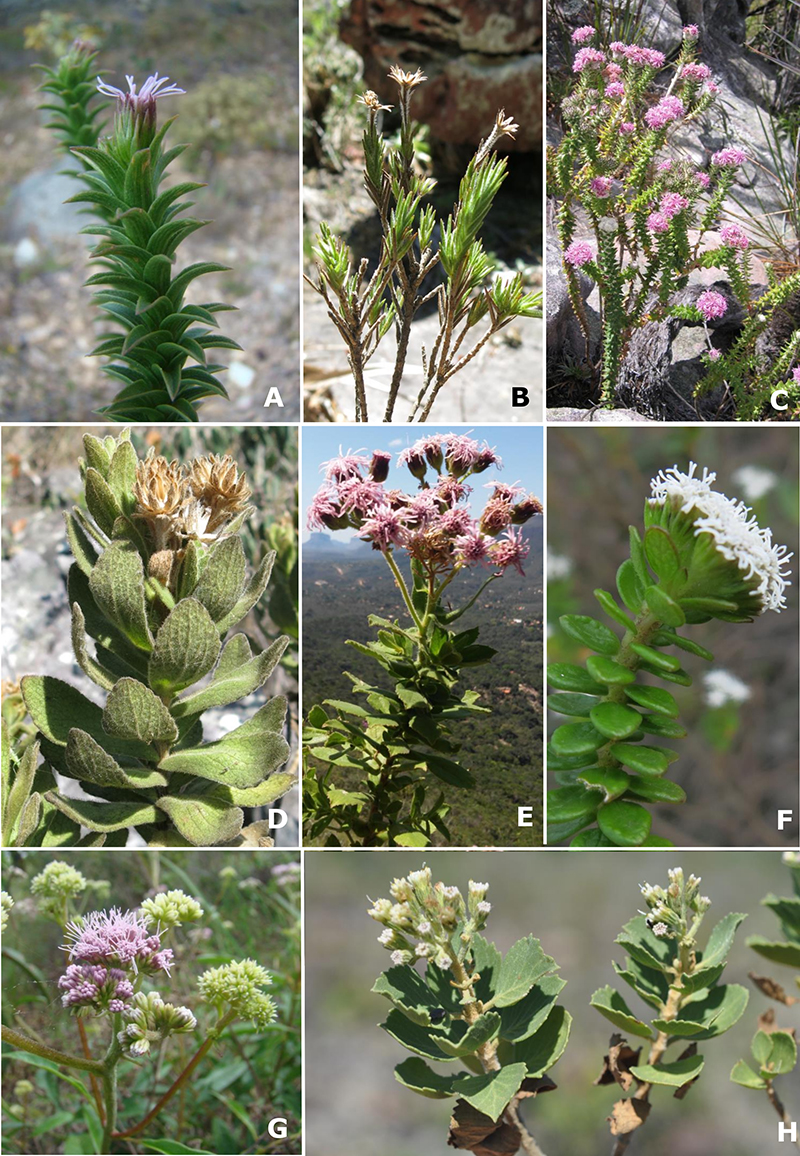
The ingroup included all six genera and 16 species (60% of the total) represented in the clades recovered by Rivera et al. (2016a) and Amorim (2019). Regarding the outgroup, two genera and two species were sampled (Fig. 1, Table 1).
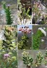
Representative species of the Chapada Diamantina Clade (A-F) and outgroups (G-H). A. Agrianthus myrtoides Matt., B. Arrojadocharis santosii, C. Lasiolaena lychnophorioides, D. Semiria viscosa, E. Semiria sp. nov., F. Stylotrichium glomeratum, G. Acritopappus confertus, H. Lapidia apicifolia. Photos: A, F, G. N. Roque, B, C, E. V.O. Amorim, D. S. Ferreira, H. L. Barres.
For each species, one individual from three different populations was collected when possible. Three fully expanded leaves were taken from the branch at the third or fourth node from the stem apex, except for Bishopiella elegans, which exhibits rosette leaves. The material collected in the field was fixed in 70% FAA - Formaldehyde, Acetic Acid, and preserved in 70% ethyl alcohol after 48 hours (Johansen, 1940). Voucher specimens were herbarium-prepared and deposited at the Alexandre Leal Costa Herbarium (ALCB) of the Universidade Federal da Bahia, with duplicates sent to the HUEFS Herbarium from the Universidade Estadual de Feira de Santana (Table 1).
Selected species from the Chapada Diamantina clade (Asteraceae, Eupatorieae) for foliar anatomy analysis. All specimens are deposited in the ALCB Herbarium. *Species that belong to the outgroup.
Sample Processing and Light Microscopy (LM) Analysis
For the anatomical study, FAA-fixed samples were used (Johansen, 1940). Herbarium materials were used when necessary, undergoing rehydration, which involved placing the leaves in 5% sodium hydroxide for two days, then washing them with distilled water five times for 20 minutes each and subsequently dehydrating them in an ascending ethanol series before storing them in 70% ethyl alcohol (Anderson, 1963).
Analyses of the epidermal surface and cross-sections were conducted at the apex, middle, and base of the leaf blade, and in the mid-region of the petiole. For epidermal analysis, samples were chemically dissociated using the Franklin method (modified by Kraus & Arduin, 1997). The isolated epidermises were then stained with 1% alcoholic Safranin (Kraus & Arduin, 1997), mounted on semi-permanent slides with 50% glycerin, and sealed with clear nail polish.
Cross-sections of the petiole and leaf blade (main vein, mesophyll, and margin) were made from dehydrated material embedded in synthetic resin (Historesin, Leica®) following the manufacturer’s instructions. The embedded material was sectioned at a thickness of 5 µm using a Carl Zeiss HM325 rotary microtome (Thermo Scientific). Histological sections were stained with 0.05% Toluidine Blue (Sakai, 1973) and mounted on permanent slides with Entellan synthetic resin (Merck®).
Anatomical analyses of the material and photographic documentation under light microscopy (LM) were performed using a Carl Zeiss Axio Scope A1 photomicroscope with an attached digital camera (EOS, Canon) installed at the Plant Anatomy and Wood Identification Laboratory (LAVIM) at the Instituto de Biologia, Universidade Federal da Bahia.
Descriptions of leaf anatomical structures followed those used by Metcalfe & Chalk (1950) and Fahn & Cutler (1992), while trichome terminology was based on Trindade et al. (2014), Budel et al. (2018), Payne (1978), Silva et al. (2019), Liesenfeld et al. (2019), and Martínez-Quezada et al. (2022). Only trichomes that were fully intact in paradermal and cross-sections were considered for description. In Table 2, ‘unassessed character’ indicates structures that could not be described due to their location, either within stomatal crypts or beneath the cuticle.
Anatomical characteristics present in the adaxial and abaxial epidermis of species from the Chapada Diamantina clade (Asteraceae, Eupatorieae). Legend: * Character not analyzed due to stomatal crypts; ani - anisocytic stomata; ano - anomocytic stomata.
Character coding and mapping
A matrix of binary and/or multistate anatomical characters was elaborated based on the analysis of the dermal and vascular systems of leaf blades. Character coding followed Sereno (2007), and hypotheses of primary homologies were proposed following De Pinna (1991).
All characters were mapped onto a majority-rule consensus tree obtained using Mesquite v.4.02 (Maddison & Maddison, 2025), adapted from the phylogenetic analysis of Amorim (2019), which was based on Bayesian inference (BI) of a concatenated dataset including seven molecular markers: three nuclear regions (ITS, gsh1, shmt) and four plastid regions (trnL-trnF, psbA-trnH, trnC-petN, trnY-rpoB).
Character mapping was performed under parsimony criteria, and figures were generated for characters recovered as synapomorphies, autapomorphies, or homoplastic traits potentially informative for circumscribing genera or clades.
Results
Anatomical Aspects of the Petiole
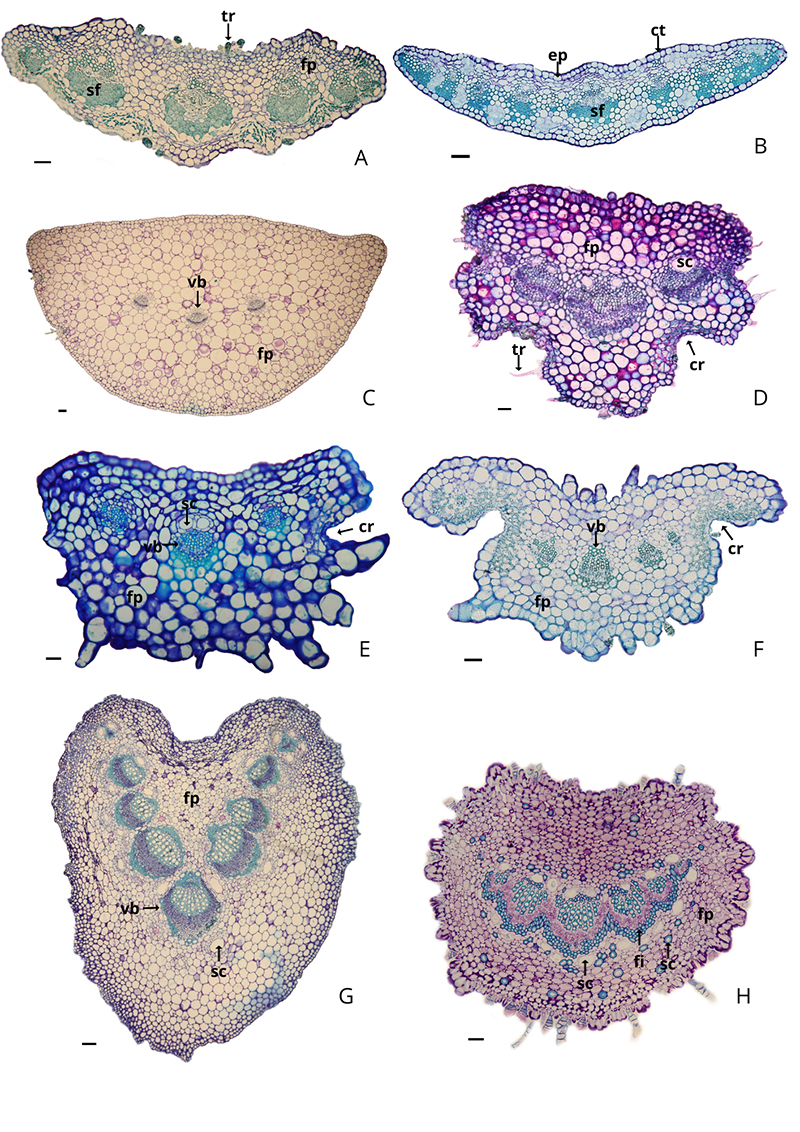
The petiole is present in Agrianthus luetzelburgii, Agrianthus microlicioides, Bishopiella elegans, Lasiolaena santosii, Stylotrichium corymbosum, Stylotrichium rotundifolium, and the outgroup species Acritopappus confertus and Lapidia apicifolia. In cross-section, it exhibits a plano-convex (Fig. 2 A-D) and concave-convex (Fig. 2E-H) shapes.
The epidermis is uniseriate with a thick cuticle (Fig. 2), containing stomata along its entire surface, as well as glandular and non-glandular trichomes of the same types present on the leaf blade (Fig. 2A, D). Lasiolaena santosii (Fig. 2D) and the genus Stylotrichium (Fig. 2E-F) exhibit stomatal crypts. The ground parenchyma consists of circular cells throughout, with few intercellular spaces. The vascular bundles are collateral, with a central bundle and accessory lateral bundles that have secretory channels exclusively associated with the xylem, displaying variations in quantity and lumen size (Fig. 2D-E).
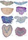
Transverse sections of the petiole with plano-convex (A-D) and concave-convex (E-H) shapes obtained by light microscopy. A. Agrianthus luetzelburgii, B. Agrianthus microlicioides, C. Bishopiella elegans, D. Lasiolaena santosii, E. Stylotrichium corymbosum, F. Stylotrichium rotundifolium, G. Acritopappus confertus, H. Lapidia apicifolia. Legend: ct - cuticle thickness; ep - uniseriate epidermis; pf - fundamental parenchyma; sc - secretory cavity; sf - sclerenchyma fibers; fi - procambial fibers; tr - trichomes; vb - vascular bundles. Scale bars: 100 µm (A-H).
In the external group species, Acritopappus confertus (Fig. 2G) and Lapidia apicifolia (Fig. 2H), secretory channels are found not only associated with the xylem but also dispersed throughout the ground parenchyma. The support function in the petiole is provided by sclerenchymatic fibers, which are more noticeable in Agrianthus species (Fig. 2B) and Acritopappus confertus. Procambial fibers (pericyclic) are observed in Lasiolaena (Fig. 2D), Stylotrichium (Fig. E-F), and Lapidia apicifolia (Fig. 2H). In Lapidia apicifolia (Fig. 2H), sclereids are present, scattered within the ground parenchyma. In Bishopiella elegans (Fig. 2C), fibers are absent, with collenchyma observed in regions adjacent to and surrounding the entire epidermis.
Anatomical Aspects of the Leaf Blade
Epidermis
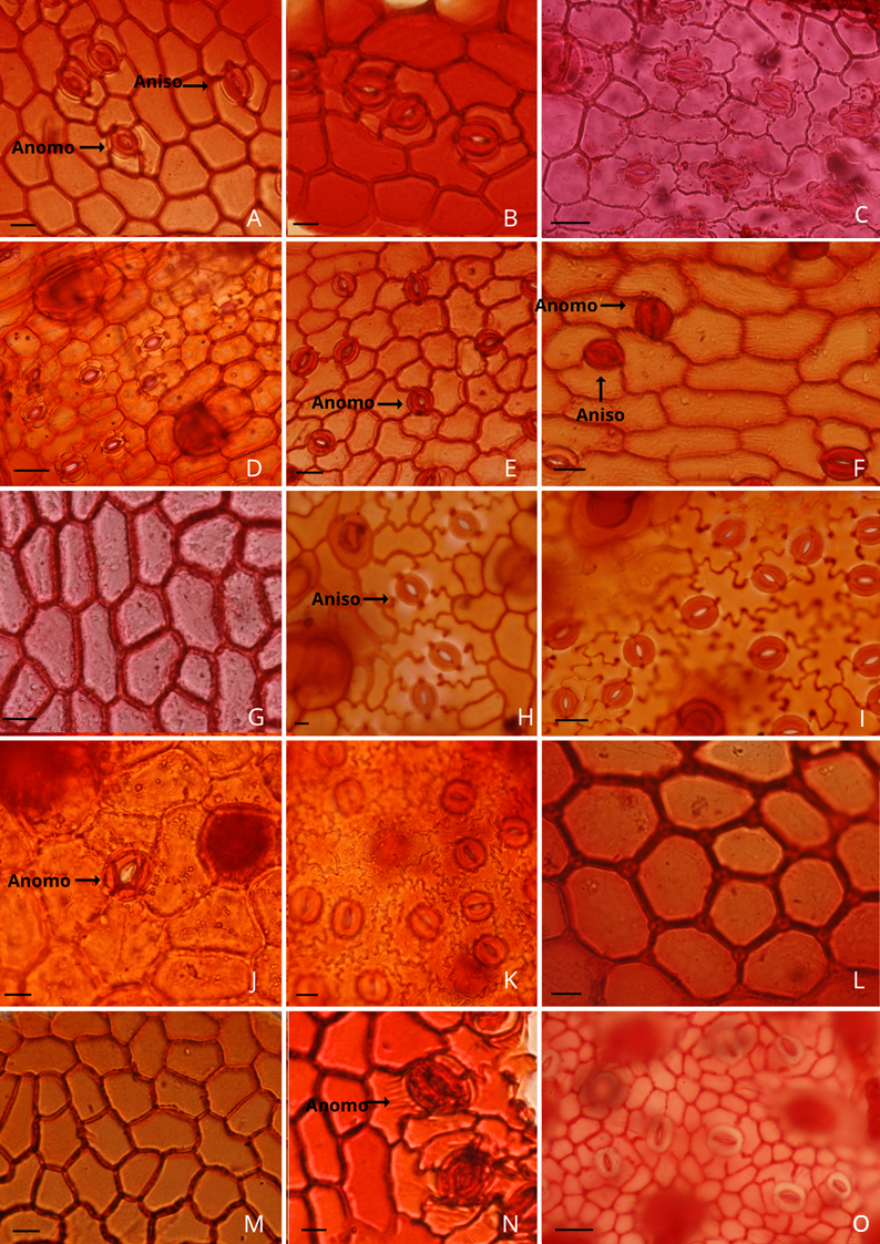
In a frontal view (Fig. 3, Table 2), the species displayed polygonal epidermal cells with straight outlines on both surfaces in Agrianthus (Fig. 3A-B), Arrojadocharis (Fig. 3C-D), Bishopiella elegans (Fig. 3E-F), as well as in the outgroup species Acritopappus confertus (Fig. 3M-N), and Lapidia apicifolia (Fig. 3O). On the other hand, Semiria sp. nov. (Fig. 3H-I) and Semiria viscosa (Fig. 3J-K) showed straight outlines on the adaxial surface and strongly sinuous outlines on the abaxial surface. In Lasiolaena (Fig. 3G) and Stylotrichium (Fig. 3L), a straight outline was only present on the adaxial surface. Regarding Lasiolaena and Stylotrichium species, the anticlinal wall outlines on the abaxial surface could not be observed due to stomatal crypts.
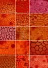
Frontal view of the leaf blade epidermis of Asteraceae species from the Chapada Diamantina Clade, obtained under light microscopy, showing cells with straight to sinuous outlines and anomocytic (A, C, D, E, I, J, K, N, O) and anisocytic (A, F, H) stomata with subsidiary cells with different outlines. Agrianthus microlicioides: A. Adaxial surface B. Abaxial surface. Arrojadocharis praxeloides: C. Adaxial surface D. Abaxial surface. Bishopiella elegans: E. Adaxial surface F. Abaxial surface. Lasiolaena duartei: G. Adaxial surface. Semiria sp. nov.: H. Adaxial surface. I. Abaxial surface. Semiria viscosa: J. Adaxial surface. K. Abaxial surface. Stylotrichium corymbosum: L. Adaxial surface. Acritopappus confertus: M. Adaxial surface. N. Abaxial surface. Lapidia apicifolia: O. Abaxial surface. Legend: Anomo - anomocytic stomata; Aniso - anisocytic stomata. Scale bars: 50 µm (C, D, G-H, I, J-K, O); 100 µm (A-B, E-F, L-M).
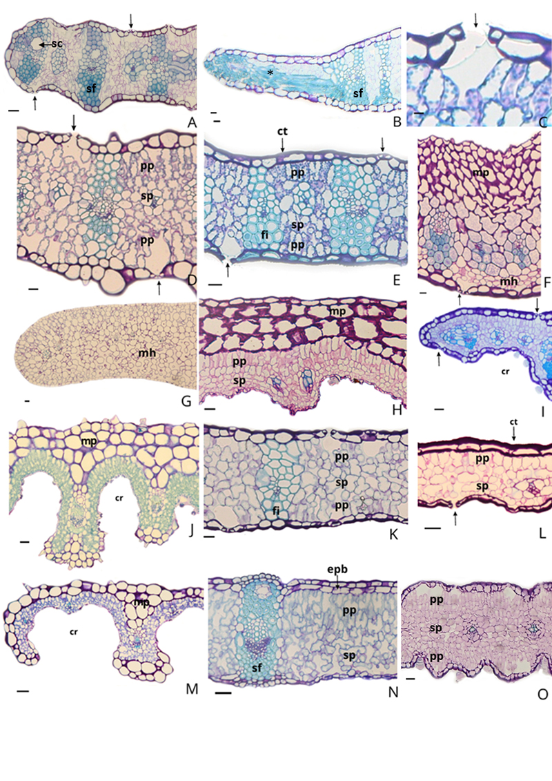
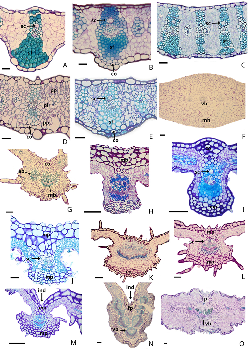
In a cross-sectional view (Figs. 4, 6; Table 2), the cuticle was thick in most species, except in Bishopiella elegans (Figs. 4G, 6F), Lasiolaena blanchetii, and Lasiolaena lychnophorioides, which exhibited thin cuticles when visually compared to other species. The epidermis was classified as uniseriate in all species (Figs. 4, 6, Table 2), except for Acritopappus confertus (Fig. 4N), which exhibited a biseriate adaxial epidermis.
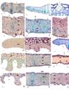
Transverse sections of the mesophyll and leaf margin with straight (A, B, I) and revolute (M) patterns obtained by light microscopy. A. Agrianthus luetzelburgii, B. Anatomy of the toothed margin with sclerenchyma fibers in Agrianthus microlicioides, C. Detail of the stomatal crest formed by cuticular projection on a stoma in the adaxial surface of Arrojadocharis santosii, D. Arrojadocharis praxeloides, E. Arrojadocharis santosii (isobilateral mesophyll), F. Arrojadocharis santosii (homogeneous mesophyll), G. Bishopiella elegans, H. Lasiolaena duartei, I. Lasiolaena pereirae, J. Lasiolaena santosii, K. Semiria sp. nov., L. Semiria viscosa, M. Stylotrichium rotundifolium, N. Acritopappus confertus, O. Lapidia apicifolia. Legend: * - toothed margin; arrow - stoma; cr - crypts; ct - cuticle thickness; ep - uniseriate epidermis; epb - biseriate epidermis; fi - procambial fibers; vb - vascular bundles; hyp - hypodermis; mh - homogeneous mesophyll; mp - multiseriate parenchyma; sf - sclerenchyma fibers; sp - spongy parenchyma; pp - palisade parenchyma. Scale bars: 100 µm (A-P).
Stomata
The predominant stomatal type in the studied species was anomocytic (stomata surrounded by four to six subsidiary cells with straight to sinuous outlines), along with the anisocytic type (stomata surrounded by three subsidiary cells), which was less common (Fig. 3, Table 2). In Arrojadocharis praxeloides (Figs. 3C-D), the outline of subsidiary cells was slightly sinuous on the adaxial surface compared to other epidermal cells, similar to Semiria sp. nov. (Fig. 3H). In Acritopappus confertus, the outline was slightly sinuous on the abaxial surface (Fig. 3N). In cross-section, stomata were at the same level as the other epidermal cells in all species, with stomatal ledges observed upon the ostioles (Fig. 4C), formed by cuticular projections.
Agrianthus (Figs. 4A-B), Arrojadocharis praxeloides (Fig. 4D), Bishopiella elegans (Fig. 4G), Lasiolaena pereirae (Fig. 4I), Semiria sp. nov. (Fig. 4K), and Semiria viscosa (Fig. 4L) were amphistomatic, while Arrojadocharis santosii displayed both amphistomatic (Fig. 4E; V.O. Amorim 465 and 450) and hypostomatic (Fig. 4F; V.O. Amorim 445) leaf blade populations. Lasiolaena and Stylotrichium are hypostomatic (Figs. 4H-J, M), with abaxial stomata located within crypts (Figs. 4J, M). Among the outgroup, Acritopappus confertus was hypostomatic (Fig. 4N), and Lapidia apicifolia was amphistomatic (Fig. 4O), lacking stomatal crypts (Table 2).
Trichomes
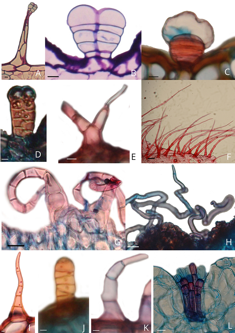
Eleven types of trichomes (Types I-XI) were identified among the studied species, including three glandular types (Types I-III, Fig. 5A-D) and eight non-glandular types (Types IV-XI, Fig. 5E-L). Glandular and non-glandular trichomes varied in occurrence and location on the leaf blades among species (Tables 3-4).
Among the glandular types, Type II (vesicular, biseriate body with 8-10 cells, globoid and unicellular head) is the most common (Figs. 5B-C), while Type III (stipitate, biseriate, multicellular body with 3-5 cells, and two apical cells with dense content forming the head) were found only in Stylotrichium rotundifolium (Fig. 5D).
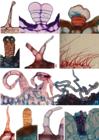
Glandular (A-D) and non-glandular (E-L) trichomes under light microscopy. A. Type I glandular trichome in Semiria sp. nov. B. Type II glandular trichome in Semiria viscosa. C. Detail of cell division in Type II glandular trichome in Stylotrichium corymbosum. D. Type III glandular trichome in Stylotrichium rotundifolium. E. Type IV tector trichome in Lasiolaena santosii. F. Type V tector trichome in Arrojadocharis santosii. G. Type VI tector trichome in Semiria sp. nov. H. Type VII tector trichome in Lasiolaena duartei. I. Type VIII tector trichome in Stylotrichium rotundifolium. J. Type IX tector trichome in Stylotrichium corymbosum. K. Type X tector trichome in Lasiolaena santosii. L. Type XI tector trichome in Bishopiella elegans. Scale bars: 100 µm (A-K) and 50 µm (L).
Among the non-glandular types, the types IV (bifid: uniseriate, multicellular body with two terminal cells, bifurcating at the terminal cell with pluricellular arms of 2-5 sharp-tipped cells) and X (uniseriate, multicellular body with 3-5 cells, with a widened basal cell and a pointed apical cell, numerous and often recurved) are restricted to Lasiolaena santosii (Fig. 5E, K, respectively). On the other hand, type VI (uniseriate, multicellular body with 3-6 cells, curved with a sharp tip) was exclusively recorded in the genus Semiria (Fig. 5G), and type IX (uniseriate, multicellular body with 6-7 cells, straight with rounded apical cell) was described exclusively for Stylotrichium corymbosum (Fig. 5J). Finally, type XI (moniliform, uniseriate, multicellular body with 3-6 cells, with a rounded apical cell) was restricted to Bishopiella elegans (Fig. 5L).
Characteristics related to the organization of fundamental and vascular tissues in the mesophyll, main veins, and leaf margin are presented in Tables 5, 6, and 7.
Anatomical characteristics present in the mesophyll of species from the Chapada Diamantina clade (Asteraceae, Eupatorieae).
Anatomical characteristics present in the mesophyll and veins of species from the Chapada Diamantina clade (Asteraceae, Eupatorieae).
Anatomical characteristics present in the veins of species from the Chapada Diamantina clade (Asteraceae, Eupatorieae).
Parenchyma
Multiseriate, composed of 1-5 layers of cells, was observed only in species of Lasiolaena and Stylotrichium (Figs. 4H-J, M). Lasiolaena blanchetii and L. duartei (Fig. 4H) showed the greatest variation regarding the number of layers in the vein region, with up to five layers exhibiting more heavily thickened cells compared to other species. Arrojadocharis did not show layers of cells (Figs. 4D, E); however, a population of A. santosii (Fig. 4F, V.O. Amorim 445) presented a five-layers cells adjacent to the adaxial epidermis.
In the mesophyll, the chlorenchyma ranged from homogeneous in Bishopiella elegans (Fig. 4G) and Arrojadocharis santosii (Fig. 4F, V.O. Amorim 445, 450), to dorsiventral and isobilateral. The latter two types displayed atypical palisade parenchyma with lobed cells irregularly distributed along the leaf area (Figs. 4A-F, H-N), except for Lapidia apicifolia (Fig. 4O), which had the typical palisade parenchyma. The mesophyll is isobilateral in Agrianthus (Fig. 4A), Arrojadocharis santosii (Figs. 4D-E), Semiria sp. nov. (Fig. 4K), and Lapidia apicifolia (Fig. 4O), formed by one layer of palisade parenchyma and 2-4 layers of spongy parenchyma. In Lasiolaena species (Figs. 4H-J), Semiria viscosa (Fig. 4L), Stylotrichium (Fig. 4M), and Acritopappus confertus (Fig. 4N), the mesophyll is dorsiventral with 1-3 layers of palisade parenchyma and 3-5 layers of spongy parenchyma.
Collenchyma
In the region adjacent to the epidermis, angular collenchyma was observed, varying from 1 to 2 layers on the abaxial face in species of Agrianthus (Fig. 6B) and Arrojadocharis (Fig. 6D-E). However, in Arrojadocharis santosii (V.O. Amorim 445), five to seven layers of this tissue were observed along the entire adaxial epidermis (Fig. 4F). In Semiria sp. nov. (Fig. 6G), S. viscosa (Fig. 6K), Acritopappus confertus (Fig. 6N), and species of Lasiolaena (Fig. 6H-J) and Stylotrichium (Fig. 6L-M), the collenchyma was presented only in the vein regions, exhibiting 3-5 layers of cells toward the upper side and 1-5 layers toward the lower side in species of Lasiolaena (Fig. 6H-J), and 1-2 layers adjacent to the upper side and 1-3 adjacent to the lower side in species of Stylotrichium (Fig. 6L-M). Bishopiella elegans (Fig. 6F) is the only species in which collenchyma was not present.
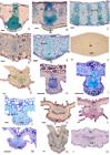
Transverse sections of the main vein obtained by light microscopy, highlighting different prominences and the tissues that fill them. A. Agrianthus giuliettiae, B. Agrianthus luetzelburgii, C. Agrianthus microlicioides, D. Arrojadocharis praxeloides, E. Arrojadocharis santosii, F. Bishopiella elegans, G. Semiria sp. nov., H. Lasiolaena duartei, I. Lasiolaena pereirae, J. Lasiolaena santosii, K. Semiria viscosa, L. Stylotrichium rotundifolium, M. Stylotrichium sucrei, N. Acritopappus confertus, O. Lapidia apicifolia. Legend: ab - accessory bundle; co - collenchyma; fp - fundamental parenchyma; ind - indentation on adaxial surface; mb - main bundle; mh - homogeneous mesophyll; mp - multiseriate parenchyma; sc - secretory channel; sf - sclerenchyma fibers; vb - vascular bundle. Scale bars: 100 µm (A-O).
Sclerenchyma
In Agrianthus (Fig. 4A-B), Arrojadocharis (Fig. 4D-F), and Acritopappus confertus (Fig. 4N), the sclerenchymatic tissue was present as fiber bundles that form sheath extensions toward the vascular bundles in the mesophyll, connecting the epidermis and vascular tissues. In Agrianthus, Arrojadocharis (Fig. 4A-B, D-E), and Acritopappus confertus (Fig. 4N), the sclerenchyma fibers, in addition to forming sheath extensions, also surrounded the vascular bundles. Lasiolaena (Fig. 4H-J), Semiria sp. nov. (Fig. 4K), Stylotrichium (Fig. 4M), and Lapidia apicifolia (Fig. 4O) showed likely pericyclic fibers surrounding or within the vascular bundles. Bishopiella elegans was the only species showing mesophyll and main vein without support tissue (Fig. 4G, 6F).
Vascularization
Vascular bundles were collateral in all species (Fig. 6A-O). The vascularization of the main vein in Agrianthus (Fig. 6A-B), Arrojadocharis (Fig. 6C-E), Semiria viscosa (Fig. 6K), and Lapidia apicifolia (Fig. 6O) consisted of a single central bundle, while in Bishopiella (Fig. 6F), Lasiolaena (Fig. 6H-J), Semiria sp. nov. (Fig. 6G), and Stylotrichium (Fig. 6L-M), in addition to the main bundle, one to three vascular bundles in the main vein region were present.
The presence of secretory channels associated with the vascular bundles of the main vein was observed in all the species within the clade, always near the xylem. Secretory channels were found in different quantities scattered in Acritopappus confertus (Fig. 6N) and Lapidia apicifolia (Fig. 6O). Agrianthus and Arrojadocharis had only one secretory channel associated with the bundle, and 1-2 channels in other genera (Fig. 6A-E).
Morphoanatomy Aspects of the Leaf Blade
The genera Agrianthus and Arrojadocharis had a main vein varying from flat on the adaxial side to slightly prominent on the abaxial side (Fig. 6A-E). In Bishopiella elegans (Fig. 6F) and Lapidia apicifolia (Fig. 6O), the main vein was slightly prominent on both sides. In genera Lasiolaena, Semiria, and Stylotrichium, the main vein was prominent on the abaxial side and flat to nearly flat on the adaxial side (Fig. 6G, H, J-L), except in Lasiolaena pereirae (Fig. 6I), which had a main vein prominent on the abaxial side and slightly prominent on the adaxial side, while Stylotrichium sucrei and Acritopappus confertus presented an indentation on the adaxial side (Fig. 6M-N).
Margin
The margin exhibited a straight pattern in Agrianthus, Arrojadocharis, Bishopiella elegans, Semiria viscosa, Semiria sp. nov., Lasiolaena pereirae, Acritopappus confertus, and Lapidia apicifolia (Fig. 4A-B, G, I). On the other hand, a revolute pattern (Fig. 4M) was observed in the remaining species of Lasiolaena and in the genus Stylotrichium. In Agrianthus microlicioides, sclerenchyma fibers are observed in its toothed margin (Fig. 4B).
Character state reconstructions
The reconstruction of twenty-seven anatomical characters of the leaf blade revealed distinct patterns of distribution within the Chapada Diamantina clade (Table 8). Among these, eighteen characters were phylogenetically informative, while nine showed no consistent phylogenetic signal or displayed only intraspecific variation.
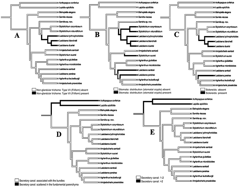
Among the informative characters, five were recovered as character states shared by species within key internal lineages and may represent synapomorphies supporting the Lasiolaena core, the Stylotrichium core, their sister group relationship, and the broader Chapada Diamantina clade: the presence of filiform non-glandular trichomes (C10), the presence of stomatal crypts (C18), the presence of sclereids (C23), the location of secretory ducts scattered in the fundamental parenchyma (C25), and the quantity of secretory ducts exceeding two (C26). Within the Lasiolaena core (L. lychnophorioides, L. blanchetii, and L. duartei), type VII filiform trichomes (character 10) was recovered as a shared derived feature (Fig. 7A). The sister-group relationship between Stylotrichium and Lasiolaena is supported by stomatal crypts (Character 18; Fig. 7B), while sclereids (Character 23) occurs in all Lasiolaena species except L. santosii (Fig. 7C), reinforcing the anatomical cohesion within the genus. Ducts associated with the bundles (character 25; Fig. 7D) and secretory duct quantity (character 26) are shared among the entire Chapada Diamantina lineage, distinguishing it from the outgroups (Fig. 7E).
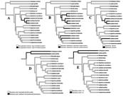
Mapping of anatomical characters recovered as synapomorphies supporting the circumscription of clades and taxa in this study. A. Character 10-Non-glandular trichome: Type VII (filiform). B. Character 18-Stomata: distribution (stomatal crypts). C. Character 23- Sclereids. D. Character 25-Secretory canal: location. E. Character 26-Secretory canal: quantity.
In contrast, nine characters were recovered as homoplastic when mapped onto the molecular phylogeny, suggesting independent origins in different lineages: the presence of glandular trichomes Type I (stipitate, biseriate, 6-8 cells; C4), glandular trichomes Type II (vesicular; C5), non-glandular trichomes Type V (uniseriate, multicellular, 3-10 cells; C8), non-glandular trichomes Type VIII (uniseriate, multicellular, 5-7 cells; C11), sinuous subsidiary cell contour on the adaxial surface (C15), amphistomatic or amphi-hypostomatic stomatal distribution (C17), dorsiventral mesophyll (C19), the presence of procambial fibers (C21), and midrib prominence (C27).
For instance, dorsiventral mesophyll (C19) is shared by Agrianthus, Lasiolaena santosii, L. pereirae, and Arrojadocharis praxeloides, but was recovered as homoplastic, providing no synapomorphic support for closer relationships among these taxa. Similarly, the presence of procambial fibers (C21) occurs in Lasiolaena taxa belonging to distinct subclades, suggesting repeated emergence of similar anatomical traits within the lineage.
Autapomorphies were restricted to terminal taxa and reflect species-specific anatomical modifications that are useful for delimitation at the species level. Examples include the presence of glandular trichomes Type III (stipitate, biseriate; C6) in Stylotrichium rotundifolium, non-glandular trichomes Type IX (straight with rounded apex; C12) in S. corymbosum, non-glandular trichomes Type X (uniseriate, multicellular, 3-5 cells; C13) in Lasiolaena santosii, and non-glandular trichomes Type XI (moniliform; C14) in Bishopiella elegans. Although phylogenetically uninformative, these features contribute to the recognition of diagnostic anatomical attributes within each lineage.
Uniseriate epidermis, absence of Type III glandular trichomes, and absence of sclerenchymatic fibers (characters 1, 6, and 22, respectively), likely represent plesiomorphic conditions retained from ancestral states. These conditions are widespread and likely represent ancestral anatomical configurations conserved within the Chapada Diamantina clade.
Finally, eleven characters were considered non-informative due to intraspecific variability or lack of consistent distribution across the phylogeny: number of epidermal layers (C1), sinuous epidermal contour on the abaxial surface (C3), non-glandular trichomes Type IV (bifid; C7), non-glandular trichomes Type V (uniseriate, multicellular, 3-10 cells; C8), non-glandular trichomes Type VII (filiform; C10), non-glandular trichomes Type VIII (uniseriate, multicellular, 5-7 cells; C11), sinuous subsidiary cell contour on the abaxial surface (C16), stomatal distribution (C17), atypical palisade parenchyma (C20), the presence of sclerenchymatous fibers (C22), and accessory bundles in the midrib (C24).
Some characters, such as epidermis layering (C1), non-glandular trichomes Type V (C8), and non-glandular trichomes Type VII (C10), appear in more than one evolutionary category depending on the clade or taxon analyzed, reflecting their potential roles as plesiomorphic, autapomorphic, or homoplastic under different phylogenetic contexts. Additionally, non-glandular trichomes Type VI (curved; C9), although restricted to Semiria species, do not support any specific hypothesis of interspecific divergence, illustrating that not all unique traits necessarily contribute to phylogenetic resolution within the group.
Discussion
Species in the Chapada Diamantina clade exhibit macro and micromorphological traits with adaptive value to the rocky fields (campo rupestre) vegetation. This ecosystem is predominant in high-altitude areas (above 900 m) on sandy, shallow soils with a high proportion of quartzite-sandstone rocky substrates. It is common in the Espinhaço Range, Brazil, where 90% of it is located (Alves & Kolbek, 2010).
The species in this clade exhibit coriaceous, imbricated leaves with reduced leaf blades, thick cuticles, high trichome density, crypted stomata, atypical palisade parenchyma, hypodermis, and sclerenchyma sheath extensions, which aid in water retention within tissues and optimize photosynthetic processes (Fahn & Cutler, 1992; Guerra & Scremin-Dias, 2017; Ariano et al., 2022). However, some of these characteristics, such as a thick cuticle, stomatal crypt, hypodermis, and support tissue, are absent in Bishopiella elegans, the only species in the clade that occurs on flooded soil during the rainy season (November-March) in the Chapada Diamantina. In other angiosperm groups, morphoanatomical adaptations in aquatic and amphibious species have been observed as evolutionary responses to environmental pressures (Bedoya & Madrinán, 2014; Leme & Scremin-Dias, 2014).
From the analysis of the foliar morphoanatomy of representatives of the Chapada Diamantina clade, characteristics that may assist in delimiting the clade and the taxa recovered by Rivera et al. (2016a), Roque et al. (2017), and Amorim (2019) were identified. The location of secretory channels in the main vein may be a synapomorphy for the Chapada Diamantina clade, since all the analyzed species showed 1-2 channels associated with vascular bundles, in contrast to outgroup species, which exhibited more than two channels dispersed in the ground parenchyma, as inferred from the phylogenetic reconstruction (Fig. 7 D-E). The differing positions of secretory channels in the leaves of Aldama (Asteraceae) helped Oliveira (2015) and Silva et al. (2014) in species distinction.
The straight outline of epidermal cells on the adaxial side was recorded for all clades and outgroup species. On the abaxial side, the outline of the cells with crypted stomata, the presence of crypted stomata (Fig. 7 B), hypostomatic leaf blades, multistratified hypodermis, and revolute margins are shared traits among the genera Lasiolaena (except L. pereirae) and Stylotrichium, supporting their grouping as probable sister groups (Amorim, 2019). In addition to its taxonomic significance, the multistratified hypodermis may represent an important environmental adaptation, as observed in Eriocaulaceae species found in campo rupestre habitats. Here, a hypodermis composed of sclerenchyma provides the plant with resistance to wind, intense light, and water stress (Mascarenhas et al., 2020). Although ontogenetic studies were not conducted, the morphological differences between parenchymatic cells and epidermal cells suggest that the former originate from the ground meristem.
Only Semiria viscosa and Semiria sp. nov. showed cells with sinuous outline on the abaxial epidermal surface (vs. straight). Similarly, in Aldama, the variability in epidermal cell outlines allowed Bombo et al. (2016) to use this trait for species differentiation, underscoring its taxonomic relevance within the genus. The restriction of sinuous epidermal cell outlines to two non sister species of Semiria suggests that this trait represents a homoplastic or species-level diagnostic character rather than a synapomorphy.
Species of Agrianthus, along with Arrojadocharis praxeloides, Bishopiella elegans, Semiria sp. nov., and S. viscosa exhibited amphistomatic leaves. Furthermore, amphistomatic leaves were also observed in Lasiolaena pereirae, a species placed within the Agrianthus clade sensu Amorim (2019). Variations in stomatal placement are common in Asteraceae species (Melo-de-Pinna, 2004; Budel et al., 2018) and may be adaptive strategies, contributing to reducing water loss and increasing carbon dioxide diffusion (Lusa et al., 2014). However, since all the studied species are restricted to campos rupestres, hypostomatic leaves with crypted stomata are believed to reflect the common evolutionary history between Lasiolaena and Stylotrichium (Fig. 7 B).
Among the 11 types of trichomes described (three glandular and eight non-glandular), six species exhibited exclusive trichomes (autapomorphies): Bishopiella elegans (type XI), Lasiolaena santosii (types IV, X), Semiria (2 spp.; type VI), Stylotrichium corymbosum (type IX), and S. rotundifolium (type III). These trichomes therefore represent reliable taxonomic markers at the species level. The diversity of trichomes was reported by Liesenfeld et al. (2019) as an important trait for Asteraceae tribe taxonomy, in addition to studies on genera (Narayana, 1979) and subtribes (Wagner et al., 2014). Glandular trichomes are the most diverse secretory structures for the family, especially for the Eupatorieae tribe (Castro et al., 1997; Martínez-Quezada et al., 2022). These structures also play crucial ecological roles, producing volatile oils and phenolic compounds that may act as defense agents against herbivores and pathogens (Fernandes et al., 2016).
Nevertheless, the greatest diversity of trichomes was observed among the non-glandular trichome types (types IV-XI). In addition to the five types mentioned above (IV, VI, IX, X, and XI), type VII is restricted to three species of Lasiolaena, excluding L. santosii and L. pereirae, and represents a derived character according to our phylogenetic reconstruction (Fig. 7A). Similarly, the presence of non-glandular trichomes was important in identifying species of Lychnophorinae (Asteraceae), suggesting a probable adaptation against water stress (Wagner et al., 2014). Trichomes can also contribute to plant protection against excessive transpiration during the dry season in the Cerrado and campos rupestres by secreting substances and protecting the plant from pathogens and herbivores (Trindade et al., 2014).
Mesophyll organization varied among the analyzed taxa, with isobilateral leaves occurring in species of Agrianthus, Arrojadocharis praxeloides, Semiria sp. nov., and the outgroup Lapidia apicifolia, whereas dorsiventral mesophyll was recorded in Semiria viscosa, Lasiolaena, Stylotrichium, and the outgroup Acritopappus confertus. Bishopiella elegans was distinguished by a homogeneous mesophyll (Table 5). The occurrence of similar mesophyll types in both ingroup and outgroup taxa suggests that this character is evolutionarily labile and likely homoplastic, limiting its value as a synapomorphy within the clade. Rather than reflecting shared ancestry, mesophyll organization appears to be more strongly influenced by ecological or functional constraints.
The species Arrojadocharis santosii showed population variation regarding stomatal location (amphi and hypostomatic) and mesophyll, being isobilateral and homogeneous, as well as the presence or absence of hypodermis. Anatomical traits variation reinforces its possible hybrid origin, as discussed by Amorim (2019), and has been observed in other angiosperm groups, such as in Poaceae (Paštová, 2017).
In all the clade species, except Bishopiella elegans, mesophyll composition included atypical palisade parenchyma with non-uniform lobed cells (Fahn, 1978), resembling the plicate parenchyma recently reported in Mikania glomerata (Milan et al., 2006). According to Fahn & Cutler (1992), atypical parenchyma can increase the area of photosynthetic tissue, benefiting species with reduced leaf areas, considered as a xeromorphic adaptation. The absence of atypical palisade parenchyma in Bishopiella elegans distinguishes this species from the remaining members of the clade, highlighting its anatomical and phylogenetic distinctiveness (Table 5).
Support tissues, derived from both the ground meristem and procambium, also proved useful for taxonomic delimitation within the group, particularly at the generic level. Procambial fibers occur in species of Lasiolaena, Semiria, and Stylotrichium, whereas sclerenchymatic fibers were recorded in Arrojadocharis and Agrianthus. Sclereids were dispersed in species of Lasiolaena, contributing to the anatomical circumscription of the genus. The absence of all support tissues in Bishopiella elegans is unique within the clade and represents a clear autapomorphy, supporting its monospecific circumscription.
In contrast, the presence of procambial or sclerenchymatic fibers and dispersed sclereids in other taxa is associated with sheath extensions that may enhance mechanical support and hydraulic continuity between vascular tissues and the mesophyll, particularly under xeric conditions typical of campos rupestres environments (Fahn & Cutler, 1992; Lusa et al., 2014). However, because similar combinations of support tissues occur across distantly related ingroup taxa and in outgroup species, these characters exhibit limited phylogenetic signal and are better interpreted as convergent, ecologically driven adaptations rather than synapomorphies.
The analysis of leaf anatomy in the Chapada Diamantina clade (Asteraceae) proved to be a valuable tool for taxonomic and evolutionary studies. The anatomical characters analyzed contributed to reinforcing or clarifying phylogenetic hypotheses previously proposed with molecular data, offering robust morphological support for distinguishing genera and understanding their relationships. These findings emphasize the relevance of anatomical traits in identifying synapomorphies and resolving taxonomic uncertainties within the group. Moreover, the integration of anatomical and molecular evidence emerges as a powerful approach for advancing the delimitation and classification of genera in the Asteraceae family, particularly within the Eupatorieae tribe.
Acknowledgments
The authors thank the management staff of the Parque Nacional da Chapada Diamantina for granting permission to conduct this study, and the local guides for their valuable support during field activities.
References
- Adedeji O, Jewoola OA. 2008. Importance of leaf epidermal characters in the asteraceae family. Notulae Botanicae Horti Agrobotanici Cluj-Napoca 36: 7-16.
- Alves RJV, Kolbek J. 2010. Can campo rupestre vegetation be floristically delimited based on vascular plant genera? Plant Ecology 207: 67-79.
- Amorim VO. 2019. Filogenia molecular do clado Chapada Diamantina (Asteraceae- Eupatorieae) e Revisão taxonômica de Agrianthus Mart. Ex DC. PhD Thesis, Universidade Estadual de Feira de Santana, Brazil.
- Anderson LC. 1963. Studies on Petradoria (Compositae): Anatomy, cytology, taxonomy. Transactions Kansas Academy of Science 66: 632-684.
- Ariano APR, Pessoa MJG, Ribeiro-Júnior NG, Eisenlohr PV, Silva IV. 2022. Structural leaf attributes indicate different degrees of xeromorphism: New discoveries in co-occurring species of savanna and forest formations. Flora 286: 151972.
- Bedoya AM, Madrinán S. 2014. Evolution of the aquatic habit in Ludwigia (Onagraceae): Morpho-anatomical adaptive strategies in the Neotropics. Aquatic Botany 120: 352-362.
- Bento JPSP, Scremin-Dias E, Alves FM, Mansano VF, Sartori ALB. 2020. Phylogenetic implications of the anatomical study of the Amburaneae clade (Fabaceae: Faboideae). Botanical Journal of the Linnean Society 194: 69-83.
- Bombo AB, Filartiga AL, Appezzato-da-Glória B. 2016. Solving taxonomic problems within the Aldama genus based on anatomical characters. Australian Journal of Botany 64: 501-512.
- Budel JM, Raman V, Monteiro LM et al 2018. Foliar anatomy and microscopy of six Brazilian species of Baccharis (Asteraceae). Microscopy Research & Technique 81: 832-842.
- Castro MM, Leitão-Filho HF, Monteiro WR. 1997. Utilização de estruturas secretoras na identificação dos gêneros de Asteraceae de uma vegetação de cerrado. Revista Brasileira de Botânica 20: 163-174.
- Castro EM, Pinto JEBP, Soares AM et al 2007. Adaptações anatômicas de folhas de Mikania glomerata Sprengel (Asteraceae), em três regiões distintas da planta, em diferentes níveis de sombreamento. Revista Brasileira de Plantas Medicinais 9: 8-16.
- Cetzal-IX W, Nogueirra-Savella E, Jáuregui D, Carvevali G. 2013. Anatomía foliar comparada y sistemática del clado-Trichocentrum con énfasis em Cohniella (Asparagales: Orchidaceae). Revista de Biologia Tropical 61: 1841-1858.
- Cruz R, Duarte M, Pirani JR, Mello-de-Pinna GFA. 2017. Phylogenetic analysis and evolution of morphological characters in Metrodorea and related species in Rutoideae (Rutaceae). Plant Systematics and Evolution 303: 927-943.
- De Pinna MGG. 1991. Concepts and tests of homology in the cladistic paradigm. Cladistics 7: 367-394.
- Dickison WC. 2000. Integrative Plant Anatomy. California, Harcourt Academic Press.
- Fahn A. 1978. Anatomía Vegetal. Madrid, Ed. Blume.
- Fahn A, Cutler DF. 1992. Xerophytes. Berlin, Encyclopedia of Plant Anatomy.
- Fernandes AC. 2013. Posição filogenética de Heterocondylus R.M. King & H. Rob. (Asteraceae-Eupatorieae), com base em marcadores do DNA nuclear e plastidial. MSc Thesis, Universidade Federal de Minas Gerais, Brazil.
- Fernandes YS, Trindade LMP, Rezende MH, Paula JR, Gonçalves LA. 2016. Trichomes and chemical composition of the volatile oil of Trichogonia cinerea (Gardner) R. M. King & H. Rob. (Eupatorieae, Asteraceae). Anais da Academia Brasileira de Ciências 88: 309-322.
- Ferraro A, Scremin-Dias E. 2018. Structural features of species of Asteraceae that arouse discussions about adaptation to seasonally dry environments of the Neotropics. Acta Botanica Brasilica 32: 113-127.
- Ferreira SC. 2010. Filogenia Molecular da subtribo Gyptidinae R.M. King & H. Rob. (Eupatorieae-Asteraceae) e Revisão taxonômica do gênero Lasiolaena R.M. King & H. Rob. PhD Thesis, Universidade Estadual de Feira de Santana, Brazil.
- Guerra A, Scremin-Dias E. 2017. Leaf traits, sclerophylly and growth habits in plant species of a semiarid environment. Brazilian Journal of Botany 41: 131-144.
- Hattori EKO. 2013. Análise filogenética molecular da subtribo Disynaphiinae (Asteraceae - Eupatorieae). PhD Thesis, Universidade Federal de Minas Gerais, Brazil.
- Janaćković P, Susanna A, Marin PD. 2019. Micromorphology and anatomy in systematics of Asteraceae. An old-fashioned approach? Biologica Nyssana 10: 77-85.
- Jeiter J, Smets E. 2024. Comparative morphology at a crossroads. American Journal of Botany 111: e16392.
- Johansen DA. 1940. Plant microtechnique. New York, McGraw-Hill Book Company.
- Kellogg EA. 2006. Progress and challenges in studies of the evolution of development. Journal of Experimental Botany 57: 3505-3516.
- King RM, Robinson H. 1987. The genera of the Eupatorieae (Asteraceae). St Louis, Missouri Botanical Garden.
- Kraus JE, Arduin M. 1997. Manual Básico de Métodos em Morfologia Vegetal. Seropédica, Editora da Universidade Federal Rural.
- Leme F, Scremin-Dias E. 2014. Ecological interpretations of the leaf anatomy of amphibious species of Aeschynomene L. (Leguminosae - Papilionoideae). Brazilian Journal of Biology 74: 41-51.
- Lersten NR, Curtis JD. 1985. Distribution and anatomy of hydathodes in Asteraceae. Botanical Gazette 146: 106-114.
- Liesenfeld V, Gentz P, Freitas EM, Martins S. 2019. Morphological diversity of foliar trichomes in Asteraceae from Sand-fields of the Pampa biome, Rio Grande do Sul State, Brazil. Hoehnea 46: e752018.
- Lusa MG, Appezzato-da-Glória B, Loeuille B, Bartoli G, Ciccarelli D. 2014. Functional groups in Lychnophorinae (Asteraceae: Vernonieae) based on morphological and anatomical traits. Australian Journal of Botany 62: 150-163.
- Lusa MG, Loeuille BFP, Ciccarelli D, Appezzato-da-Glória B. 2018. Evolution of stem and leaf structural diversity: A case study in Lychnophorinae (Asteraceae). The Botanical Review 84: 203-241.
-
Maddison WP, Maddison DR. 2025. Mesquite: A modular system for evolutionary analysis. Version 4.02. http://www.mesquiteproject.org 15 Nov. 2025.
» http://www.mesquiteproject.org - Matos RR, Araújo JS. 2021. Morfoanatomia das glândulas foliares e falicinais de Stigmaphyllon A.Juss. (Malpighiaceae): evidências funcionais, contribuições taxonômicas e evolutivas. Hoehnea 48: e282021.
- Martínez-Sagarra G, Abad P, Antonio Devesa JA. 2017. Study of the leaf anatomy in cross-section in the Iberian species of Festuca L. (Poaceae) and its systematic significance. PhytoKeys 83: 43-74.
- Martínez-Quezada DM, Rivera P, Rojas-Leal A., Villaseñor JL, Terrazas T. 2022. Leaf secretory structures in Asteraceae: A synthesis of their diversity and evolution. The Botanical Review 89: 59-90.
- Mascarenhas AAS, Harley AMG, Scatena VL. 2020. Leaf and scape anatomy of Leiothrix Ruhland (Eriocaulaceae) from a taxonomic and ecological perspective. Flora 262: 151518.
- Mello ACMP, Almeida RF, Amorim AMA, Oliveira DMT. 2019. Leaf structure in Amorimia and closely related neotropical genera and implications for their systematics and leaf evolution in Malpighiaceae. Botanical Journal of the Linnean Society 191: 102-127.
- Melo-de-Pinna GF. 2004. Anatomia foliar de Richterago Kuntze (Multisieae, Asteraceae). Acta Botânica Brasilica 18: 591-600.
- Metcalfe CF, Chalk L. 1950. Anatomy of the Dicotyledons: Leaves, stems and wood in relation to taxonomy with notes on economic uses. Oxford, Clarendon Press.
- Milan P, Hayashi AH, Appezzato-da-Glória B. 2006. Comparative leaf morphology and anatomy of three asteraceae species. Brazilian Archives of Biology and Technology 49: 135-144.
- Muniz LF, Bombo AB, Filartiga AL, Appezado-da-Glória B. 2018. Can climate and soil conditions change the morpho-anatomy among individuals from different localities? A case study in Aldama grandiflora (Asteraceae). Brazilian Journal of Biology 78: 706-717.
- Muravnik LE, Kostina OV, Shavarda AL. 2016. Glandular trichomes of Tussilago farfara (Senecioneae, Asteraceae). Planta 244: 737-752.
- Narayana BM. 1979. Taxonomic value of trichomes in Vernonia Schreb. (Asteraceae). Proceedings Indian Academy of Sciences 88: 347-357.
- Oliveira T, Bombo AB, Appezzato-da-Glória B. 2013. Anatomy of vegetative organs with an emphasis on the secretory structures of two species of Aldama (Asteraceae Heliantheae). Botany 91: 335-342.
- Oliveira CT. 2015. Sistemática de Mikania Willd. (Eupatorieae-Asteraceae). PhD Thesis, Universidade de São Paulo, Brazil.
- Paštová L. 2017. Variation in leaf anatomy within the Elytrigia intermedia - E. ×mucronata - E. repens (Poaceae) hybrid complex. Phytologia Balcanica 23: 187-198.
- Payne WW. 1978. A glossary of plant hair terminology. Brittonia 30: 239-255.
- Rivera VL, Panero JL, Schilling EE, Crozier BS, Moraes MD. 2016a. Origins and recent radiation of Brazilian Eupatorieae (Asteraceae) in the eastern Cerrado and Atlantic Forest. Molecular Phylogenetics and Evolution 97: 90-100.
- Rivera VL, Ferreira SC, Panero JL. 2016b. Trichogoniinae, a new subtribe of Eupatorieae (Asteraceae). Phytotaxa 260: 296-300.
- Robinson H, Schilling E, Panero JL. 2009. Eupatorieae. In: Funk VA, Susanna A, Stuessy T, Bayer RJ (eds.). Systematics, Evolution and Biogeography of Compositae. Vienna, International Association of Plant Taxonomists.
- Roque N, Ferreira SC, Van Den Berg C. 2017. Lapidia, a new monotypic genus of Asteraceae (Eupatorieae) from Brazil, and its phylogenetic placement. Phytotaxa 291: 1-16.
- Ruiz AI, Mercado MI, Guatany ME, Ponessa GI. 2016. Arquitectura y morfoanatomia foliar de Dinoseris salicifolia (Asteraceae). Lilloa 53: 112-121.
- Sakai WS. 1973. Simple Method for Differential Staining of Paraffin Embedded Plant Material Using Toluidine Blue O. Stain Technology 48: 247-249.
- Scatena V, Giulietti A, Borba E, Van Den Berg C. 2005. Anatomy of Brazilian Eriocaulaceae: Correlation with taxonomy and habitat using multivariate analyses. Plant Systematics and Evolution 253: 1-22.
- Sereno PC. 2007. Logical basis for morphological characters in phylogenetics. Cladistics 23: 565-587.
- Silva, EMS, Hayashi, AH, Appezato-da-Glória, B. 2014. Anatomy of vegetative organs in Aldama tenuifolia and A. kunthiana (Asteraceae: Heliantheae). Brazilian Journal of Botany 37: 505-517.
- Silva CCS, Leite KRB, Roque N. 2019. Leaf morphology and anatomy of Richterago discoidea (Asteraceae): A taxonomic and ecology approach. Feddes Repertorium 130: 1-16.
- Siniscalchi CM, Loeuille B, Roque N. 2021. Asteraceae in a megadiverse flora: Results from the Flora of Brazil 2020. Capitulum 1: 54-60.
- Solereder H. 1908. Systematic Anatomy of the Dicotyledons: A Handbook for Laboratories of Pure and Applied Botany. Oxford, Clarendon Press , vol. II.
- Trindade LMP, Fernandes YS, Gonçalves LA. 2014. Diversidade e desenvolvimento dos tricomas glandulares de Lomatozona artemisiifolia Baker (Asteraceae - Eupatorieae) - uma planta endêmica do Cerrado de Goiás. Iheringia, Série Botânica 69: 235-243.
- Wagner MA, Loueuille BFP, Siniscalchi CM, Melo-de-Pinna GF, Pirani JR. 2014. Diversity of non-glandular trichomes in subtribe Lychnophorinae (Asteraceae: Vernonieae) and taxonomic implications. Plant Systematics and Evolution 300: 1219-1233.
-
Data Availability
The primary datasets generated and analyzed, specifically the character matrix and the phylogenetic tree files, are openly available in editable format in the Figshare repository under the identifier [doi: 10.6084/m9.figshare.30489671.v2]. All other data supporting the findings are included within the article.
-
Funding Information
This work was supported by the Smithsonian Institution (US Herbarium) through the Harold E. Robinson Fellowship Award granted to Vivian Oliveira Amorim, and by the Coordenação de Aperfeiçoamento de Pessoal de Nível Superior - CAPES (Finance Code 001). Bárbara Oliveira acknowledges support from the Coordenação de Aperfeiçoamento de Pessoal de Nível Superior - CAPES, through a scholarship.
The primary datasets generated and analyzed, specifically the character matrix and the phylogenetic tree files, are openly available in editable format in the Figshare repository under the identifier [doi: 10.6084/m9.figshare.30489671.v2]. All other data supporting the findings are included within the article.