 Linking pollen traits to wind pollination in nettles (Urticaceae)
Abstract
Nettles (tribe Urticeae) are characterized by stinging glands and wind-dispersed pollen. However, despite the prevalence of anemophily in Urticeae and across Urticaceae, the pollen traits associated with wind dispersal remain poorly understood. Here, we investigate pollen traits in distinct evolutionary lineages of Urticeae, represented by Laportea aestuans, Urera baccifera, and Urtica dioica. Staminate flowers were processed for light, scanning electron, and transmission electron microscopy to assess pollen morphology, viability, and reserve substances. Pollen grains are released at the bicellular stage, are small, isopolar, oblate-spheroidal, with a nanoechinate exine. Laportea aestuans exhibits pantoporate pollen, whereas U. baccifera and U. dioica are 3-porate. All species present opercula covering the pores, and annuli in L. aestuans. Starch predominates in the vegetative cell across all species, with lipids additionally detected in L. aestuans. Proteins, also detected in the pollen of all species, may form glycoproteins associated with allergic responses common in this group. Pollen viability was high across species. Pollen traits in these species indicate only partial conformity to the anemophilous syndrome, combining typical wind-pollination features, such as small size, high viability, and amyliferous reserves, with deviations (lipid-rich pollen, a higher number of apertures, and a nanoechinate exine) that suggest functional and evolutionary complexity.
Keywords:
anemophily; cytology; cytochemistry; morphology; Urticeae
Introduction
Urticaceae is a plant family that comprises approximately 54 genera and over 2,000 species (Wu et al., 2013). Its members exhibit a cosmopolitan distribution, primarily in moist habitats and tropical regions (Friis, 1993). The family holds significant economic, ecological, and medicinal importance. Vegetable fibers extracted from species of Urtica L., Girardinia Gaudich., and particularly Boehmeria Jacq. are the main economically relevant products (Friis, 1993). In the medicinal field, species of Urtica are reported to possess analgesic, antimicrobial, and antioxidant properties (Gülçin et al., 2004; Farag et al., 2013). Ecologically, genera such as Urtica, Elatostema J.R.Forst & G.Forst, Pilea Lindl., and Pellionia Gaudich. are dominant components of their respective habitats (Wu et al., 2013; Große-Veldmann, 2017). The genus Cecropia Loefl. is notable for being a group of pioneer tree species in forest regeneration post-disturbance, as well as for its mutualistic interactions with ants (Berg et al., 2005; Dutra et al., 2006).
Among the six tribes within Urticaceae (Conn & Hadiah, 2009), Urticeae is distinguished by the presence of stinging glands in nearly all of its 12 genera and 220 species (Friis, 1993). These structures are external, tapering, and produce substances such as acetylcholine and histamine, and cause perforation upon contact with human skin, leading to allergic reactions characterized by a sensation of burning and itching (Emmelin & Feldberg, 1947). Allergenic pollen is also common among members of the Urticeae tribe, particularly those of the genus Urtica (D’Amato & Spieksma, 1990; Negrini et al., 1992; Agashe & Caulton, 2009; Weger et al., 2013).
Species of the Urticeae tribe, like other members of the Urticaceae family, are typically monoecious or dioecious and wind-pollinated (Berg, 1990; Pedersoli et al., 2019). Floral traits such as inflexed stamens in the floral bud and basifixed anthers with longitudinal and explosive dehiscence are considered adaptive features for wind pollination (Friis, 1993; Pedersoli et al., 2019). Pollen may also exhibit various adaptations to wind pollination, including the production of numerous small grains with few apertures, a smooth exine or minimal ornamentation, and the absence of pollenkitt, traits that remain poorly studied in Urticaceae species (Whitehead, 1983; Culley et al., 2002; Agashe & Caulton, 2009).
Additionally, pollen of anemophilous species is often characterized by the accumulation of starch as a reserve substance in the vegetative cell, in contrast to that of entomophilous species, which typically contain oil reserves (Baker & Baker, 1979; Culley et al., 2002; Lu et al., 2021). However, some studies challenge the direct correlation between starch content and anemophily (Roulston & Buchmann, 2000) or suggest that the relationship between exine ornamentation and pollination mode may vary across taxa (Sannier et al., 2009).
The aim of this study was to assess whether pollen traits in Urticeae conform to patterns typically associated with anemophily by comparing species representing distinct evolutionary lineages within the tribe: Laportea aestuans (L.) Chew, Urera baccifera (L.) Gaudich. ex Wedd., and Urtica dioica L. (Fig. 1). Specifically, we evaluate pollen morphology (shape, size, aperture, and exine features), cytochemical composition, and viability, as well as the extent to which these traits deviate from the expected anemophilous patterns.

Photographs of staminate inflorescences of Laportea aestuans (A) and Urera baccifera (B). Scale bars: 5 cm.
Materials and Methods
Samples of staminate flowers of L. aestuans and U. baccifera were collected from specimens located at the University of São Paulo campus in Ribeirão Preto, Brazil, and at the Ecological Station of Ribeirão Preto (EERP), respectively, between November and March from 2014 to 2023. Samples of staminate flowers of U. dioica were collected in Pérouges, France, in 2016. The vouchers of L. aestuans and U. baccifera were deposited in the SPFR herbarium (FFCLRP, University of São Paulo, Ribeirão Preto, Brazil) under the following records: L. aestuans - S. P. Teixeira nº 89 & G.D. Pedersoli; U. baccifera - S.P. Teixeira nº 92 & G.D. Pedersoli, S.P. Teixeira nº 93 & G.D. Pedersoli. Samples of all species were also stored in the spirit collection of the LabBot (FCFRP-USP) under the numbers: L. aestuans - 212; U. baccifera - 216, 217; U. dioica - 218.
For morphological analysis, the pollen grains were acetolyzed following the protocol proposed by Erdtman (1960) and modified by Melhem et al. (2003) . Diameters were randomly measured for 25 pollen grains up to seven days after the acetolysis protocol preparation (Salgado-Labouriau et al., 1965). Apertures and exine layers (sexine, nexine, and tectum) were also randomly measured for ten pollen grains. The measurements were performed in an OLYMPUS CX22LED light microscope (Tokyo, Japan), and photomicrographs were obtained using a LEICA DM1000LED light microscope (Wetzlar, Germany). The pollen morphology terminology used in the descriptions follows Punt et al. (2007) , Halbritter et al. (2018), and Bellonzi et al. (2020) . The exine thickness classes follow Faegri & Iversen (1966) .
For the quantitative data acquired, a descriptive statistical analysis was conducted to obtain the arithmetic means (x), standard deviation (Sx), standard error (S), 95% confidence intervals (CI), coefficient of variability (V), and range (R), according to Zar (2010) , Vieira (2011) and Triola et al. (2017) . In addition, a multivariate analysis was performed to evaluate patterns of similarity based on quantitative pollen traits of 10 specimens of each species. Prior to the analysis, all variables were standardized using a z-score transformation in order to minimize the influence of differences in measurement scales among variables. A principal component analysis (PCA) was then carried out based on the correlation matrix of five quantitative variables that are common to all species: diameter 1 or polar diameter in equatorial view, diameter 2 or equatorial diameter in equatorial view, polar/equatorial diameter ratio, pore diameter, and exine thickness. The results and biplot of the PCA were obtained using the R software (R Core Team, 2026)
The surface of the pollen grains was examined using scanning electron microscopy. Anthers were dehydrated in an ethanol series, CO2 critical-point dried in a Balzers CPD 030 dryer (Balzers, Liechtenstein), and mounted on aluminum stubs. The pollen grains were exposed using a thin needle and coated with gold in a Bal-Tec SCD 050 sputter coater (Ashford, UK). Samples were observed and registered under a JEOL JSM 6610LV scanning electron microscope (Tokyo, Japan), located at FMRP-USP, at 25kV.
For cytochemistry analysis, anthers with pollen grains were fixed in buffered formalin (Lillie, 1954) or Karnovsky’s solution (Karnovsky, 1965; McDowell & Trump, 1976), dehydrated in an ethanol series, embedded in Leica Historesin, and cut into 3-4 µm sections with a Leica RM 2245 rotary microtome. The sections obtained were then submitted to the following reagents: toluidine blue O pH 6.8, for general staining (O’Brien et al., 1964); sudan black B for lipids (Pearse, 1972); lugol for starch (Johansen, 1940); periodic acid-Schiff for polysaccharides (O’Brien & McCully, 1981); and xilidine Ponceau for proteins (Vidal, 1970). Photomicrographs were obtained using a Leica DM5000B light microscope and a Leica DFC295 digital camera.
The pollen ultrastructure was analyzed in anthers fixed in Karnovsky’s solution for 24h (Karnovsky, 1965), post-fixed in osmium tetroxide in 0.1 M phosphate buffer (pH 7.2), dehydrated in acetone solutions, and embedded in Araldite resin (EMS 6005). Ultrathin sections (70nm) were obtained using a Leica Reichert Ultracut S ultramicrotome and contrasted with 2% uranyl acetate and lead citrate for 15 min. The pollen grains were then observed and registered under a JEOL 100CX II transmission electron microscope.
Pollen viability was estimated using the colorimetric tests 3,3′-diaminobenzidine and acetic carmine. Diaminobenzidine indicates the presence of peroxidases in pollen (Dafni et al., 2005), whereas acetic carmine assesses chromosomal integrity (Kearns & Inouye, 1993). Both reagents were applied to fixed samples; diaminobenzidine was additionally applied to fresh samples of L. aestuans and U. baccifera, following standard protocols. Ten floral buds were sampled from different inflorescences and, when possible, from different individuals. Each bud was crushed on a slide with a drop of the respective reagent, and 500 pollen grains were counted per slide.
Result
Pollen traits
Laportea aestuans (Table 1, Figs 2-3): The pollen grains are dispersed as monads, apolar, small-sized, with circular amb, and spheroidal (Figs 2A-C). Diameter 1 measures, on average, 12.40 ± 0.36 μm, varying between 10 and 15 μm, while diameter 2 measures 12.50 ± 0.38 μm on average, extending from 8.75 to 15 μm. The pollen is pantoporate, with an average pore diameter of 1,36 μm, often presenting an operculum and an annulus (Figs 2D-F). The intine forms an oncus beneath the pore, consisting of a system of ramifying lamellae or tubules adjacent to the cellulosic layer beneath the exine (Fig. 2F). Mitochondria, starch grains, and oleosomes are frequently observed in proximity to the oncus. The exine is tectate with nanoechinate ornamentation, with irregularly distributed nano-spines along the surface (Figs 2G-I). The spines have an average height of 0.15 μm when measured using scanning electron microscopy. On average, the exine is thin, measuring 0.81 μm, and the sexine is thicker than the nexine (Fig. 2J).
Comparison of biological characteristics of pollen of the three sampled Urticeae species. PDEV = polar diameter in equatorial view; D1 = diameter 1; EDEV = equatorial diameter in equatorial view; D2 = diameter 2; EDPV = equatorial diameter in polar view; P/E = ratio between polar and equatorial diameter. Numbered entries indicate characteristics that show variation among previously published studies: 1Sorsa & Huttunen (1975) ; 2Barth (1976) ; 3Punt & Malotaux (1984) ; 4Corrêa et al. (2016) .

Pollen grains of Laportea aestuans in light microscopy after acetolysis (A, D, G), in scanning electron microscopy (B, E, H) and in transmission electron microscopy (C, F, I, J). A-C. Overview of pollen in each microscopy. D-F. View of the pores, showing the operculum in E and the annulus (arrowheads) and oncus in F. G-I. Details of the nanoechinate ornamentation on the exine, highlighting the spines in I (arrows). J. Approximate view of the exine layers (tectum, sexine and nexine) and of the intine. Abbreviations: in = intine, ne = nexine, on = oncus, se = sexine, te = tectum. Scale bars: A, D, G = 5 cm; B, C = 2 µm; E, H, I = 1 µm; F, J = 500 nm.

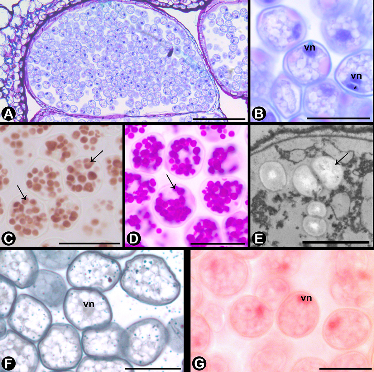
Pollen grains of Laportea aestuans in light microscopy (A-D, F, H) after treatment with different reagents and in transmission electron microscopy (E, G). A. Overview of the pollen grains inside the anther locule after toluidine blue staining. B. Approximate view of the pollen, showing the vegetative cell nucleus after staining with toluidine blue. C-D. Pollen grains stained with lugol reagent (C) and periodic acid-Schiff (D) showing starch grains (arrows). E. Large starch grains (arrows) in the vegetative cell cytoplasm. F. Pollen grains stained with sudan black B. G. Oleosome (red arrowhead) in the vegetative cell cytoplasm. H. Pollen stained with xylidine Ponceau, highlighting the vegetative cell nucleus and the generative cell (*). Abbreviations: vn = vegetative cell nucleus. Scale bars: A-D, F, H = 20 µm; E, G = 1 µm.
The pollen grains are arranged unsystematically in the anther locules. (Fig. 3A) and released from the anthers in a bicellular form (Figs 3B, I). The main reserve substance observed in the vegetative cell is starch (Figs 3C-E), lipids (Figs 3F-G), and proteins (Fig. 3H). Pollen coat substances were not observed on the pollen wall (Figs 2J, 3F).
All viability tests executed on the pollen indicated viability values above 95%. Diaminobenzidine did not work on fixed material but indicated 98.4 ± 0.85% viability on fresh samples. Meanwhile, acetic carmine indicated 98.9 ± 0.17% of viability in tests on fixed material.
Urera baccifera (Table 1, Figs 4-5): The pollen grains are dispersed as monads and isopolar, small-sized, with circular amb, and often oblate-spheroidal, ranging between suboblate and subprolate (Figs 4A-B). In the equatorial view, the polar axis measures, on average, 12.80 ± 0.21 μm, ranging from 11.25 to 15 μm, while the equatorial diameter measures 13.55 ± 0.25 μm on average, ranging between 11.25 and 15 μm. In polar view, the equatorial diameter measures between 10 and 16.25 μm, with an average of 13.50 ± 0.26 μm. The pollen is 3-porate, and the pores have an average diameter of 1.20 μm, often presenting an operculum (Figs 4C-D). The exine is tectate with nanoechinate ornamentation, with irregularly distributed nano-spines along the surface (Figs 4E-F). The spines have an average height of 0.14 μm when measured using scanning electron microscopy. On average, the exine is thin, measuring 0.86 μm, and the sexine is thicker than the nexine (Fig. 4G).

Pollen grains of Urera baccifera in light microscopy after acetolysis (A, C, E), in scanning electron microscopy (B, D, F) and in transmission electron microscopy (G). A-B. Overview of the pollen grains. C-D. View of the pores, showing the operculum in D. E-F. Details of the nanoechinate ornamentation on the exine. G. Approximate view of the exine layers (tectum, sexine and nexine). Abbreviations: ne = nexine, se = sexine, te = tectum. Scale bars: A, C, E = 5 cm; B = 2 µm; D, F = 1 µm; G = 200 nm.

Pollen grains of Urera baccifera in light microscopy (A-D, F, G) after treatment with different reagents and in transmission electron microscopy (E). A. Overview of the pollen grains inside the anther locule after toluidine blue staining. B. Approximate view of the pollen, showing the vegetative cell nucleus and the generative cell (*) after staining with toluidine blue. C-D. Pollen grains stained with Lugol reagent (C) and periodic acid-Schiff (D) showing starch grains (arrows). E. Starch grains (arrows) in the vegetative cell cytoplasm. F. Pollen grains stained with Sudan black B, with a negative reaction for lipid reserves. G. Pollen stained with xylidine Ponceau, highlighting the vegetative cell nucleus and the generative cell (*). Abbreviations: vn = vegetative cell nucleus. Scale bars: A-D, F, G = 20 µm; E = 1 µm.
The pollen grains are arranged asystematically in the anther locules (Fig. 5A) and released from the anthers in a bicellular form; the generative cell is fusiform and is found close to the nucleus of the vegetative cell (Fig. 5B). The main reserve substances of the vegetative cell are starch (Figs 5C-E) and proteins (Fig. 5G). Lipid reserves were not detected in the vegetative cell (Fig. 5F). Pollen coat substances also were not observed on the pollen wall (Figs 4G, 5F).
The pollen viability of samples showed greater differences between tests. In this species, there was no ambiguity between viable and non-viable pollen grains with acetic carmine, which indicated a 98.22 ± 0.45% viability. Similar to L. aestuans, tests with diaminobenzidine did not yield results in fixed material. Still, the tests with fresh samples indicated viability percentages of 52.10 ± 13.50%, with significant variability in the results of each sample.
Urtica dioica (Table 1, Figs 6-7): The pollen grains are monads and isopolar, small sized, with circular amb, and frequently oblate-spheroidal, ranging between oblate and subprolate (Figs 6A-B). On the equatorial view, the polar axis measures, on average, 15.65 ± 0.35 μm, extending from 12.50 to 17.50 μm, while the equatorial diameter varies between 12.50 and 20 μm, with 17.00 ± 0.40 μm on average. On the polar view, the equatorial diameter measures, on average, 17.05 ± 0.26 μm, fluctuating between 15 and 20 μm. The pollen is 3-porate, and the pores have an average diameter of 1.53 μm, often presenting an operculum (Figs 6C-D). The exine is tectate with nanoechinate ornamentation, with irregularly distributed nano-spines along the surface (Figs 6E-F). The spines have an average height of 0.22 μm when measured using scanning electron microscopy. On average, the exine is thin, measuring 0.83 μm, and the sexine is thicker than the nexine (Fig. 6G).

Pollen grains of Urtica dioica in light microscopy after acetolysis (A, C, E), in scanning electron microscopy (B, D, F) and in transmission electron microscopy (G). A-B. Overview of the pollen grains. C-D. View of the pores, showing the operculum in D. E-F. Details of the nanoechinate ornamentation on the exine. G. Approximate view of the exine layers (tectum, sexine and nexine). Abbreviations: ne = nexine, se = sexine, te = tectum. Scale bars: A, C, E = 5 cm; B = 2 µm; D, F = 1 µm; G = 200 nm.

Pollen grains of Urtica dioica in light microscopy (A-D, F, G) after treatment with different reagents and in transmission electron microscopy (E). A. Overview of the pollen grains inside the anther locule after toluidine blue staining. B. Approximate view of the pollen, showing the vegetative cell nucleus and the generative cell (*) after staining with toluidine blue. C-D. Pollen grains stained with lugol reagent (C) and periodic acid-Schiff (D) showing starch grains (arrows). E. Starch grains (arrows) in the vegetative cell cytoplasm. F. Pollen grains stained with sudan black B, with a negative reaction for lipid reserves. G. Pollen stained with xylidine Ponceau, highlighting the vegetative cell nucleus. Abbreviations: vn = vegetative cell nucleus. Scale bars: A-D, F, G = 20 µm; E = 2 µm.
The pollen grains are arranged asistematically in the anther locules (Fig. 7A) and released from the anthers in a bicellular form (Fig. 7B). The main reserve substances observed in the vegetative cell are starch (Figs 7C-E) and proteins (Fig. 7G). Lipid reserves were not detected in the vegetative cell (Figs 7E, F). Pollen coat substances also were not observed on the pollen wall (Figs 6G, 7F).
The pollen viability values were similar between different tests. Diaminobenzidine also did not work on the fixed material of this species. Acetic carmine indicated a viability of 98.8 ± 0.16%.
Analysis of quantitative data
The Principal Component Analysis revealed multivariate differentiation among the three analyzed species based on five quantitative pollen traits (Fig. 8). The first two principal components explained 61.92% of the total variance, with PC1 accounting for 40.5% and PC2 for 21.42%. PC1 was mainly influenced by pollen diameter variables (D1 and D2) and the P/E ratio, whereas PC2 was primarily associated with pore diameter and exine thickness. Along PC1, U. dioica specimens were predominantly separated from those of L. aestuans and U. baccifera and were associated with higher pollen diameter values. In contrast, L. aestuans was positively associated with the P/E ratio, and U. baccifera showed partial overlap with the other two species. No clear pattern of species distribution was observed along PC2.

Principal Component Analysis performed with pollen quantitative variables from Urticeae species. Abbreviations: D1 = Diameter 1 or polar diameter in equatorial view; D2 = Diameter 2 or equatorial diameter in equatorial view; Exine = Exine thickness; PC1 = First Principal Component; PC2 = Second Principal Component; PD = Pore diameter; P/E = Polar/equatorial diameter ratio.
Discussion
The pollen of Laportea aestuans, Urera baccifera, and Urtica dioica, three closely related species of the tribe Urticeae, reveals a largely conserved set of morphological traits. This overall similarity, however, contrasts with variation in aperture number, as U. baccifera and U. dioica are typically 3-porate, whereas L. aestuans exhibits a higher number of pores, suggesting potential differences in pollen performance. Likewise, the predominance of starch as a reserve substance in the pollen vegetative cell is another similarity, although lipids are only found in L. aestuans. Together, these features highlight a pattern of general conservatism with specific deviations that may be functionally relevant and are explored in the following sections.
Pollen traits in Urticeae: a partial fit to the anemophilous syndrome
The pollen grains of Laportea aestuans, Urera baccifera, and Urtica dioica exhibit traits commonly associated with wind pollination, including relatively small size, a reduced number of apertures (except in L. aestuans), a thin, weakly ornamented exine sometimes described as psilate (Punt & Malotaux, 1984), absence of pollenkitt, high pollen viability, and vegetative cell reserves predominantly composed of starch (Whitehead, 1983; Culley et al., 2002; Rosbakh et al., 2026).
The pollen ornamentation of L. aestuans, U. baccifera, and U. dioica is not psilate, but nanoechinate, as typically expected for wind-pollinated taxa. Even so, their exine spines are much smaller than those commonly observed in entomophilous groups, such as Asteraceae. For example, spines in some Praxelis Cass. (Asteraceae) species range from 1.6 to 3.9 µm in height (Abreu et al., 2014), while in the subtribe Inulinae they can reach 2 to 5.5 µm (Coutinho & Dinis, 2007). Interestingly, the numerous small spines on the exine surface are thought to reduce settling velocity by increasing low-density volume, thereby promoting greater dispersal distances in anemophilous pollen (Niklas, 1985; Rosbakh et al., 2026). The thin exine observed in these species may likewise contribute to reduced pollen mass, further facilitating long-distance dispersal (Rosbakh et al., 2026).
Additionally, ornamented pollen in anemophilous species may reduce drag forces acting on pollen grains (Grega et al., 2013), thereby lowering resistance to movement and facilitating dispersal. However, this effect seems to become significant only when the ornamentation occupies a substantial proportion of pollen diameter, such as approximately 12% in experiments with the gymnosperms Pinus strobus L. and Falcatifolium taxoides (Brongn. & Gris) de Laub. (Grega et al., 2013). In contrast, in the three Urticaceae species analyzed here, ornamentation corresponds to less than 2% of the mean pollen diameter (see Figs 2, 4, and 6), suggesting that it likely has a negligible effect on the drag forces in pollen dispersal. Thus, the effects of pollen shape and ornamentation on pollen aerodynamics remain poorly understood and warrant further investigation, as irregular grain shapes and varying degrees of surface roughness may influence dispersal dynamics (Timerman & Barrett, 2021).
The high pollen viability observed in the studied species is consistent with expectations for wind-pollinated taxa. However, because these values were obtained using colorimetric tests, they should be interpreted with caution. Such assays are known to potentially overestimate pollen viability compared to in vivo and in vitro germination tests (Rodriguez-Riano & Dafni, 2000; De Jesus et al., 2018). The colorimetric tests employed in this study, together with the histochemical analyses applied to both anther sections and intact pollen grains, yielded generally consistent results, except in U. baccifera. In this species, diaminobenzidine staining indicated markedly lower viability than acetic carmine, a pattern corroborated by transmission electron microscopy, which revealed that many pollen grains in this species lacked cytoplasmic content. This suggests that diaminobenzidine did not overestimate pollen viability in this case. As reported by Beck-Pay (2012) and supported by our observations, diaminobenzidine is less effective for assessing viability in fixed pollen samples, which may reduce the risk of overestimation. In contrast, acetic carmine may have overestimated pollen viability, at least in U. baccifera. Additionally, in Acacia podalyriifolia A.Cunn. ex G.Don and A. mearnsii De Wild. (Fabaceae), some colorimetric tests showed greater agreement with in vivo pollen germination on the stigma than with in vitro assays (Beck-Pay, 2012). Taken together, these results support the interpretation of high pollen viability as a trait associated with anemophily, while also indicating that colorimetric tests, particularly diaminobenzidine, can provide useful, albeit indirect, estimates of pollen viability when interpreted with appropriate caution (De Jesus et al., 2018).
The predominance of starch as the main reserve substance in all studied species conforms to classical expectations for anemophilous pollen syndromes (Baker & Baker, 1979; Culley et al., 2002). However, the presence of lipids in L. aestuans pollen differs from typical patterns reported for wind-pollinated taxa (Baker & Baker, 1979; Culley et al., 2002; Lu et al., 2021), in which lipids are generally associated with entomophily, serving as a nutritive resource for pollinators (Manning, 2001; Vaudo et al., 2016). This unexpected occurrence of lipids, together with starch, may nevertheless play important roles in pollen survival and germination. This supports the view of Roulston & Buchmann (2000) that reserve substances do not necessarily correlate with pollination mode. Starch and other carbohydrates contribute to osmotic regulation in the vegetative cell and to the maintenance of cellular water content, thereby increasing pollen longevity (Speranza et al., 1997; Nepi et al., 2001), whereas lipids function as energy reserves and structural components for pollen tube growth (Piffanelli et al., 1998; Ischebeck, 2016).
In addition to their role in pollen physiology, reserve substances may also be linked to allergenic potential. L. aestuans, U. baccifera, and U. dioica belong to a group well known for inducing allergic reactions via stinging trichomes and emergences on leaves and stems (Tiotiu et al., 2016). Pollen may also accumulate allergenic compounds (D’Amato & Spieksma, 1990; Negrini et al., 1992; Agashe & Caulton, 2009; Weger et al., 2013), many of which belong to panallergen protein classes capable of triggering cross-reactive allergic responses (Finkina & Ovchinnikova, 2018; Guryanova et al., 2022). Our cytochemical analyses detected proteins and carbohydrates in the vegetative cell cytoplasm of all three Urticaceae species. These proteins, particularly when conjugated with carbohydrates as glycoproteins, are recognized as allergens (Grote, 1999; Tiotiu et al., 2016; Finkina & Ovchinnikova, 2018; Guryanova et al., 2022) and may occur intracellularly, within the pollen wall, or as components of pollenkitt (Roulston et al., 2000; Pacini & Hesse, 2005). Therefore, the pollen of the studied nettle species exhibits potential allergenic properties and warrants further pharmacological investigation.
Pollen morphology of Urticaceae: similarities and discrepancies
The studied species share several pollen morphological characteristics, including pollen grains within the small-size class, as well as similar pollen shape and exine features. However, despite their overall morphological similarity, the studied species exhibited quantitative differences in pollen traits, as evidenced by the multivariate distribution revealed by the PCA (see Fig. 8). In particular, U. dioica was distinguished from L. aestuans and U. baccifera mainly by pollen size, indicating that pollen diameter represents the primary source of quantitative distinction among the analyzed taxa. Nevertheless, the partial overlap observed between species suggests a conserved pollen morphology within Urticeae.
Notably, some quantitative pollen traits observed in the studied species differ from previous palynotaxonomic reports for these taxa. The average pollen size of Urtica dioica observed here was similar, but slightly larger than the 15 μm reported by Sorsa & Huttunen (1975) , reaching up to 17 μm. In contrast, pollen grains of Urera baccifera were previously classified as small, with diameters ranging from 10 to 13.5 μm (Barth, 1976), values that are lower than those obtained in the present study (ca. 16 μm). Nevertheless, most of the observed size values fall within the broader range of 9-29 μm reported by Sorsa & Huttunen (1975), except for Laportea aestuans, which may exhibit slightly smaller pollen grains (ca. 8.75 μm). Additionally, the ranges of polar and equatorial diameters in U. dioica are consistent with those described by Punt & Malotaux (1984) , who reported values of 12.5-17.5 μm and 14-19 μm, respectively.
There are also discrepancies between the number and diameter of pores recorded in the present study and those reported for Urticaceae in the literature. A higher number of pores has previously been described for U. baccifera (10-12 pores: Barth, 1976; 3-4-porate to 6-8-pantoporate grains: Corrêa et al., 2016) and for Urtica species (8-11 pores: Barth, 1976). In contrast, the mean pore diameters observed here for L. aestuans and U. baccifera are smaller than those reported by Corrêa et al. (2016) (2.7 μm and 2 μm, respectively). Nevertheless, the overall range of pore diameters in the three species is consistent with previous reports, including values between 0.5 and 3.1 μm for U. dioica and U. baccifera (Sorsa & Huttunen, 1975), and between 1 and 2.5 μm for U. dioica (Punt & Malotaux, 1984). The presence of an operculum covering the pores, as observed in the present study and also reported by Barth (1976), Punt & Malotaux (1984), and Corrêa et al. (2016), remains debated, as it was not recorded for U. dioica or U. baccifera by Sorsa & Huttunen (1975). Particular attention should be given to the pore structure in L. aestuans, in which an intine-derived oncus was observed beneath the pores. This feature, previously described in Parietaria judaica (Parietarieae; Vega-Maray et al., 2003), is here reported for the first time in the tribe Urticeae and may be associated with pollen osmoregulation and the accommodation of volume changes in the vegetative cell, as proposed for Corylus avellana L. (Heslop-Harrison et al., 1986).
Pollen grains with three pores, frequently observed in the present study for Urera baccifera, were previously reported as a rare condition (Corrêa et al., 2016). Moreover, the exine of this species has been described as granulate under light microscopy (Barth, 1976) and as spinulose when examined using both light and scanning electron microscopy (Sorsa & Huttunen, 1975; Corrêa et al., 2016). These differences highlight the importance of employing multiple microscopy techniques in pollen studies. Most of the remaining morphological discrepancies may be attributed to differences in sampling effort (e.g., larger samples for Urtica dioica and smaller ones for U. baccifera in Sorsa & Huttunen, 1975, compared to the present study), geographic variation (see Kordofani & Ingrouille, 1992), or intraspecific variation among populations or individuals (see Maciejewska-Rutkowska et al., 2021). A synthesis of the pollen characteristics and quantitative data of the three studied species of Urticaceaecan be seen in Table 1 and S1, respectively.
The multi-technique approach applied here to three Urticaceae species enabled a more comprehensive understanding of their pollen biology. Novel features for the tribe are reported, including the presence of an oncus beneath the apertures. Colorimetric assays, particularly diaminobenzidine staining, proved to be a valuable alternative for estimating pollen viability in these species. While most pollen traits are consistent with those typically associated with anemophilous taxa, several notable differences were identified compared to previously published data on Urticaceae, including variation in pollen size, aperture number and type, and exine ornamentation. Contrary to conventional descriptions of anemophilous pollen, our results demonstrate that nettle pollen can store both starch and lipids as reserve substances. In addition to carbohydrates, nettle pollen also contains proteins in the vegetative cell cytoplasm, which may form glycoproteins, compounds known to trigger allergic responses.
Supplementary Material
The following online material is available for this article:
Table S1.
Acknowledgments
We thank Maria Tuca Dolores S. Ferreira, José Augusto Maulin (Department of Cell Biology and Bioagents, FMRP-USP) and Edimárcio S. Campos (FCFRP/USP) for technical assistance.
References
-
Abreu VHR, Santos JC, Esteves RL, Gonçalves-Esteves V. 2014. Pollen morphology of Praxelis (Asteraceae, Eupatorieae, Praxelinae) in Brazil. Plant Systematics and Evolution 301: 599-608. doi: 10.1007/s00606-014-1098-5
» https://doi.org/10.1007/s00606-014-1098-5 - Agashe SN, Caulton E. 2009. Pollen and spores: Applications with special emphasis on aerobiology and allergy. Boca Raton, CRC Press.
-
Baker HG, Baker I. 1979. Starch in angiosperm pollen grains and its evolutionary significance. American Journal of Botany 66: 591-600. doi: 10.1002/j.1537-2197.1979.tb06262.x
» https://doi.org/10.1002/j.1537-2197.1979.tb06262.x -
Barth OM. 1976. Catálogo sistemático dos pólens das plantas arbóreas do Brasil meridional XXIV: Urticaceae. Memórias do Instituto Oswaldo Cruz 74: 341-346. doi: 10.1590/S0074-02761976000300012
» https://doi.org/10.1590/S0074-02761976000300012 -
Beck-Pay SL. 2012. Optimisation of pollen viability tests for Acacia podalyriifolia and two ploidys of Acacia mearnsii South African Journal of Botany 78: 285-289. doi: 10.1016/j.sajb.2011.08.001
» https://doi.org/10.1016/j.sajb.2011.08.001 -
Bellonzi TK, Dutra FV, Sousa CN, Rezende AA, Gasparino EC. 2020. Pollen types of Sapindaceae from Brazilian forest fragments: apertural variation. Acta Botanica Brasilica 34: 327-341. doi: 10.1590/0102-33062020abb0022
» https://doi.org/10.1590/0102-33062020abb0022 - Berg CC. 1990. Differentiation of flowers and inflorescences of Urticales in relation to their protection against breeding insects and to pollination. Botanical Garden and Museum 11: 13-34.
- Berg CC, Rossi PF, Davidson DW. 2005. Flora Neotropica, Volume 94: Cecropia. New York, New York Botanical Garden Press.
-
Conn BJ, Hadiah JT. 2009. Nomenclature of tribes within the Urticaceae. Kew Bulletin 64: 349-352. doi: 10.1007/s12225-009-9108-4
» https://doi.org/10.1007/s12225-009-9108-4 -
Coutinho AP, Dinis AM. 2007. A contribution to the ultrastructural knowledge of the pollen exine in subtribe Inulinae (Inuleae, Asteraceae). Plant Systematics and Evolution 269: 159-170. doi: 10.1007/s00606-007-0585-3
» https://doi.org/10.1007/s00606-007-0585-3 -
Corrêa AMS, Ferreira LG, Ribeiro VBS, Gaglioti AL, Romaniuc Neto S, Guido BMO. 2016. Flora polínica da reserva do Parque Estadual das Fontes do Ipiranga (São Paulo, Brasil). Família: Urticaceae. Hoehnea 43: 479-485. doi: 10.1590/2236-8906-46/2016
» https://doi.org/10.1590/2236-8906-46/2016 -
Culley TM, Weller SG, Sakai AK. 2002. The evolution of wind pollination in angiosperms. Trends in Ecology & Evolution 17: 361-369. doi: 10.1016/S0169-5347(02)02540-5
» https://doi.org/10.1016/S0169-5347(02)02540-5 - Dafni A, Pacini E, Nepi M. 2005. Pollen and stigma biology. In: Dafni A, Kevan PG, Husband BG. (eds.). Practical Pollination Biology. Cambridge, Enviroquest.
-
D’Amato G, Spieksma FTM. 1990. Allergenic pollen in Europe. Grana 30: 67-70. doi: 10.1080/00173139109427772
» https://doi.org/10.1080/00173139109427772 -
De Jesus LDGA, Silva RNO, Gomes MFC et al 2018. Efficiency of colorimetric tests to determine pollen viability in peppers. Revista Brasileira de Agropecuária Sustentável 8: 77-82. doi: 10.21206/rbas.v8i2.497
» https://doi.org/10.21206/rbas.v8i2.497 -
Dutra HP, Freitas AVL, Oliveira PS. 2006. Dual ant attraction in the Neotropical shrub Urera baccifera (Urticaceae): The role of ant visitation to pearl bodies and fruits in herbivore deterrence and leaf longevity. Functional Ecology 20: 252-260. doi: 10.1111/j.1365-2435.2006.01096.x
» https://doi.org/10.1111/j.1365-2435.2006.01096.x -
Emmelin N, Feldberg W. 1947. The mechanism of the sting of the common nettle (Urtica urens). The Journal of Physiology 106: 440-455. doi: 10.1113/jphysiol.1947.sp004225
» https://doi.org/10.1113/jphysiol.1947.sp004225 - Erdtman G. 1960. The acetolysis method - A revised description. Svensk Botanisk Tidksrift 54: 561-564.
- Faegri G, Iversen J. 1966. Textbook of Modern Pollen Analysis. Oslo, Scandinavian University Books.
-
Farag MA, Weigend M, Luebert F, Brokamp G, Wessjohann LA. 2013. Phytochemical, phylogenetic, and anti-inflammatory evaluation of 43 Urtica accessions (stinging nettle) based on UPLC-Q-TOF-MS metabolomic profiles. Phytochemistry 96: 170-183. doi: 10.1016/j.phytochem.2013.09.016
» https://doi.org/10.1016/j.phytochem.2013.09.016 -
Finkina EI, Ovchinnikova TV. 2018. Plant Defensins: Structure, Functions, Biosynthesis, and the Role in the Immune Response. Russian Journal of Bioorganic Chemistry 44: 261-278. doi: 10.1134/S1068162018030056
» https://doi.org/10.1134/S1068162018030056 -
Friis I. (1993) Urticaceae. In: Kubitzki K, Rohwer JG, Bittrich V. (eds.). Flowering Plants: Dicotyledons. New York, Springer. p. 612-630. doi: 10.1007/978-3-662-02899-5_76
» https://doi.org/10.1007/978-3-662-02899-5_76 -
Grega L, Anderson S, Cheetham M et al 2013. Aerodynamic characteristics of saccate pollen grains. International Journal of Plant Sciences 174: 499-510. doi: 10.1086/668694
» https://doi.org/10.1086/668694 - Große-Veldmann B. 2017. Systematics, taxonomy, and evolution of Urtica L. (Urticaceae) . Ph.D. Thesis. University of Bonn, Germany. 2017.
-
Grote M. 1999. In situ localization of pollen allergens by immunogold electron microscopy: allergens at unexpected sites. International Archives of Allergy and Immunology 118: 1-6. doi: 10.1159/000024024
» https://doi.org/10.1159/000024024 -
Guryanova SV, Finkina EI, Melnikova DN, Bogdanov IV, Bohle B, Ovchinnikova TV. 2022. Frontiers in Molecular Sciences 9: 900533. doi: 10.3389/fmolb.2022.900533
» https://doi.org/10.3389/fmolb.2022.900533 -
Gülçin I, Küfrevioglu OI, Oktay M, Büyükokuroglu ME. 2004. Antioxidant, antimicrobial, antiulcer and analgesic activities of nettle (Urtica dioica L.). Journal of Ethnopharmacology 90: 205-215. doi: 10.1016/j.jep.2003.09.028
» https://doi.org/10.1016/j.jep.2003.09.028 -
Halbritter H, Ulrich S, Grímsson F et al 2018. Illustrated Pollen Terminology. New York, Springer . doi: 10.1007/978-3-319-71365-6
» https://doi.org/10.1007/978-3-319-71365-6 -
Heslop-Harrison Y, Heslop-Harrison JS, Heslop-Harrison J. 1986. Germination of Corylus avellana L. (Hazel) pollen: Hydration and the function of the oncus. Acta Botanica Neerlandica 35: 265-284. doi: 10.1111/j.1438-8677.1986.tb01289.x
» https://doi.org/10.1111/j.1438-8677.1986.tb01289.x - Ischebeck T. 2016. Lipids in pollen - They are different. Biochimica et Biophysica Acta 1861: 1315-1328.
- Johansen DA. 1940. Plant microtechnique. New York, McGraw-Hill Book Company.
- Karnovsky MJ. 1965. A formaldehyde glutaraldehyde fixative of high osmolality for use in electron microscopy. Journal of Cell Biology 27: 1-149.
- Kearns CA, Inouye DW. 1993. Techniques for pollination biologists. Louisville, University Press of Colorado.
-
Kordofani M, Ingrouille M. 1992. Geographical variation in the pollen of Acacia (Mimosaceae) in Sudan. Grana 31: 113-118. doi: 10.1080/00173139209430730
» https://doi.org/10.1080/00173139209430730 - Lillie RD. 1954. Histopathology technic and practical histochemistry. New York, McGraw-Hill Book Company .
-
Lu X, Ye X, Liu J. 2021. Morphological differences between anemophilous and entomophilous pollen. Microscopy Research and Technique 85: 1056-1064. doi: 10.1002/jemt.23975
» https://doi.org/10.1002/jemt.23975 -
Maciejewska-Rutkowska I, Bocianowski J, Wronska-Pilarek D. 2021. Pollen morphology and variability of Polish native species from genus Salix L. Plos One 16: e0252253. doi: 10.1371/journal.pone.0243993
» https://doi.org/10.1371/journal.pone.0243993 - McDowell EM, Trump BF. 1976. Histologic fixatives suitable for diagnostic light and electron microscopy. Archives of Pathology & Laboratory Medicine 100: 405-411.
- Melhem TS, Cruz-Barros MAV, Corrêa AS, Makino-Watanabe H, Silvestre-Capelato MSF, Gonçalves-Esteves VL. 2003. Variabilidade polínica em plantas de Campos do Jordão, São Paulo, Brazil. Boletim do Instituto de Botânica 16: 9-104.
-
Negrini AC, Ariano R, Delbono G, Ebbli A, Quaglia A, Arobba D. 1992. Incidence of sensitisation to the pollens of Urticaceae (Parietaria), Poaceae and Oleaceae (Olea europea) and pollen rain in Liguria (Italy). Aerobiologia 8: 355-358. doi: 10.1007/BF02272899
» https://doi.org/10.1007/BF02272899 -
Nepi M, Franchi GG, Pacini E. 2001. Pollen hydration status at dispersal: cytophysiological features and strategies. Protoplasma 216: 171-180. doi: 10.1007/BF02673869
» https://doi.org/10.1007/BF02673869 -
Niklas KJ. 1985. The Aerodynamics of Wind Pollination. The Botanical Review 51: 328-386. doi: 10.1007/BF02861079
» https://doi.org/10.1007/BF02861079 -
O’Brien TP, Feder N, McCully ME. 1964. Polychromatic staining of plant cell walls by toluidine blue O. Protoplasma 59: 368-373. doi: 10.1007/BF01248568
» https://doi.org/10.1007/BF01248568 - O’Brien TP, McCully ME. 1981. The study of plant structure principles and selected methods. Melbourne, Termarcarphi Pty. Ltd.
-
Pacini E, Hesse M. 2005. Pollenkitt - its composition, forms and functions. Flora 200: 399-415. doi: 10.1016/j.flora.2005.02.006
» https://doi.org/10.1016/j.flora.2005.02.006 - Pearse AGE. 1972. Histochemistry: theoretical and applied. Philadelphia, The Williams & Wilkins Company.
-
Pedersoli GD, Leme FM, Leite VG, Teixeira SP. 2019. Anatomy solves the puzzle of explosive pollen release in wind-pollinated urticalean rosids. American Journal of Botany 106: 489-506. doi: 10.1002/ajb2.1254
» https://doi.org/10.1002/ajb2.1254 -
Piffanelli P, Ross JHE, Murphy DJ. 1998. Biogenesis and function of the lipidic structures of pollen grains. Sexual Plant Reproduction 11: 65-80. doi: 10.1007/s004970050122
» https://doi.org/10.1007/s004970050122 -
Punt W, Malotaux M. 1984. Cannabaceae, Moraceae and Urticaceae. Review of Palaeobotany and Palynology 42: 23-44. doi: 10.1016/0034-6667(84)90061-7
» https://doi.org/10.1016/0034-6667(84)90061-7 -
Punt W, Hoen PP, Blackmore S, Nilsson S, Thomas AL. 2007. Glossary of pollen and spore terminology. Review of Palaeobotany and Palynology 143: 1-81. doi: 10.1016/j.revpalbo.2006.06.008
» https://doi.org/10.1016/j.revpalbo.2006.06.008 -
R Core Team. 2026. R: A language and environment for statistical computing (Version 4.5.3) [Computer software]. R Foundation for Statistical Computing. https://www.r-project.org 28 apr 2026
» https://www.r-project.org -
Rodriguez-Riano T, Dafni A. 2000. A new procedure to assess pollen viability. Sexual Plant Reproduction 12: 241-244. doi: 10.1007/s004970050008
» https://doi.org/10.1007/s004970050008 -
Rosbakh S, Nepi M, Pichler M. 2026. Does pollen form follow function? Effects of stigma type, pollination mode and habitat on pollen morphological traits. Journal of Ecology 114: e70290. doi: 10.1111/1365-2745.70290
» https://doi.org/10.1111/1365-2745.70290 - Roulston TH, Buchmann SL. 2000. A phylogenetic reconsideration of the pollen starch-pollination correlation. Evolutionary Ecology Research 2: 627-643.
-
Roulston TH, Cane JH, Buchmann SL. 2000. What governs protein content of pollen: pollinator preferences, pollen-pistil interactions, or phylogeny? Ecological Monographs 70: 617-643. doi: 10.1890/0012-9615(2000)070[0617:WGPCOP]2.0.CO;2
» https://doi.org/10.1890/0012-9615(2000)070[0617:WGPCOP]2.0.CO;2 -
Salgado-Labouriau ML, Vanzolini PE, Melhem TS. 1965. Variation of polar axes and equatorial diameters in pollen grains of the two species of Cassia Grana Palynologica 6: 166-176. doi: 10.1080/00173136509429142
» https://doi.org/10.1080/00173136509429142 -
Sannier J, Baker WJ, Anstett MC, Nadot S. 2009. A comparative analysis of pollinator type and pollen ornamentation in the Araceae and the Arecaceae, two unrelated families of the monocots. BMC Research Notes 2: 1-11. doi: 10.1186/1756-0500-2-145
» https://doi.org/10.1186/1756-0500-2-145 - Sorsa P, Huttunen P. 1975. On the pollen morphology of the Urticaceae. Annals Botanici Fennici 12: 165-182.
-
Speranza A, Calzoni GL, Pacini E. 1997. Occurrence of mono- or disaccharides and polysaccharide reserves in mature pollen grains. Sexual Plant Reproduction 10: 110-115. doi: 10.1007/s004970050076
» https://doi.org/10.1007/s004970050076 -
Timerman D, Barret SCH. 2021. The biomechanics of pollen release: new perspectives on the evolution of wind pollination in angiosperms. Biological Reviews 96: 2146-2163. doi: 10.1111/brv.12745
» https://doi.org/10.1111/brv.12745 -
Tiotiu A, Brazdova A, Longé C et al 2016. Urtica dioica pollen allergy: Clinical, biological, and allergonomics analysis. Annals of Allergy, Asthma & Immunology 117: 527-534. doi: 10.1016/j.anai.2016.09.426
» https://doi.org/10.1016/j.anai.2016.09.426 - Triola MF, Triola CM, Roy J. 2017. Biostatistics for the Biological and Health Sciences. 2 ed. London, Pearson.
-
Vega-Maray AM, Fernández-González D, Valencia-Barrera R, Seoane-Camba JA, Suárez-Cervera M. 2003. Ultrastructural modifications in the apertural intine of Parietaria judaica L. (Urticaceae) pollen during the early stages of hydration. Grana 42: 220-226. doi: 10.1080/00173130310016446
» https://doi.org/10.1080/00173130310016446 - Vidal BC. 1970. Dichroism in collagen bundles stained with Xylidine-Ponceau 2R. Annales d’Histochimie. 15: 289-296.
- Vieira S. 2011. Introdução à Bioestatística. Amsterdam, Elsevier.
-
Weger LA, Bergmann KC, Rantio-Lehtimäki A et al 2013. Impact of pollen. In: Sofiev M, Bergmann KC. (eds.). Allergenic pollen: A review of the production, release, distribution and health impacts. New York, Springer . p. 161-215. doi: 10.1007/978-94-007-4881-1_6
» https://doi.org/10.1007/978-94-007-4881-1_6 - Whitehead DR. 1983. Wind pollination: Some ecological and evolutionary perspectives. In: Real L. (ed.). Pollination Biology. Amsterdam, Elsevier . p. 97-108
-
Wu ZY, Monro AK, Milne RI et al 2013. Molecular phylogeny of the nettle family (Urticaceae) inferred from multiple loci of three genomes and extensive generic sampling. Molecular Phylogenetics and Evolution 69: 814-827. doi: 10.1016/j.ympev.2013.06.022
» https://doi.org/10.1016/j.ympev.2013.06.022 - Zar JH. 2010. Biostatistical Analysis. New Jersey, Prentice-Hall.