Clinicopathologic Session
Clinicopathologic Session
Case 5/2001 Heart failure and insufficiency of the aortic and mitral valves in a 68-year-old woman with rheumatoid arthritis
(Instituto do Coração do Hospital das Clínicas FMUSP - São Paulo)
Luciano Campos Killinger, Paulo Sampaio Gutierrez
São Paulo, SP - Brazil
A 68-year-old woman sought medical care complaining of dyspnea on minimum exertion and pain in the anterior part of the thorax, which worsened with breathing movements.
The patient had rheumatoid arthritis and used 10mg of methotrexate and 5mg of prednisone daily. At the time of preoperative assessment for repair of a joint deformity due to arthritis, a cardiac murmur was diagnosed. The patient was then referred for evaluation at the Instituto do Coração.
On the medical visit (1/22/99), the patient complained of dyspnea when walking quickly or going up a slope. In addition, she also reported 4 episodes of syncope, which were preceded by tachycardic palpitations, during the 3 months prior to the medical visit. On the occasion of 1 of the episodes of syncope, the patient was admitted to an intensive care unit. She denied hypertension and reported hypercholesterolemia. In addition to methotrexate and prednisone, the patient also used 40 mg of furosemide and 0.25mg of digoxin daily.
On physical examination, the patient was in good general condition. Her pulse was regular and 78bpm, and her blood pressure was 130/70mmHg. The examination of her lungs revealed crepitant rales in the base of her thorax. The the heart apex was palpated on the 6th intercostal space on the left midclavicular line. A systolic murmur (++/4+) irradiating to the neck and a diastolic murmur (++/4+) could be heard in the aortic area. The examination of the abdomen and lower limbs was normal. Ulnar deviation of the metacarpophalangeal joints existed.
The laboratory tests (1/19/1999) were as follows: hemoglobin, 13g/dL; hematocrit, 40%; mean corpuscular volume, 83 m³; mean corpuscular hemoglobin concentration, 33g/dL; 12,700 leukocytes/mm³ (74% neutrophils, 1% eosinophils, 19% lymphocytes, and 5% monocytes), 396,000 platelets/mm³; and serum levels of creatinine 0.8mg/dL, of sodium 142mEq/L, and of potassium 4.7mEq/L.
The chest X-ray revealed an enlargement (++/4+) of the cardiac silhouette, and an elongated aorta.
The electrocardiogram (1/19/99) revealed sinus rhythm, heart rate of 79bpm, P axis of +60° forward, QRS axis of 0° backward, hypertrophy of the left ventricle, and secondary alterations in ventricular repolarization (fig. 1).
The echocardiogram (4/23/99) showed the following measures: thickness of the interventricular septum and of the posterior wall of the left ventricle, 9mm; left ventricular diastolic diameter, 76mm; left ventricular systolic diameter, 60 mm; left ventricular ejection fraction, 50%. The diameter of the initial part of the aorta was 37mm, the diameter of the left atrium was 40mm, and the diameter of the right ventricle was 20mm. The aortic valve was calcified, the left ventricular-aortic pressure gradient was estimated as 34mmHg, and the valvar area was estimated as 1.5cm². There were moderate aortic and mitral insufficiency.
The hemodynamic and cineangiocardiographic studies (5/26/99) showed the following: left ventricular pressures, 130/0/25 (systolic/ initial diastolic/ end diastolic); aortic pressures, 130/50 (systolic/diastolic), and irregularities in the coronary arteries. Left ventricular dilation and moderate and diffuse hypokinesia could be seen, also marked aortic valve and moderate mitral valve insufficiencies.
Surgical treatment for the aortic and mitral valve insufficiencies was indicated (6/2/99).
During evolution, the patient needed hospitalization due to intense dyspnea and had undergone cardiopulmonary arrest in another service. Once again (6/23/99), the patient sought medical care complaining of dyspnea triggered by minimum exertion and intense precordial pain.
The physical examination then revealed an intensely dyspneic patient. The pulse was 120bpm, blood pressure 130/70mmHg. Deformation, edema, erythema, and pain existed on palpation and compression of the sternum and left costochondral junctions. Her lung examination revealed subcrepitant rales in the bases, and the heart examination revealed systolic and diastolic (++/4+) murmurs in the aortic area, and a systolic murmur (++/4+) in the mitral area.
The diagnosis of trauma of the sternum and costal arches was established and attributed to the maneuvers of cardiopulmonary resuscitation.
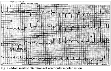
The electrocardiogram (7/23/99) revealed sinus rhythm, a heart rate of 107bpm, P axis of +30° parallel, QRS axis of 10° backward, hypertrophy of the left chambers, and alterations of the ventricular repolarization more marked than those in the first electrocardiogram (fig. 2).
Laboratory findings at that time (16h; 7/23/99) were as follows: hemoglobin of 11.6g/dL; hematocrit of 36%; mean corpuscular volume of 90 µm³; mean corpuscular hemoglobin concentration of 32g/dL; 34,400 leukocytes/mm³ (1% metamyelocytes, 19% band neutrophils, 76% segmented neutrophils, 2% lymphocytes, and 2% monocytes), 66,5000 platelets/mm³; prothrombin time of 13.7s (1.24 INR), activated partial thromboplastin time of 29.7s (time ratio of 1.12). The following serum levels were observed: creatinine, 0.8mg/dL; glucose, 125mg/dL; urea, 65mg/dL; sodium, 136mEq/L; and potassium, 4.8mEq/L.
The patient was admitted to the hospital to have her heart failure compensated and to undergo surgical treatment for her cardiac valve disease.
Her dyspnea improved. On her 2nd day of hospital stay, however, she experienced an episode of hyperthermia.
Ceftriaxone at the dosage of 2g per day was started. The patient also received 37.5mg of captopril, 40mg of furosemide, 0.25mg of digoxin, 5mg of prednisone, 40mg of enoxaparin, and analgesic drugs (tramadol or the paracetamol/codeine association).
On the 3rd night of hospitalization, the patient experienced mental confusion, intense dyspnea, arterial hypotension (60/40mmHg), and cyanosis. Her lung examination was normal, the cardiac sounds had reduced intensity, and the systolic murmur remained audible in the aortic area. Her abdominal examination revealed no visceromegaly. A sore wound existed in the sacral region, and the lower limbs showed no edema or signs of deep thrombosis. Orotracheal intubation was required for ventilatory support and administration of vasoactive drugs. The O2 saturation of the hemoglobin was 93% in a device of mechanical ventilation and O2 inspired fraction of 60%. The patient remained in shock and in the dawn of the following day she had bradycardia, followed by asystolia and death.
Discussion
Clinical findings Our patient is a 68-year-old female with dyspnea on exertion, thoracic pain, and episodes of syncope preceded by tachycardic palpitations she had been previously diagnosed with rheumatoid arthritis with joint deformity, and to whom surgical treatment had been indicated.
We believe that the patient's dyspnea may have had a cardiac origin, because pulmonary disease seems not to have played a role in it. No other respiratory complaints, such as cough, expectoration, history of asthma, bronchitis, tobacco use, or other risk factors for pulmonary diseases existed. The hematocrit was normal. The physical examination revealed rales in the pulmonary bases. The chest X-ray did not show any pulmonary alteration. The echocardiogram revealed a right ventricle with normal dimensions, and no signs of hypertension in the pulmonary artery could be seen.
The patient had valvar heart disease. Her heart examination showed a diastolic (2+/4+) and a systolic (2+/4+) murmur, the latter with irradiation to the neck, both in the aortic area, suggesting possible aortic stenosis. Other clinical conditions, such as congenital supra, sub, and valvar aortic stenoses, and hypertrophic subaortic stenosis (characteristic of hypertrophic cardiomyopathy), may account for a similar auscultation. However, these conditions are not applicable to our case. The patient's aortic stenosis seemed to be functional. The echocardiogram showed a valvar gradient of 34mmHg, and a valvar area of 1.5cm2; these values do not usually suggest critical obstruction to the left ventricular flow.
No evidence suggesting ischemic heart disease existed in this case, despite the patient's hypercholesterolemia, sex, and age (postmenopausal woman). The coronary angiography showed some irregularities in the coronary arteries, not compatible with the moderate degree of dysfunction.
Other causes of heart disease less likely to contribute to left ventricular dysfunction could not be remembered.
Rheumatoid disease may cause a number of heart manifestations, pericarditis being the most common (11 to 50%). Pericarditis may have accounted for the thoracic pain of our patient, which became worse during the breathing movements. Rheumatoid disease may cause myocarditis (20% of the cases), but rarely causes significant ventricular dysfunction. Valvar impairment has been reported in rheumatoid disease and has a prevalence of 3 to 5% in postmortem examinations. Of the rheumatoid diseases, 13% have abnormalities in the mitral valve, and cases of progressive aortic insufficiency have been reported. Other manifestations are: coronary arteritis, disorders of the conducting system of the heart (atrial blocks and arrhythmias). Pleural affections occurring in rheumatoid arthritis may cause similar pain. Pleuritis may be undetectable on chest X-ray and causes pain.
The patient's echocardiogram did not show pericardial effusion or thickening, but in patients with rheumatoid disease, the effusions are evidenced in only 30% of the cases. Usually the electrocardiogram is not diagnostic in such conditions.
The use of methotrexate and other modulators of the immune system, which are used in the treatment of patients with rheumatoid disease, may cause cardiomyopathy, but it is not common.
The valvar heart disease of our patient may have had a rheumatic origin, despite its long evolution. The findings of rheumatoid disease (arthritis + fever) may have been masked, and this may have made the early diagnosis difficult. The aortic rheumatic lesion results from adhesions and fusions of the commissures and cuspides, leading to retraction and stiffness of the free margins of the cuspides. Another stigma of rheumatic disease that may be found in the heart is mitral involvement, which in our case comprises fusion of the commissures and moderate insufficiency. In our patient, the aortic insufficiency seems to be primary, because no evidence of dilation of the ascending aorta existed on echocardiography or on catheterization.
The patient's aortic valve was calcified, also suggesting a degenerative etiology. Diabetes and hypercholesterolemia are risk factors for degenerative lesions of the aortic valve, which is usually accompanied by calcification of the mitral ring and of the coronary arteries, but rarely by aortic valve insufficiency. Mitral insufficiency, in our case, can be secondary to left ventricular dilation.
After 1 month and 21 days, the patient sought medical care once again, complaining of dyspnea on minimum exertion and intense precordial pain. On that occasion, the patient reported an episode of intense dyspnea with cardiopulmonary arrest in another service. Among several possibilities, the intense dyspnea could have resulted from a greater decompensation of the heart failure, infection, or pulmonary thromboembolism.
Clinically, the patient had more decompensated heart failure, hemodynamic instability, and trauma of the sternum and costal arches with evident signs of inflammation. The patient evolved with hyperthermia and received an antibiotic, ceftriaxone, due to the presumed diagnosis of a focus of infection. Based on the last events, one may infer the existence of a pulmonary focus (orotracheal intubation during the cardiopulmonary arrest, aspiration of oropharyngeal content, and thoracic trauma with fracture).
On the 3rd day of hospitalization, the patient evolved with hemodynamic instability. Based on the physical examination, causes, such as acute left ventricular dysfunction and rupture of the mitral or aortic valve, were less likely, because the pulmonary auscultation was normal. Mental confusion, intense dyspnea, and cyanosis are among the findings of cardiovascular collapse, and do not help in the differential diagnosis of our patient. Cardiac sounds with reduced intensity suggest cardiac tamponade as the possible cause of shock. Pulmonary thromboembolism and septic shock are other possibilities with the same level of importance. Chest X-ray and echocardiography could have been useful at that time.
The triple finding of drop in systemic blood pressure, elevation in systemic venous pressure, and quiet heart was reported in 1935 by Beck, and it is typical of cardiac tamponade. In not immediately fatal cases, a drop in cardiac output and blood pressure occurs followed by tachycardia and tachypnea. The patient may become torpid or agitated, and the additional important finding of paradoxical pulse may be difficult to identify in severe hypotension. Venous jugular pressure is usually increased, and the cardiac sounds are usually reduced in intensity or not audible at all. Cold limbs and anuria may be found.
Of the possible causes of cardiac tamponade, the most common are as follows: malignant disease, idiopathic pericarditis, uremia, postacute myocardial infarction with the use of heparin, iatrogenic (following surgical or diagnostic procedures), and bacterial. In our case, the latter is the most probable cause of cardiac tamponade and the fatal evolution.
Purulent pericarditis occurs as a major complication of pneumonia due to contiguous infection: hematogenous (following endocarditis), following surgery, or traumatic (most common agents: staphylococcus and pneumococcus). Pericardial effusion and immunosuppression are predisposing factors.
Bacterial pericarditis is usually a fulminating acute disease that lasts a few days. High fever, nocturnal sweating, and dyspnea are common findings. Most patients do not have the typical pericardial pain.
Survival is low and reaches 30%. The poor prognosis is mainly caused by a failure in clinical diagnosis prior to death. In patients treated only with antibiotics with no pericardial drainage, the rapid development of voluminous pericardial effusion may result in sudden cardiovascular collapse and death due to cardiac tamponade. Mortality may be reduced with the parenteral use of antibiotics and early complete surgical drainage, which also prevent constrictive pericarditis. In a recent series, surgical mortality was 8%, and the 5-year survival was 91%, with no late pericardial constriction. The use of antibiotics should, if possible, be guided by the microbiological findings in the surgically drained fluid.
(Dr. Luciano Campos Killinger)
Diagnostic hypothesis Septic shock of unknown etiology: a) bacterial pericarditis; b) pneumonia.
(Dr. Luciano Campos Killinger)
Autopsy
The postmortem examination revealed moderate double mitral valve lesion, with questionable fusion of the tendinous cords, and double lesion of the aortic valve, with predominance of insufficiency; the right coronary leaflet was partially filled with a friable and bloody material (fig. 3). The histopathologic examination revealed the presence of rheumatoid nodules in both valves (fig. 4), and bacterial endocarditis, with Gram-positive colonies of cocci in the right coronary leaflet of the aortic valve (fig. 5).
These lesions led to moderate dilation of the left ventricle, chronic passive congestion of the lungs and liver, and signs of septic and cardiogenic shock, which caused the patient's death. In regard to the other organs, interstitial nephritis was the only finding, possibly a secondary focus of the infectious process.
Two points are particularly interesting in this case: the etiology of the heart valve disease and the aspects related to the patient's decompensation and death.
Rheumatic disease is the most common cause of heart valve disease, mainly when concomitant involvement of the mitral and aortic valves exists; rheumatoid arthritis, however, may also account for this affection 1,2. We could prove that this happened in this case, not only based on clinical findings, but mainly because of the histopathologic aspect of both valves, which was very characteristic of a rheumatoid nodule (fig. 4).
The cause of cardiac decompensation, which was not clinically clarified, was bacterial endocarditis overlapping the changes in the aortic valve caused by arthritis. Infection may be a complication of any heart valve disease, but it is worth noting that previous reports on endocarditis in cases of heart valve disease due to rheumatoid arthritis are rare 3.
(Dr. Paulo Sampaio Gutierrez)
Session editor: Alfredo José Mansur (ajmansur@incor.usp.br)
Associate editors: Desiderio Favarato (dclfavarato@incor.usp.br)
Vera Demarchi Aiello (anpvera@incor.usp.br)
Mailing address: Alfredo José Mansur - InCor - Av. Dr. Enéas C. Aguiar, 44 - 05403-000 - São Paulo, SP, Brazil
English version by Stela Maris C. e Gandour
- 1. Correlaçăo anatomoclínica - Caso 4/99 - Homem de 63 anos de idade, com insuficięncia cardíaca tręs anos depois de implante de bioprótese em posiçőes aórtica e mitral - Instituto do Coraçăo do Hospital das Clinicas- FMUSP, Săo Paulo). Arq Bras Cardiol 1999; 73: 225-35.
- 2. Shimaya K, Kurihashi A, Masago R, Kasanuki H. Rheumatoid arthritis and simultaneous aortic, mitral, and tricuspid valve incompetence. Int J Cardiol 1999; 71: 181-3.
- 3. Rowe IF, Deans AC, Keat AC. Pyogenic infection and rheumatoid arthritis. Postgrad Med J 1987; 63: 19-22.
Publication Dates
-
Publication in this collection
28 Nov 2001 -
Date of issue
Oct 2001


