Abstracts
OBJECTIVE: To study the clinical pattern of subaortic stenosis associated with perimembranous ventricular septal defect. METHODS: From January 1979 to June 2000, 36 children with perimembranous ventricular septal defect and fixed subaortic stenosis were followed-up regarding anatomic characteristics, evolvement, and clinical events. RESULTS: Age at diagnosis of subaortic stenosis ranged from 6 months to 170 months, and it was less than 1 year in only 2 children. Regarding sex, the distribution was 2:1 with a greater predominance of males. Ventricular septal defect was small in 61.0% of cases, medium in 30.56%, and large in 8.40%; the size of the septal defect decreased during follow-up in 30.56% (11 cases). In all patients, subaortic stenosis was membranous and fixed. During follow-up, 23 patients experienced evolvement of the stenosis. Surgical treatment was performed in 21 cases, and one patient underwent surgery for restenosis. Infectious endocarditis occurred in 2 patients; one of the patients died. CONCLUSION: Subaortic stenosis occurs in the natural history of ventricular septal defect usually after the first year of life, and it is progressive and requires surgery in most cases.
ventricular septal defect; subaortic stenosis; congenital heart disease; endocarditis
OBJETIVO: Estudar o comportamento clínico da estenose subaórtica associada a comunicação interventricular perimembranosa. MÉTODOS: Foram acompanhadas, de janeiro 1979 a junho 2000, quanto às características anatômicas, caráter evolutivo e eventos clínicos, 36 crianças com comunicação interventricular perimembranosa e estenose subaórtica fixa. RESULTADOS: A idade de diagnóstico da estenose subaórtica fixa variou de seis meses a 170 meses, sendo abaixo de 1 ano apenas em duas crianças. Quanto ao sexo a distribuição foi de 2:1 com grande predomínio do masculino. A comunicação interventricular era de tamanho pequeno em 61,00% dos casos, médio em 30,56% e grande em 8,40%, apresentando diminuição do tamanho da comunicação durante o acompanhamento em 30,56% (11 casos). Em todos os pacientes a estenose subaórtica era fixa, em membrana. Durante o tempo de acompanhamento, 23 pacientes apresentaram progressão da estenose. Foi realizado tratamento cirúrgico em 21 casos, sendo um paciente reoperado por reestenose. Endocardite bacteriana ocorreu em dois casos, um deles faleceu. CONCLUSÃO: A estenose subaórtica ocorre na história natural da comunicação interventricular geralmente após o 1º ano de vida, apresentando caráter progressivo e necessitando de cirurgia na maioria dos casos.
comunicação interventricular; estenose subaórtica; cardiopatia congênita; endocardite
ORIGINAL ARTICLE
Subaortic stenosis associated with perimembranous ventricular septal defect. Clinical follow-up of 36 patients
Maria da Gloria Cruvinel Horta; Carlos Alberto Franco Faria; Dilermando Fazito Rezende; Tereza Lucia Masci; Cathia Costa C. Rabelo; Tamara Katina; Marly de Oliveira; Luciana Paulino Oliveira
Belo Horizonte, MG - Brazil
Faculdade de Medicina da UFMG e Santa Casa de Misericórdia de Belo Horizonte
Correspondence Correspondence to Maria da Glória Cruvinel Horta Rua Modesto Carvalho Araújo, 428 - Belo Horizonte, MG Cep 30320-410 E-mail: mgchorta@hotmail.com
ABSTRACT
OBJECTIVE: To study the clinical pattern of subaortic stenosis associated with perimembranous ventricular septal defect.
METHODS: From January 1979 to June 2000, 36 children with perimembranous ventricular septal defect and fixed subaortic stenosis were followed-up regarding anatomic characteristics, evolvement, and clinical events.
RESULTS: Age at diagnosis of subaortic stenosis ranged from 6 months to 170 months, and it was less than 1 year in only 2 children. Regarding sex, the distribution was 2:1 with a greater predominance of males. Ventricular septal defect was small in 61.0% of cases, medium in 30.56%, and large in 8.40%; the size of the septal defect decreased during follow-up in 30.56% (11 cases). In all patients, subaortic stenosis was membranous and fixed. During follow-up, 23 patients experienced evolvement of the stenosis. Surgical treatment was performed in 21 cases, and one patient underwent surgery for restenosis. Infectious endocarditis occurred in 2 patients; one of the patients died.
CONCLUSION: Subaortic stenosis occurs in the natural history of ventricular septal defect usually after the first year of life, and it is progressive and requires surgery in most cases.
Key words: ventricular septal defect, subaortic stenosis, congenital heart disease, endocarditis
Subaortic stenosis may occur as a complication of the natural evolvement of several congenital heart diseases; however, its etiology is still unclear1,2. Ventricular septal defect may develop into subaortic stenosis in 20% of cases2-4. The subaortic stenosis developed in these cases is frequently membranous and fixed, and it may occur when the diameter of the ventricular septal defect decreases, after spontaneous closure5-7, or after surgical correction8,9.
In this study, we assessed the clinical evolvement, the echocardiographic, cineangiographic, and surgical characteristics of children with perimembranous ventricular septal defect who developed subaortic stenosis during follow-up.
Methods
From January 1979 to June 2000, 36 children with perimembranous ventricular septal defect who developed fixed subaortic stenosis were followed-up. Age ranged from 6 to 170 months; 12 children were females, and 24 were males. Children with a diagnosis of subaortic stenosis were included in the study.
Cases of subaortic stenosis associated with other types of septal defect and other heart diseases that might interfere with the clinical evolvement and the indication of surgical correction were excluded.
Data about clinical evolvement of this group were retrospectively collected. With the necessary revisions, the following data from bidimensional Doppler echocardiograms and/or surgery and/or cineangiography were assessed: identification of the anatomical type10 and size11 of the ventricular septal defect, anatomic characteristics of the fixed subaortic stenosis12, evolvement assessment of the left ventricular outflow pressure gradient; symptomatology; events occurring during clinical evolvement: infectious endocarditis, surgical treatment, duration of follow-up, and death.
Doppler echocardiography was recorded during previous examinations, by using bidimensional examinations, pulsed Doppler, and continuous wave Doppler. Maximum and medium transvalvular aortic gradient were determined using Bernoulli's simplified equation.
Statistical analysis was performed using ANOVA - One Way - tables with comparison between the average demonstrated in a numerical scale, the chi-square test or Fisher's exact test (when indicated), Fisher's exact test to determine the level of statistical significance of the differences observed, using the Kruskal-Wallis test whenever Bartlett's test was significant. The level of statistical significance adopted was 5%.
Results
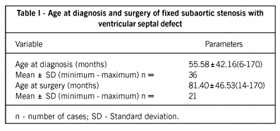
Table I presents the distribution of ages at diagnosis and subaortic stenosis surgery.
Diagnosis of subaortic stenosis by echocardiogram was performed in 29 (80.6%) children and by cineangiography in 7 (19.4%). Males were predominant over females, accounting for 66.67% (24) and 33.33% 12 of the cases, respectively. In 5 patients (13.90%), the diagnosis of subaortic stenosis was simultaneous with that of ventricular septal defect, with all 5 patients over 2 years old. None of the diagnoses of subaortic stenosis occurred after surgical closure of the septal defect, and no reports of familial incidence occurred in the cases studied.
Table II describes the sizes of ventricular septal defects in the sample.
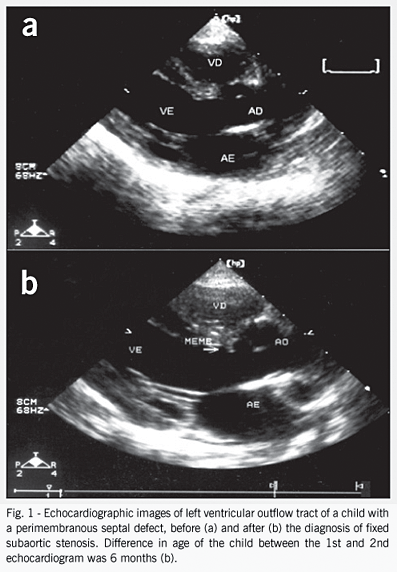
We observed a trend toward development of subaortic stenosis following the decrease in size of the ventricular septal defect in 11 (30.56%) children. Figure 1 demonstrates serial echocardiograms of the same children, with the appearance of subaortic stenosis by 6 months.
The pattern of the ventricular septal defect during follow-up is described in table III.
We identified other associated heart diseases in 6 patients (16.67%), described in table IV.
Table V demonstrates the subaortic gradient at diagnosis and surgery in 23 children (63.80%). We have observed a progressive increase in the left ventricular outflow pressure gradient during follow-up (mean, 20.90 ±6.81 mmHg), whereas in 5 children (13.89%) significant progression of the gradient (>5 mmHg) did not occur in the follow-up period of 36 and 127 months (mean, 81.60 ± 45.60). In 13 patients (36.11%), significant progression of the gradient occurred during follow-up, and the patients underwent surgery.
None of the patients had symptoms related to the development of subaortic stenosis.
The characteristics of evolvement and treatment are found in table VI.
In 15 cases (41.67%), children were followed-up only through clinical treatment for a period of 12 to 130 months (mean, 58.38 ± 40.17). Surgical treatment was performed in 21 children (58.33%), and surgery was indicated when the subaortic gradient was higher than 40 mmHg except for a patient with endocarditis who had a 35 mmHg gradient and underwent. In 8 children (22.22%), the surgery was indicated immediately after the diagnosis because they had a subaortic gradient above 40 mmHg. The 21 children (58.33%) undergoing surgery were followed-up for a mean time of 23.41 ± 34.34 months, with one case of reoperation for subaortic stenosis recurrence during that period.
Two patients (5.56%) had infectious endocarditis at 48 and 60 months of age, respectively, and underwent surgery. One of the patients evolved to death in the immediate postoperative period.
Discussion
Diagnosis of subaortic stenosis occurred after the first year of life in 94.40% of cases, with a predominance of males in a 2:1 ratio, in agreement with other observations 13-18.
We have not identified a family history in the anamneses in our study group. Petsas and cols 19. reported 4 cases of different anatomic types of subaortic stenosis in one family.
Seven children underwent cardiac catheterization before the use of echocardiography.
The first reports on subaortic stenosis in patients with ventricular septal defect and left ventricular pressure outflow previously reported as normal through cineangiography occurred in the 70s and the 80s. Some authors attributed these findings to the inaccuracy of the method stating that cineangiography-the procedure routinely performed at that time-was not suitable for the diagnosis of subaortic stenosis 20.
The advent of echocardiography, a safe and noninvasive method of follow-up of patients with ventricular septal defect, enableld us to understand that the obstruction may not be present in the first year of life in children, but arises generally when the ventricular septal defect shows signs of a decrease in size and spontaneous closure 21-23. We have observed its appearance before one year of age in only 2 patients.
We have clearly identified the tendency of subaortic stenosis to occur especially in perimembranous ventricular septal defects with favorable characteristics for spontaneous closure, because most of them were small and had formation of subtricuspid tissue in their borders. The mechanism of spontaneous closure of the ventricular septal defect was regarded by some authors as responsible for the formation of the obstruction in the left ventricular outflow tract 5,7.
Subaortic stenosis did not cause symptoms, even in those patients with up to 90 mmHg of left ventricular outflow tract gradient, and its appearance did not lead to electrocardiographic or laboratory alterations, therefore being an occasional finding in control echocardiograms as in other studies 24-26, although some authors report symptoms in adulthood.
Eight children presented at the first examination with high gradients in left ventricular outflow tract. These late diagnoses may be explained through the absence of symptoms and the lower social level of our patients.
Twenty-three children (63.90%) had subaortic stenosis progression with a mild increase in the gradient over the years, whereas others evolved in a few months to surgical treatment. This finding confirms the evolving characteristic of the disease 27,28, and demonstrates the need for prolonged follow-up of these patients29,30.
Usually, surgery was indicated when the subaortic gradient was > 40 mmHg. Some physicians indicate surgery immediately after diagnosis, regardless of the gradient, because it is not a benign progressive disease 31,32. Other physicians indicate it with gradients between 20 to 80 mmHg 33,34. It is safer to indicate surgery with higher gradients, because many children observed for years did not experience progression of the subaortic stenosis35,36.
It is interesting to observe that, in our series, surgical indication occurred due to the development of stenosis rather than to clinical repercussions from ventricular septal defect, which was small in most cases, therefore explaining the absence of a correlation between size of ventricular septal defect and surgical indication. We agree that the need for surgery is determined by the subaortic gradient rather than by the size of the ventricular septal defect in most cases 37,38, in several studied series.
Restenosis occurred in one child who was operated on, in the 80s, when surface resection of the stenotic membrane was common. Later, prevention of restenosis was performed through myectomy deeper in the left ventricular outflow tract 39,40.
Two of 36 children (5.50%) had infectious endocarditis, and one child died. The incidence of endocarditis, a frequent complication in this disease, has decreased 41,42. The low social level of our sample, with poor access to dental treatment, may explain its recent occurrence in one of our children.
In conclusion, we have observed in our sample that the diagnosis of fixed subaortic stenosis occurred in the majority of cases after the first year of life, with a predominance of males in a ratio of 2:1. All patients were asymptomatic, with the diagnosis made by echocardiographic follow-up. In the majority of cases, subaortic stenosis with ventricular septal defect developed together with a small perimembranous septal defect. A progression in the subaortic gradient in 63.89% of cases during clinical follow-up, and this gradient, rather than the size of the ventricular septal defect, was the main determinant of surgical indication. Infectious endocarditis occurred in 2 children, leading to death in one.
References
Received for publication: 25/05/2003
Accepted for publication: 04/06/2004
- 1. Kitchiner, D. Subaortic stenosis: still more questions than answers. Heart 1999; 82:647-648.
- 2. Somerville, J. Fixed subaortic stenosis: a frequently misunderstood lesion. Int J Cardiol 1985; 8:145-148.
- 3. Otterstad JE, Erikssen J, Michelsen S, Nitter Hauge S. Long- term follow-up in isolated ventricular septal defect considered too small to warrant operation. J Intern Med 1990; 228:305-309.
- 4. Vogel M, Smallhorn JF, Freedom RM, Coles J, Williams WG, Trusler GA. An echocardiographic study of the association of ventricular septal defect and right ventricular muscle bundles with a fixed subaortic abnormality. Am J Cardiol 1988; 1,61:857-860.
- 5. Zielinsky P, Rossi M, Haertel JC, Vitola D, Lucchese FA, Rodrigues R. Subaortic fibrous ridge and ventricular septal defect: role of septal malalignment. Circulation 1987; 75:1124-9.
- 6. Chung KJ, Fulton DR, Kreidberg MB, Payne DD, Cleveland RJ. Combined discrete subaortic stenosis and ventricular septal defect in infants and children. Am J Cardiol 1984; 53:1429-32.
- 7. Zielinsky P. Correlação morfológico-ecocardiográfica bidimensional na detecção dos mecanismos responsáveis pela diminuição do diâmetro da comunicação interventricular perimembranosa. Tese de doutorado. Universidade Federal do Rio Grande do Sul, Porto Alegre, 1988.
- 8. Alden HD, Anderson RC, Noren GR, Moller JH. Post-operative follow-up of patients with ventricular septal defect. Circulation 1974; 50:465-71.
- 9. Cicini MP, Giannico S, Marino B, Iorio FS, Corno A, Marcelletti C. "Acquired" subvalvular aortic stenosis after repair of a ventricular septal defect. Chest 1992; 101:115-8.
- 10. Becker A, Anderson RH. Classification of Ventricular Septal Defects: A matter of precision. Heart Vessels 1985; 1: 120-1.
- 11. Sharef DS, Huhta JC, Marantz P, Hawkins HK, Yoon GY. Two-dimensional echocardiographic determination of ventricular septal defect size: correlation with autopsy. Am Heart J 1989; 117:1333-6.
- 12. Becker A, Anderson R. Pathology of Congenital Heart Disease. London: Britterworths. 1981; p. 93-117.
- 13. Graham TP, Gutgesell MD. Ventricular septal defect. In: Moss AJ, Adams FH. Heart disease in infants, children and adolescents including the fethus and young adult. 5 ed. Baltimore: Emmanouelides 1995; v. 1: n. 53, 724-46.
- 14. Kitchiner D, Jackson M, Malaiya N, et al. Incidence and prognosis of obstruction of the left ventricular outflow tract in Liverpool (1960-91): a study of 313 patients. Br Heart J 1994; 71:588-95.
- 15. Moss AJ, Adonis FH, Emmanouilides GC. Heart disease in infants, children and adolescents. London: Williams and Wilkins, 1995.
- 16. Newfeld EA, Muster AJ, Paul MH. Discrete subvalvular aortic stenosis in childhood. Study of 51 patients. Am J Cardiol 1976; 38:53-61.
- 17. Shem-Tov A, Schneeweiss A, Motro M, Neufeld HN. Clinical presentation and natural history of mild discrete subaortic stenosis. Follow-up of 1-17 years. Circulation 1982; 66:509-12.
- 18. Somerville J. Congenital heart disease: changes in form and function. Br Heart J 1979; 41:1-22.
- 19. Petsas AA, Anastassiades LC, Constantinou EC, Antonopoulos AG. Familial discrete subaortic stenosis. Clin Cardiol 1998; 21:63-5.
- 20. Grenadier E, Keidar S, Alpan G, Milo S, Palant A. Discrete membranous sub-aortic stenosis in adult patient obtained by echocardiography and not proved by catheterization. Angiology 1982; 33:800-5.
- 21. Leichter DA, Sullivan I, Gersony WM. "Acquired" discrete subvalvular aortic stenosis: natural history and hemodynamics. J Am Coll Cardiol 1989;15:14:1539-44.
- 22. Vogel M, Freedom RM, Brand A, Trusler GA, Williams WG, Rowe RD. Ventricular septal defect and subaortic stenosis: na analysis of 41 patients Am J Cardiol 1983; 52:1258-63.
- 23. Lampros TD, Cobanoglu A. Discrete subaortic stenosis: an acquired heart disease. European J Cardio-Thorac Surg 1998; 14: 296-303.
- 24. Newfeld EA, Muster AJ, Paul MH. Discrete subvalvular aortic stenosis in childhood. Study of 51 patients. Am J Cardiol. 1976; 38:53-61.
- 25. Katz NM, Buckley MJ, Liberthson RR. Discrete membrane subaortic stenosis. Report of 31 patients, review of the literature, and delineation of management. Circulation. 1977; 56:1034-8.
- 26. De Vries AG, Hess J, Witsemburg M, et al. Management of fixed subaortic stenosis- a retrospective study of 57 cases. J Am Coll Cardiol. 1992; 19:1013-7.
- 27. Wright GB, Keane JF, Nadas AS, Bernhard WF, Castaneda R. Fixed subaortic stenosis in the young: medical and surgical course in 83 patients. Am J Cardiol. 1983; 1:52:830-5.
- 28. Somerville J, Stone S, Ross D. Fate of pacients with fixed subaortic stenosis after surgical removal. Br Heart J. 1980; 43:629-47.
- 29. Reis RL, Peterson LM, Mason DT, Simon AL, Morrow AG. Congenital fixed subaortic stenosis: a anatomical classification and correlations with operative results. Circulation. 1971; 43-44:111-8.
- 30. Brown Stevens L, Lynch L, Caldwell R, et al. Surgery of discrete subvalvular aortic stenosis actuarial survival, hemodynamic results, and acquired aortic regurgitation. Ann Thor Surg. 1985; 40:151-4.
- 31. Vouhé PR, Neveux JI. Surgical management of diffuse subaortic stenosis: an integrated approach. Ann Thorac Surg. 1991; 52:654-62.
- 32. Douville EC, Sade RM, Crawford FAJ, Wiles HB. Subvalvar aortic stenosis: Timing of operation. Ann Thorac Surg. 1990; 50:29-34.
- 33. Rohlicek CV, Del Pino SF, Hosking M, et al. Natural history and surgical outcomes for isolated discrete subaortic stenosis in children. Heart. 1999; 82:6, 708-13.
- 34. Brauner R, Laks H, Drinkwater DC, JR, et al. Benefits of early surgical repair in fixed aortic stenosis. JACC. 1997; 30:35-42.
- 35. Ivert T, Astudillo R, Brodin LA, Wranne B. Late results after resection of fixed subaortic stenosis. Scand J Thorac Cardiovasc Surg. 1989; 23:211-8.
- 36. Moses RD, Barnhart GR, Jones M. The late prognosis after localized resection for fixed (discrete and tunnel) left ventricular outflow tract obstruction. J Thorac Cardiovasc Surg. 1984; 87:410-20.
- 37. Newfeld EA, Muster AJ, Paul MH. Discrete subvalvular aortic stenosis in childhood. Study of 51 patients. Am J Cardiol. 1976; 38:53-61.
- 38. Hardesty RL, Griffith BP, Matews RA. Discrete subvalvular aortic stenosis. An evaluation of operative therapy. J Thorac Card Surg. 1977; 74:352-60.
- 39. Keralay E, Ozal E, Bingol H, Cingoz F, Tatar H. Discrete subaortic stenosis: assessing adequacy of myectomy by transesophageal echocardiography. J Card Surg. 1999; 14:348-53.
- 40. Lavee J, Porat L, Smolinsky A, Hegesh J, Neufeld HN, Goor DA. Myectomy versus myotomy as an adjunct to membranectomy in the surgical repair of discrete and tunnel subaortic stenosis. J Thorac Cardiovasc Surg. 1986; 92:944-9.
- 41. Kondo N; Ono Y; Onozuka N; Koyama M; Fukui K; Takaya S; Suzuki S.Surgical treatment of infectious endocarditis complicated by subaortic stenosis. Kyobu Geka. 2001; 54:777-9.
- 42. Sharma BD, Mittal S, Kasliwal RR, Trehan N, Kohli V. Discrete subvalvular aortic stenosis. J Assoc Physicians India. 2000;48:1103-6.
Publication Dates
-
Publication in this collection
09 Mar 2005 -
Date of issue
Feb 2005
History
-
Accepted
04 June 2004 -
Received
25 May 2003