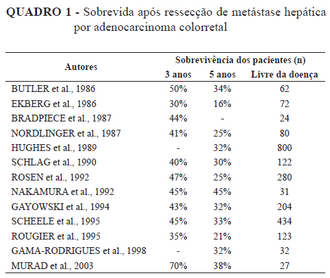
BACKGROUND: Colorectal cancer belongs to the most frequent malignant neoplasia in the world and responsible for the cause of death among other types of cancer; ranked second behind lung cancer. Metastasis frequently occurs and disease worsening leads to patient death. AIM: To analyze if radical surgical resection for colorectal cancer liver metastases with resection margin greater than 10 mm promotes better survival rates and the factors that might predict prognosis. METHODS: Retrospective analysis of 49 patients presenting colorectal adenocarcinoma liver metastases without evidence of concomitant disease and submitted to surgical treatment. Epidemiologic parameters were: age, gender, size of liver metastasis and or the largest lesion, number of regional lymph nodes dissection and involvement, neoplasia-free margin resection. Patients were evaluated clinically, undergoing laboratory exams analysis and imaging studies for disease follow-up. Exclusion criteria were non-histological proof of liver metastasis and evidence of disease in sites other than colon and liver, at the time of surgical treatment and liver metastasis. RESULTS: Casuistic group consisted of 24 female and 25 male patients. Mean and standard deviation for age was 55,9 + 11,9 years, median of 56 years. Surgical procedures included 15 right hepatectomy and 11 left hepatectomy; 13 right and left segmentectomy; 9 nodulectomy and 1 biopsy. Additionally, 2 alcoholization, 4 chemoembolization, 1 thermoablative therapy, 1 selective portal vein block with later right hepatectomy and thermoablative thereapy on segments III and IV were performed. Liver weighted 555,71 + 261,96 g, median of 600g. Median of lymph nodes resection was 2. The mean lesion size consisted in 4,45 + 2,8. Resection margin greater than 10 mm was observed in 32 cases. Serum CEA value before surgical procedure was 68,13 + 105,65 ng/ml, median of 22,2 ng/ml. Death occurred in 22 cases (44,89%). Predominant histological diagnoses was moderate differentiated tubular adenocarcinoma in 65,96%, 17,02% poorly and 17,02% well differentiated. Factors such as undifferentiated histological type, less inflammatory peritumor infiltration, greater desmoplastic reaction and the absence of capsule around the tumor seem to reflect worse prognosis, although none of the factors being statistic significantly isolated. Significant association was noticed between CEA serum level under 7 ng/mg and synchronic hepatic metastases. CONCLUSION: Radical surgical resection for colorectal cancer liver metastases with a resection margin greater than 10 mm promotes better survival rates; elevated serum CEA levels were related to recurrence after hepatic resection for metastatic colorectal cancer and worse clinical outcome; undifferentiated histological type, less inflammatory peritumor infiltration, greater desmoplastic reaction and the absence of capsule around the tumor suggested worse prognosis.
Colorectal neoplasms; Liver neoplasms; Prognosis