Abstracts
BACKGROUND: Immune-mediated skin diseases encompass a variety of pathologies that present in different forms in the body. OBJECTIVE: The objective of this study was to establish the prevalence of the principal immune-mediated skin diseases affecting the oral cavity. METHODS: A total of 10,292 histopathology reports stored in the archives of the Anatomical Pathology Laboratory, Department of Oral Pathology, Federal University of Rio Grande do Norte, covering the period from 1988 to 2009, were evaluated. For the cases diagnosed with some type of disease relevant to the study, clinical data such as the gender, age and ethnicity of the patient, the anatomical site of the disease and its symptomatology were collected. RESULTS: Of all the cases registered at the above-mentioned service, 82 (0.8%) corresponded to immune-media ted skin diseases with symptoms affecting the oral cavity. The diseases found in this study were: oral lichen planus, pemphigus vulgaris and benign mucous membrane pemphigoid. Oral lichen planus was the most common lesion, comprising 68.05% of the cases analyzed. Of these cases, 64.3% were women and the cheek mucosa was the anatomical site most commonly affected (46.8%). CONCLUSION: Immune-mediated skin diseases affecting the oral cavity continue to be rare, the prevalence found in this study being similar to that reported for the majority of regions worldwide. Nevertheless, early diagnosis is indispensable in the treatment of these diseases, bearing in mind that systemic involvement is possible in these patients.
Lichen planus; oral; Pemphigoid; benign mucous membrane; Pemphigus
FUNDAMENTO: As doenças dermatológicas imunologicamente mediadas compõem diversas patologias que apresentam formas variadas de manifestação no organismo. OBJETIVO: Foi proposição desta pesquisa, estabelecer a prevalência das principais doenças dermatológicas imunologicamente mediadas que apresentam manifestação oral. MÉTODOS: Foram avaliados laudos histopatológicos de 10.292 casos arquivados no Serviço de Anatomia Patológica da Disciplina de Patologia Oral da Universidade Federal do Rio Grande do Norte, no período de 1988 a 2009. Dos casos diagnosticados como algum tipo de doença em estudo, coletaram-se dados clínicos como sexo, idade, raça, sítio anatômico e sintomatologia das doenças. RESULTADOS: Do total de casos registrados, no serviço supracitado, 82 (0,8%) corresponderam a doenças dermato lógicas imunologicamente mediadas com manifestação na cavidade oral. As doenças encontradas neste estudo foram: líquen plano oral, pênfigo vulgar e penfigoide benigno das membranas mucosas, sendo o líquen plano oral a lesão mais prevalente, representando 68,05% dos casos analisados, dos quais 64,3% apresentavam-se em mu lheres, sendo a mucosa jugal o sítio anatômico mais acometido (46,8%). CONCLUSÃO: A ocorrência de doenças dermatológicas imunologicamente mediadas que apresentam manifestação oral ainda é um fato incomum, semelhante ao observado na maioria das regiões mundiais. No entanto, a busca pelo diagnóstico precoce é um requisito essencial para a condução do tratamento dessas doenças, tendo em vista o possível comprometimento sistêmico do organismo nos pacientes.
Líquen plano bucal; Penfigoide mucomembranoso benigno; Pênfigo
INVESTIGATION
An epidemiological study of immune-mediated skin diseases affecting the oral cavity*
Cyntia Helena Pereira de CarvalhoI; Bruna Rafaela Martins dos SantosII; Camila de Castro VieiraIII; Emeline das Neves de Araújo LimaII; Pedro Paulo de Andrade SantosII; Roseana de Almeida FreitasIV
IMaster's degree. Professor of Oral Microbiology, School of Dentistry, Federal University of Campina Grande (UFCG), Patos, Paraíba, Brazil
IIMaster's degree. Doctoral student in Oral Pathology at the Federal University of Rio Grande do Norte (UFRN), Natal, Rio Grande do Norte, Brazil
IIIUndergraduate student, School of Dentistry, Federal University of Rio Grande do Norte (UFRN), Natal, Rio Grande do Norte, Brazil
IVPhD. Professor of the Postgraduate Program in Oral Pathology, Federal University of Rio Grande do Norte (UFRN), Natal, Rio Grande do Norte, Brazil
Mailing address
ABSTRACT
BACKGROUND: Immune-mediated skin diseases encompass a variety of pathologies that present in different forms in the body.
OBJECTIVE: The objective of this study was to establish the prevalence of the principal immune-mediated skin diseases affecting the oral cavity.
METHODS: A total of 10,292 histopathology reports stored in the archives of the Anatomical Pathology Laboratory, Department of Oral Pathology, Federal University of Rio Grande do Norte, covering the period from 1988 to 2009, were evaluated. For the cases diagnosed with some type of disease relevant to the study, clinical data such as the gender, age and ethnicity of the patient, the anatomical site of the disease and its symptomatology were collected.
RESULTS: Of all the cases registered at the above-mentioned service, 82 (0.8%) corresponded to immune-media ted skin diseases with symptoms affecting the oral cavity. The diseases found in this study were: oral lichen planus, pemphigus vulgaris and benign mucous membrane pemphigoid. Oral lichen planus was the most common lesion, comprising 68.05% of the cases analyzed. Of these cases, 64.3% were women and the cheek mucosa was the anatomical site most commonly affected (46.8%).
CONCLUSION: Immune-mediated skin diseases affecting the oral cavity continue to be rare, the prevalence found in this study being similar to that reported for the majority of regions worldwide. Nevertheless, early diagnosis is indispensable in the treatment of these diseases, bearing in mind that systemic involvement is possible in these patients.
Keywords: Lichen planus, oral; Pemphigoid, benign mucous membrane; Pemphigus
INTRODUCTION
Immune-mediated skin diseases are pathological conditions that occur when the immune system becomes activated against components of the organism itself. The mucocutaneous lesions are characterized by an inadequate production of antibodies directed against adhesion molecules such as the desmosomes and the hemidesmosomes, which are responsible for adhesion between the epithelial cells, as well as between the epithelium and the underlying connective tissue, through components of the basal membrane. These interactions between the autoantibodies and the host tissue result in tissue damage visible clinically as a pathological process often referred to as autoimmune vesiculobullous disease. 1-4
The most common immune-mediated skin diseases are: pemphigus vulgaris, benign mucous membrane pemphigoid, lichen planus, erythema multiforme, systemic lupus erythematosus and graft-versushost disease. 2,4
Pemphigus vulgaris is characterized by the formation of intraepithelial blisters that result from the disintegration or loss of cellular adhesion, with the consequent loss of intercellular connections known as acantholysis. 5,6 After the blisters have burst, diffuse ulceration follows, leading to debilitating pain, fluid loss and electrolyte imbalance. Its pathogenesis is characterized by the abnormal production of autoantibodies against the surface glycoproteins of the epithelial cells known as desmoglein 1 and desmoglein 3, which are components of the desmosomes. 7
Cicatricial pemphigoid was referred to by Lo Russo et al. as a group of chronic autoimmune bullous diseases in which autoantibodies are directed against one or more components of the basal membrane. 3,8,9,10
Bascones et al. defined lichen planus as a chronic mucocutaneous disease of an inflammatory nature and undefined etiology in which lymphocytes attack the basal cells of the epithelium in the oral mucosa. 11 Scully and Carrozzo called attention to the fact that various clinical forms of lichen planus have been identified: reticular, papular, plaque-like, erosive (ulcerative) and bullous, and that these forms may occur separately or simultaneously. 4
Various studies have been published in the literature worldwide in which the possible etiologic factors, means of diagnosis and recognized treatments for immune-mediated skin diseases affecting the mouth are discussed. Nevertheless, few studies have been conducted on the prevalence of these diseases affecting the oral cavity, particularly in South America and more specifically in Brazil.
Therefore the objective of the present study was to establish the prevalence of the principal immunemediated skin diseases affecting the oral cavity.
MATERIALS AND METHODS
The prevalence of the principal immune-mediated skin diseases affecting the oral cavity was obtained by analyzing the histopathology reports and clinical charts at the laboratory of anatomical pathology of the Department of Oral Pathology, Federal University of Rio Grande do Norte with respect to the period ranging from 1988 to 2009. Based on the histopathology reports, the other clinical data such as the patient's gender, age and ethnicity, the anatomical site of the lesions and the symptoms of the diseases were then investigated.
RESULTS
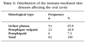
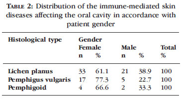
Of a total of 10,292 cases registered between 1988 and 2009, 82 (0.8%) corresponded to systemic skin diseases affecting the oral cavity. The diseases evaluated in the study were: oral lichen planus, pemphigus vulgaris and benign mucous membrane pemphigoid (Table 1). During the period analyzed in this study, there were no records of any other skin diseases in which the oral cavity was affected. The majority of the patients were women (64.3%) and the most common anatomical site affected was the cheek mucosa, with 46.8% of cases (Table 2). With respect to age, the majority of patients were middle-aged (Table 3). Regarding ethnicity, the largest ethnic group consisted of white individuals (44% of cases) (Table 4).
Oral lichen planus (OLP)
The results show that OLP was the most common disease in this sample, with a total of 54 registered cases (65.8%). The condition occurred most commonly in women and in individuals in the 5th to 6th decades of life. With respect to ethnicity, 26 cases were registered in white patients, 16 in brownskinned patients and only 5 in black patients. Data on ethnicity was missing in seven cases. The anatomical site most commonly affected was the cheek mucosa (46.8%) followed by the tongue, the alveolar border, lip and soft palate (Table 4). Data on symptoms were often missing from the charts; nevertheless, the majority of lesions were classified as asymptomatic. The duration of lesions ranged from 1 month to 4 years.
Pemphigus vulgaris (PV)
Pemphigus vulgaris was the second most prevalent immune-mediated skin disease of the oral cavity. Prevalence was higher in women and brown-skinned individuals, with 17 (77.8%) and 9 (44.5%) cases, respectively. The most common site affected was the cheek mucosa, corresponding to around 58.3% of cases. The most common time for this condition to develop was during the third to the fifth decades of life. With respect to symptomatology, 11 cases (78.6%) were registered as symptomatic. Duration of the lesions ranged from 4 months to 2 years.
Benign mucous membrane pemphigoid (BMMP)
The individuals most affected by BMMP were in the third to fifth decades of life. The cheek mucosa and the gingiva were the anatomical sites found to be affected by the disease and the disease was more prevalent in women (80% of cases). All the individuals affected by this disease were white. Painful symptoms were reported in 90% of cases and the duration of the lesions ranged from two months to two years.
DISCUSSION
Various mucocutaneous diseases of immunological origin affect the oral cavity. These diseases are characterized by epithelial shedding, erythema, the formation of vesicles or blisters followed by ulceration and an intense inflammatory reaction.
Arhmed defines these diseases as morbid processes, the etiopathogeny of which is related to activation of the immune system by components of the organism itself, which then acquire immunogenic properties. 12 Therefore, there is antigenic recognition of this same organism by the immunocompetent cells, consequently generating an immune response directed against components of the body itself.
These findings are common in benign diseases such as benign mucous membrane pemphigoid, oral lichen planus and pemphigus vulgaris. Each one of these conditions is characterized by the production of specific types of autoantibodies, which can generate macro- and microscopically distinct characteristics. 13,14
Accurate diagnosis of these conditions results from analysis of the combination of clinical, histopathological and immunological findings. With respect to the immunological tests, the literature recommends the use of direct immunofluorescence as a routine procedure, particularly when other diseases of an autoimmune nature form part of the diagnostic hypothesis. 15
The prevalence of mucocutaneous diseases in the oral mucosa varies in accordance with the type of dermatosis. The present results are in agreement with those reported by Arizawa et al. and Leão, who reported oral lichen planus as being the most common lesion found in the oral cavity, followed by pemphigus vulgaris and benign mucous membrane pemphigoid. 13,16
In the present study, the most common site of these lesions was the cheek mucosa, with the gingiva being affected in only six of the reported cases. The low prevalence in the gingiva may be explained by the fact that many skin diseases in this region remain unnoticed or are confused with chronic gingivitis, the etiology of which is dental plaque. 16 Moreover, one limitation of the present study concerns difficulties that are inherent to a retrospective study in which the data are obtained from charts. Many of the charts were incomplete, with data missing on the exact location of the lesion, its symptomatology and, principally, on the form of presentation of the lesion within the oral cavity. This latter variable was excluded from the present study due to the lack of data on the charts, which could lead to underestimated results in studies of this nature.
Oral lichen planus is a relatively common dermatological disease, the prevalence of which is estimated at 1-2% of the population. 17 Its etiology remains unknown; however, it is believed that factors such as stress, the ingestion of acidic food and seasonings, systemic diseases including hepatitis C infection 9 and excessive alcohol intake and tobacco consumption are associated with periods of exacerbation of the disease. 10,18 In the present study, it was impossible to relate this dermatosis with any of the above-mentioned habits because of the absence of the relevant data on the charts consulted.
According to Bermejo-Fenoll et al., lichen planus usually affects more women than men. 18 In the present study, 61.1% of the patients were women. Also in agreement with the literature, the disease was found to be more common in whites and in individuals in the fifth and sixth decades of life, as reported by Eisen et al. (Tables 3 and 5 ). 9 According to Souza et al., the prevalence of the condition in whites may be related to genetic factors and cases with a family history of the disease have been reported in the literature in which an increase was found in the expression of the histocompatibility molecules HLA-3 and HLA-5. 19
Pemphigus vulgaris is a chronic vesiculobullous disease of an autoimmune nature that affects the skin and mucosae. It is characterized by the presence of autoantibodies against the desmosomal proteins (desmoglein 1 and 3) found in the epithelial junctions of the lining tissues. 17 In general, the disease begins with the development of oral lesions, which later go on to affect the skin. In the majority of cases (7080%), the first signs of the disease present in the oral mucosa. 20 Diagnosis is confirmed by histopathology, which reveals intraepidermal acantholytic blisters just above the basal layer. 6,14,21
With respect to frequency, this disease is characterized by being rare. According to Bystryn et al., it occurs in 0.75 to 5 cases per million individuals per year; however, these figures also depend on geographical location. 22 In the present study, only 18 cases were found within a 21-year period, making it lower than the rate found in a similar study conducted by Miziara et al., who reported 23 cases within 12 years. 21
Regarding the higher prevalence in women and the age group most affected by the disease (between the third and fifth decades of life), the results of the present study are in agreement with the findings of studies conducted by Arisawa et al., Scully et al. and Shamin et al. 13,17,23 Concerning the site of the lesions, there is no consensus among authors, and any area of the oral mucosa may be affected. Nevertheless, the most commonly affected regions are those subjected to frictional trauma such as the cheek mucosa followed by the palate, lower lip and tongue. 20 In the present case, a greater prevalence was found in the cheek mucosa, a finding that is in agreement with the literature reviewed. The majority of patients in the present sample reported painful symptomatology, a characteristic that was also seen in the studies conducted by Shamin et al. and Lo Russo et al. in which pain was present in the majority of the cases analyzed. 8,23
Benign mucous membrane pemphigoid is a rare disease of unknown etiology that is characterized by the loss of adhesion between the connective tissue and the epithelium. The participation of autoimmune phenomena has been demonstrated, among other aspects, by the presence of antibodies that react against components of the hemidesmosomes of the basal membrane, affecting elderly individuals in the majority of cases. Clinically, the blisters are taut, with serous and/or hematic contents and are situated over erythematous, urticarious or normal skin. 6,8,17,19,24 Schellinck et al. estimate the incidence of BMMP at 1.5 to 9.6 new cases per 100,000 inhabitants per year. 25 In the present study, only 5 cases were found, with a greater prevalence in women and in individuals over 50 years of age, confirming reports found in the literature. 1,3 Lo Russo et al. analyzed 125 cases of desquamative gingivitis and found that BMMP was the second most prevalent disease with 11 cases (9%). 8 In a previous study, Lo Russo et al. found that BMMP was the disease most commonly associated with desquamative gingivitis, with women and individuals of 50-70 years of age being the groups most commonly affected. 8 Schellinck et al. also reported that BMMP appeared to affect more women than men (a ratio of 2:1), and estimated that the oral cavity was affected in 83-100% of cases. 25 With respect to the site affected, data in the literature vary. Bagan reported that the gingiva, the hard and soft palate, the cheek mucosa and the tongue are the areas most affected by the disease and that lesions on the lips are the least common. 1 Arisawa et al. evaluated 5,770 cases of skin diseases and found only 6 cases of BMMP, the majority of which were located in the alveolar mucosa, followed by the lip and soft palate mucosa. 13 In the present study, the only sites affected were the cheek mucosa and the gingiva.
CONCLUSION
In accordance with the results obtained, it is possible to conclude that the prevalence of immunemediated skin diseases affecting the mouth is low.
Oral lichen planus was the most common dermatosis found.
Dental surgeons should be made aware of these diseases, since they represent oral symptoms of systemic diseases and may constitute the first sign of the dermatoses in question. Early diagnosis and appropriate treatment minimize dissemination of these diseases, leading to a better prognosis and quality of life for the patient.
REFERENCES
- 1. Bagan JM. Mucosal disease series number III: mucous membrane pemphigoid. Oral Dis. 2005;11:197-218.
- 2. Bermejo-Fenoll A, Lópes-Jornet P. Líquen plano oral. Naturaleza, aspectos clínicos y tratamiento. RCOE. 2004;9:395-408.
- 3. Cazal CM, Moraes ES, Costa LJ, Marchi M. Pênfigo Vulgar e Penfigóide Benigno de Mucosa: Considerações gerais e relato de casos. Rev Bras Patol Oral. 2003;2:8-13.
- 4. Scully C, Carrozzo M Oral mucosal disease: lichen planus. Br J Oral Maxillofac Surg. 2008;46:15-21.
- 5. Farias ABL, Lucas Neto A, Silva JJC, Brito HBS, Oka SCR, Figueiredo EQG, et al. Pênfigo: Revisão da literatura e relato de um caso. Rev Bras Patol Oral. 2004;3:145-50.
- 6. Gonçalves LM, Bezerra Júnior JRS, Cruz MCFN. Avaliação clínica das lesões orais associadas a doenças dermatológicas. An Bras Dermatol. 2010;85:150-6.
- 7. Rodriguez Calzadilla OL. Manifestaciones mucocutáneas del liquen plano: Revisión bibliográfica. Rev Cubana Estomatol. 2002;39:157-86.
- 8. Lo Russo L, Fedele S, Guiglia R, Ciavarella D, Lo Muzio L, Gallo P, et al. Diagnostic pathways and clinical significance of desquamative gingivitis. J Periodontol. 2008;79:4-24.
- 9. Eisen D, Carrozzo M, Bagan Sebastian J-V, Thongprasom K. Oral lichen planus: clinical features and management. Oral Dis. 2005;11:338-49.
- 10. Sousa FACG, Rosa LEB. Líquen plano bucal: considerações clínicas e histopatológicas. Rev. Bra. Otorrinolaringol. 2008;74:284-92.
- 11. Bascones-Ilundain C, Gonzáles Moles MA, Campo-Trapero J, Bascones-Martínez A. Liquen plano oral (II). Mecanismos apoptóticos e posible malignización. Av Odontoestomatol. 2006;22:21-31.
- 12. Ahmed AR. Recent Advances in the Treatment of Autoimmune Mucocutaneous Blistering Diseases. Int J Periodontics Restorative Dent. 2007;27:309-10.
- 13. Arisawa EAL. Clinicopathological analysis of mucous autoimmune disease: A 27-year study. Med Oral Patol Cir Bucal. 2008;1:94-7.
- 14. Neville BW, Damm DD, Allen CM, Bouquot JE. Patologia Oral e Maxilofacial. 2 ed. Rio de Janeiro: Guanabara Koogan; 2004. p.617-70.
- 15. Kazatchkine MD, Michel D, Kaveri SV. Advances in Immunology: Immunomodulation of Autoimmune and Inflammatory Diseases with Intravenous Immune Globulin. N Engl J Med. 2001;345:747-55.
- 16. Leao JC. Desquamative gingivitis: retrospective analyses of disease associations of a large cohort. Oral Dis. 2008;14:556-60.
- 17. Scully C, Paes De Almeida O, Porter SR, Gilkes JJ. Pemphigus vulgaris: The manifestations and long- term management of 55 pacients with oral lesions. Br J Dermatol. 1999;140:84-9.
- 18. Bermejo-Fenoll A, Sanchez-Siles M, López-Jornet P, Camacho-Alonso F, Salazar-Sanchez N. Premalignant nature of oral lichen planus. A retrospective study of 550 oral lichen planus patients from south-eastern Spain. Oral Oncol. 2009;45:e54-e56.
- 19. Sousa FACG, Rosa LEB. Perfil epidemiológico dos casos de líquen plano oral pertencentes aos arquivos da disciplina de patologia bucal da Faculdade de Odontologia de São José dos Campos - UNESP. Cienc Odontal Bras. 2005;8:96-100.
- 20. Dağistan S, Goregen M, Miloğlu O, Çakur B. Oral pemphigus vulgaris: a case report with review of the literature. J Oral Sci. 2008;50:359-362.
- 21. Miziara ID. Acometimento oral no pênfigo vulgar. Rev Bras Otorrinolaringol. 2003;69:27-31.
- 22. Bystryn JC, Rudolph JL. Pemphigus. Lancet. 2005;366:61-73.
- 23. Shamim T, Varghese VI, Shameena PM, Sudha S. Pemphigus vulgaris in oral cavity: Clinical analysis of 71 cases. Med Oral Pathol Oral Cir Bucal. 2008;13:622-6.
- 24. Darling MR, Daley T. Blistering mucocutaneous diseases of the oral mucosa- a review: part. 1 Mucous membrane pemphigoid. J Can Dent Assoc. 2005;71:841-4.
- 25. Schellinck AE, Rees TD, Plemons JM,Kessler HP, Rivera-Hidalgo F, Solomon ES. A comparison of the periodontal status in patients with mucous membrane pemphigoid: A 5-year follow-up. J Periodontol. 2009;80:1765-73.
Publication Dates
-
Publication in this collection
01 Dec 2011 -
Date of issue
Oct 2011
History
-
Received
01 Sept 2010 -
Accepted
17 Oct 2010


