 Perifollicular fibrosis and inflammation in androgenetic alopecia and seborrheic dermatitis: diagnostic challenges in differentiation from fibrosing alopecia in a pattern distribution
Abstract
Background: Androgenetic Alopecia (AGA) and Seborrheic Dermatitis (SD) are scalp conditions that may show histopathological features overlapping with Fibrosing Alopecia in a Pattern Dis-tribution (FAPD), potentially leading to diagnostic confusion between non-scarring and scarring alopecias.
Objective: To identify histopathological features that distinguish AGA from SD and to evalu-ate findings overlapping with those described in FAPD, focusing on perifollicular fibrosis and inflammation.
Methods: Transverse scalp biopsy sections from 56 Caucasian male patients (21 AGA, 35 SD) were blindly evaluated. Quantitative follicular parameters and qualitative epidermal, follicular, inflammatory, and adnexal features were assessed and statistically compared.
Results: AGA showed higher vellus follicle counts (p = 0.029) and lower terminal follicle and anagen hair counts (p = 0.002; p = 0.045), confirming follicular miniaturization. Perifollicular fibrosis (infundibular and/or isthmic) was frequent in both conditions, limiting its specificity for scarring alopecias. Mild basal vacuolization (14.3%; p = 0.048) and eccrine duct dilatation (23.8%; p = 0.006) occurred exclusively in AGA, whereas polytrichia was observed only in SD (17.1%; p = 0.050). Perifollicular inflammation was common and non-specific in both groups, occasionally mimicking lichenoid patterns. Demodex spp. was more prevalent in AGA (p = 0.007). Necrotic keratinocytes were rare, with no significant differences. Study limitations: The study included only male patients and had a limited sample size, a cross-sectional design, and no FAPD comparison. Nonetheless, substantial histopathological overlap exists between AGA, SD, and features attributed to FAPD, particularly perifollicular fibrosis and inflammation. Careful clinicopathological correlation is essential to avoid misclassification, with prognostic and therapeutic implications.
KEYWORDS
Androgenetic alopecia; Dermatitis; seborrheic; Fibrosis; Inflammation; Cicatricial alopecia
Introduction
Androgenetic Alopecia (AGA) is primarily diagnosed clini-cally. Trichoscopy typically demonstrates hair shaft diam-eter variability and yellow dots.1-7 Histopathologically, AGA is characterized by follicular miniaturization associated with variable degrees of perifollicular lymphocytic inflammation and fibroplasia.7-9 Although these inflammatory and fibrotic changes are usually mild, they may overlap with features traditionally associated with scarring alopecias, particularly when interpreted in isolation.
Fibrosing Alopecia in a Pattern Distribution (FAPD) has classically been regarded as a variant of Lichen Planopi-laris (LPP), presenting with patterned hair loss involving the central scalp and vertex.10,11 Clinically, patients may report pruritus or trichodynia and show perifollicular ery-thema, hyperkeratosis, loss of follicular openings, tufting, and broken hairs. Histologically, FAPD combines features of AGA, such as follicular miniaturization, with perifollicular lichenoid inflammation and fibrosis typical of LPP.12,13 Never-theless, the nosologic status of FAPD remains controversial. Recent studies have questioned its classification as a true variant of LPP, proposing instead that it represents a clin-icopathologic pattern characterized by features of diffuse alopecias accompanied by perifollicular inflammation and fibrosis.14
Seborrheic Dermatitis (SD) of the scalp, although not primarily an alopecia, may further complicate this diag-nostic spectrum. SD is a chronic, relapsing inflammatory dermatosis that may coexist with or exacerbate other hair disorders, including telogen effluvium and AGA.15,16 Tricho-scopic findings typically include yellow-white scales and arborizing vessels, while histopathology may demonstrate psoriasiform epidermal hyperplasia, parakeratosis, spongio-sis, and inflammatory changes involving follicular ostia.17 Despite its frequent coexistence with AGA, with a reported prevalence of approximately 27.9%,18 the histopathological impact of SD on the hair follicle, particularly regarding per-ifollicular inflammation and fibrosis, remains insufficiently explored.
In this context, differentiating non-scarring conditions such as AGA and SD from FAPD based solely on histopatho-logical findings may be particularly challenging. Certain features commonly associated with cicatricial alopecias, such as perifollicular fibrosis, basal layer vacuolization, or lichenoid-appearing inflammation, may also be encountered in non-scarring disorders, increasing the risk of misdiagnosis.
This study uses transverse scalp biopsy sections to sys-tematically compare the histopathological features of AGA and SD, with special emphasis on perifollicular fibrosis and inflammation that may mimic the changes described in FAPD. By clarifying areas of overlap and distinction among these entities, this work aims to support more accurate clinico-pathological correlation and reduce the risk of misclassifying common non-scarring scalp disorders as scarring alopecias, which carry markedly different prognostic and therapeutic implications.
Materials and methods
This descriptive, cross-sectional study evaluated Caucasian male patients aged 18-65 years with clinically confirmed SD or AGA at a university hospital’s dermatology outpa-tient service. Participants, randomly selected based on unambiguous clinical and trichoscopic diagnoses, had SD diagnosed via scalp erythema and fine, white interfollicular scaling in mild cases or intense erythema with coarse, yel-lowish scaling in severe cases, per established criteria. AGA was diagnosed using the Hamilton-Norwood classification (stages I-IV), characterized by symmetrical, progressive hair shaft thinning in the parietal and vertex regions with occip-ital area preservation, showing trichoscopic features like hair shaft diameter variability and yellow dots. Exclusion criteria encompassed prior treatment within six months, including systemic or topical medications, microneedling, mesotherapy, or laser therapies, as well as other alopecias, scalp disorders such as atopic dermatitis, contact dermati-tis, tinea capitis, or psoriasis, systemic conditions like HIV or neurological disorders, including cerebrovascular disease or Parkinson’s disease, and hypersensitivity to 2% lidocaine used for biopsies. All participants provided written informed consent, and the study was approved by the institutional review board (Research Ethics Committee, from Hospital de Clínicas [Federal University of Paraná], Curitiba - Paraná. Certificate of Presentation for Ethical Appreciation [CAAE 75757223.4.0000.0096] approval no6.624.101).
Each participant underwent a single 4-mm punch biopsy from the vertex scalp, fixed in formalin, and processed for transverse histological sectioning with hematoxylin and eosin staining. Slides, anonymized with preassigned codes, were evaluated in a blinded manner across all histological levels from epidermis to hypodermis, assessing 35 param-eters, including seven quantitative measures, such as total follicular units, total hair follicles, terminal follicles, vel-lus follicles, follicles in anagen and telogen phases, and sebaceous gland size, defined as the maximal uninterrupted diameter of the largest lobe, and 28 qualitative mea-sures, such as epidermal and follicular spongiosis, defined as edema within epithelial layers, intrafollicular paraker-atosis at the ostium or upper follicular canal, basal layer spongiosis/vacuolization, necrotic keratinocytes, microbial elements like yeasts, bacterial colonies, or Demodex spp. in follicular ostia or sebaceous ducts, inflammatory features including perifollicular inflammation, eosinophils, plasma cells, acute neutrophilic folliculitis, foreign body-type gran-ulomatous reactions, and structural or adnexal changes like infundibular dilatation, cicatricial fibrous tracts, perifollicu-lar fibrosis, sebaceous gland atrophy, polytrichia (5 or more hair shafts in the same follicular canal) and eccrine duc-tal dilatation. For cases with perifollicular inflammation, additional details included affected follicle type, whether terminal or vellus, anatomical location, either isthmus or infundibulum, and lichenoid features, defined as obscura-tion of the follicular dermoepidermal junction, significant basal vacuolization and lymphocyte exocytosis, and ker-atinocyte necrosis (necrotic keratinocytes and perifollicular fibrosis further classified by anatomical level and follicle type).
Histopathological data were recorded in Microsoft Excel for analysis, with quantitative variables summarized using mean, standard deviation, and range, and categorical variables analyzed via frequency distributions in oneand two-way contingency tables. Data normality, assessed using the Shapiro-Wilk test, guided the selection of parametric tests, such as t-tests, or non-parametric tests, such as Mann-Whitney U-tests, for group comparisons, with significance set at p < 0.05. Statistical analyses, performed using Statis-tica software, compared microscopic findings between SD and AGA groups based on tabulated results.
Results
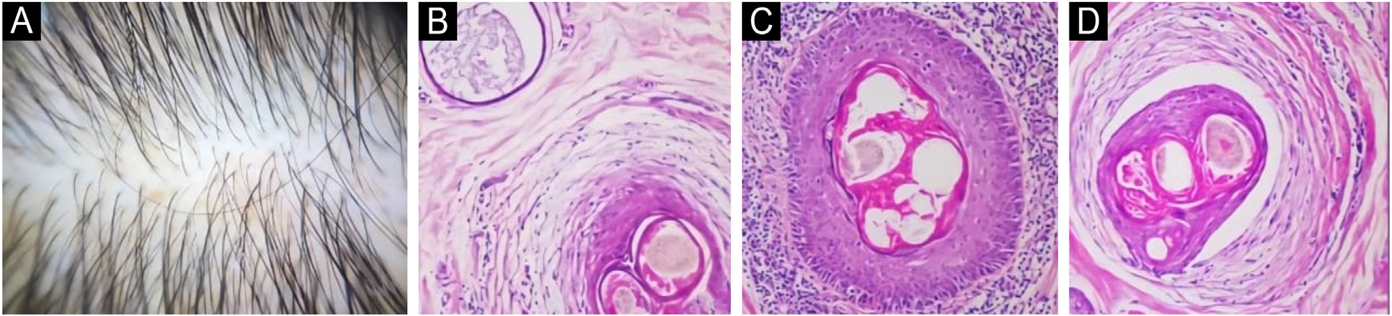
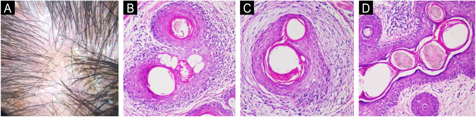
Transverse scalp biopsy specimens from 56 Caucasian male participants aged 18-65 years, comprising 35 (62.5%) with Seborrheic Dermatitis (SD) and 21 (37.5%) with AGA, were histologically evaluated, with mean age showing no signifi-cant difference between groups (p = 0.16) (Figs. 1 and 2).

This figure presents a clinical picture and photomicrographs of patients from the androgenetic alopecia group. (A) Tri-choscopic view of Patient 58 showing hair shaft diameter variability. (B) Corresponding transverse histological section from Patient 58 showing perifollicular fibrosis, lymphocytic infiltrate, and spongiosis in the follicular epithelium with lymphocyte exocytosis; a dilated eccrine gland is visible in the upper left corner (Hematoxylin & eosin, 200x). (C) Severe lymphocytic inflammation surround-ing a hair follicle with mild epithelial spongiosis in Patient 15 (Hematoxylin & eosin, 200x). (D) Prominent perifollicular fibrosis, epithelial thinning, and lymphocytic infiltration in the adjacent dermis in Patient 28 (Hematoxylin & eosin, 200x).

This figure presents a clinical picture and photomicrographs of patients from the seborrheic dermatitis group. (A) Tricho-scopic view of Patient 32 showing perifollicular and interfollicular yellowish-white scales. (B) Transverse histological section from Patient 32 depicting the isthmus area of a follicle surrounded by lymphocytic inflammation, with epithelial spongiosis, vacuolar interface change, and lymphocyte exocytosis (Hematoxylin & eosin, 200x). (C) Patient 53 showing prominent perifollicular fibrosis, blurred interface, and lymphocytic infiltration in the follicular epithelium (Hematoxylin & eosin, 200x). (D) Histological section from Patient 30 showing multiple hair shafts in a common follicular canal (polytrichia) (Hematoxylin & eosin, 200x).
Quantitative analysis revealed that SD had significan-tly higher mean counts of total hair follicles (p = 0.047), terminal follicles (p = 0.002), and anagen hairs (p = 0.045) compared to AGA, which exhibited a significantly greater number of vellus follicles (p = 0.029), consistent with fol-licular miniaturization in AGA. The anagen-to-telogen ratio was lower in AGA, approaching statistical significance (p = 0.051), while other quantitative measures, such as follicular units, telogen hairs, and sebaceous gland size, defined as the maximal uninterrupted diameter of the largest lobe, showed no significant differences (p > 0.05).
Qualitative analysis identified significant differences favoring AGA for Demodex spp. presence in the follic-ular canal (42.9% vs. 11.4%; p = 0.007) and sebaceous duct (38.1% vs. 8.6%; p = 0.007), basal layer spongio-sis/vacuolization (14.3% vs. 0%; p = 0.048), and eccrine ductal dilatation (23.8% vs. 0%; p = 0.006). Perifollicular fibrosis was prevalent in both conditions, observed in over 60% of cases, with isthmic fibrosis more frequent in AGA (38.1% vs. 14.3%; p = 0.044) and trends toward higher infundibular fibrosis (42.9% vs. 20.0%; p = 0.067) and fibrosis around terminal follicles (p = 0.067) in AGA, while fibrosis around vellus follicles was rare and not significantly dif-ferent (p = 0.14). Perifollicular inflammation was observed in over 70% of AGA and 60% of SD cases, with no sta-tistically significant difference between the conditions.
Necrotic keratinocytes were uncommon, with no signifi-cant differences in the infundibulum (p = 0.61) or isthmus (p = 0.27) between groups. Polytrichia, observed exclusively in SD (17.1%), approached significance (p = 0.050). Other qualitative parameters, including epidermal and follicular spongiosis, intrafollicular parakeratosis, bacterial or fun-gal colonization, perifollicular inflammation, and sebaceous gland size, showed no significant differences (p > 0.14). Histopathologic findings are summarized in Table 1.
Comparison of 7 quantitative and 28 qualitative histopathologic parameters between patients with androgenetic alopecia (n = 21) and seborrheic dermatitis (n = 35).
Discussion
The diagnosis of diffuse alopecia often presents clinical and histopathologic challenges. Fibrosing Alopecia in a Pattern Distribution (FAPD) is traditionally regarded as a variant of Lichen Planopilaris (LPP), characterized by diffuse central scalp involvement and overlapping features of Androge-netic Alopecia (AGA) and classic LPP.19-23 However, recent viewpoints suggest that FAPD may not represent a dis-tinct nosologic entity, but rather a clinicopathologic pattern encompassing early or diffuse LPP, fibrosing AGA, and AGA associated with inflammatory scalp conditions such as Seb-orrheic Dermatitis (SD).14,24,25 Its classification as a primary cicatricial alopecia therefore remains debated,26 particu-larly in light of the growing recognition of ‘‘scarring AGA’’ in the literature.27 Notably, previous reviews, including that of Griggs et al.,3 have not systematically examined the histopathologic criteria that distinguish AGA with perifol-licular fibrosis from FAPD.
Although AGA is not classically defined as an inflam-matory or scarring alopecia, chronic cases frequently exhibit mild perifollicular lymphocytic inflammation and fibrosis.28-30 This inflammatory component has been pro-posed as a contributor to disease progression, although it differs in intensity and distribution from the dense lichenoid infiltrates characteristic of LPP, a progressive cicatricial alopecia that may also present diffusely.30 In the present study, perifollicular fibrosis was commonly observed in both AGA and SD, but was more prevalent in AGA. Isthmic fibro-sis was significantly more frequent in AGA (38.1%) than in SD (14.3%; p = 0.044), while infundibular fibrosis occurred in 42.9% of AGA cases versus 20.0% of SD cases (p = 0.067). In both groups, fibrosis predominantly involved terminal fol-licles. These findings reinforce that perifollicular fibrosis, when considered in isolation, lacks specificity for the diag-nosis of scarring alopecias.
Other inflammatory features, including perifollicular inflammation involving terminal and vellus follicles, follicu-lar spongiosis, and epidermal spongiosis, were frequently observed in both groups without significant differences. Basal layer vacuolization was identified exclusively in AGA cases (14.3%; p = 0.048), potentially reflecting chronic follic-ular injury associated with long-standing disease. Necrotic keratinocytes were uncommon in both groups and did not reach statistical significance. Importantly, basal vacuoliza-tion and necrotic keratinocytes are classically associated with cicatricial alopecias such as LPP and Frontal Fibrosing Alopecia (FFA).31-36 Their occasional presence in non-scarring conditions such as AGA and SD reveals a key diagnostic pitfall and highlights the risk of misclassifying these entities as FAPD.
Misdiagnosis in this context carries important clinical consequences. Erroneously labeling common non-scarring conditions as cicatricial alopecias implies a poor and irre-versible prognosis, may increase patient anxiety, and can lead to inappropriate therapeutic decisions. This risk is fur-ther amplified when AGA and SD coexist, a frequent clinical scenario, as the combination of follicular miniaturization, perifollicular inflammation, and fibrosis may falsely sug-gest a scarring process. The overlapping histopathologic features observed in this study, when contextualized with those reported for FAPD in the literature, are summarized in Table 2.10,11,23,32,37
Comparison of histopathological features in androgenetic alopecia, seborrheic dermatitis, and fibrosing alopecia in a pattern distribution (literature-based).
A distinctive finding in this study was the presence of eccrine duct dilatation, observed exclusively in AGA cases (23.8%; p = 0.006). Previous studies have shown that eccrine duct dilatation is more commonly encountered in lymphocytic cicatricial alopecias,38 is less frequent in non-scarring alopecias, and is particularly prominent in neutrophilic scarring subtypes.39 In scarring alopecias, this change is thought to result from dermal fibrosis and duc-tal obstruction, whereas in non-scarring disorders, it may represent a reactive change related to a chronic inflamma-tory microenvironment.39 In only one AGA case, a fibrous scar tract was identified, likely representing follicular ‘‘dropout’’ occasionally seen in advanced stages of the dis-ease rather than true cicatricial alopecia.
An unexpected and novel observation was the presence of polytrichia, identified exclusively in SD cases (17.1%; p = 0.050). According to Griggs et al.,3 perifollicular casts surrounding a tuft of emerging hairs are considered a charac-teristic feature of FAPD and often guide biopsy site selection. Although polytrichia has traditionally been associated with scarring alopecias such as LPP, its occurrence in SD suggests that this feature may also arise in non-scarring inflamma-tory scalp conditions, potentially contributing to diagnostic ambiguity.
Taken together, these findings support the hypothesis proposed by Werner et al.,14 suggesting that FAPD may rep-resent a shared histopathologic pattern observed in early LPP, fibrosing AGA, and inflammatory scalp disorders such as SD, often superimposed on AGA, as summarized by compari-son with published FAPD data (Table 2). This interpretation is further supported by the recognized overlap between FAPD and diffuse variants of LPP, including LPP in a pattern distri-bution and other scarring patterns of hair loss, which share clinical, trichoscopic, and histologic features.3,11,14,32
As expected, follicular counts confirmed that SD, although an inflammatory scalp condition, is not intrinsically associated with alopecia. Patients with SD demonstrated higher total follicle counts and a predominance of termi-nal hairs, whereas AGA was characterized by a significantly greater proportion of vellus follicles and reduced terminal hair counts, consistent with follicular miniaturization. These quantitative findings allowed a clear histopathologic distinc-tion between AGA and SD, validating the appropriateness of clinical stratification and reinforcing that diagnostic over-lap arises primarily from inflammatory and fibrotic features rather than from follicular architecture.
Regarding microbial findings, no statistically significant differences were observed between AGA and SD with respect to bacterial or fungal colonization. In contrast, Demodex spp. was significantly more prevalent in AGA, in agree-ment with previous studies demonstrating an association between Demodex density and advanced AGA, predomi-nantly based on non-invasive imaging methods.40 In contrast to reports suggesting an absence of Malassezia spp. in scar-ring alopecias,37 fungal elements were rarely identified in the present cohort, highlighting the limited diagnostic util-ity of microbial detection for differentiating scarring from non-scarring alopecias.
This study has limitations. The relatively small sam-ple size restricts generalizability, particularly for rare histopathologic findings. The inclusion of male patients only ensured sample homogeneity but limits extrapolation to female populations. The cross-sectional design precludes assessment of temporal relationships between inflamma-tion, fibrosis, and disease progression. Finally, the absence of FAPD biopsy specimens prevents direct histopathologic comparison, and interobserver variability was not assessed.
Conclusions
This study pioneers the comparative evaluation of transverse scalp biopsy sections from 56 patients with non-scarring scalp disorders, providing novel histopathological insights into their overlap with Fibrosing Alopecia in a Pattern Distribution (FAPD). Perifollicular fibrosis was a rela-tively frequent finding, challenging traditional distinctions between scarring and non-scarring processes. Perifollicular inflammation was common in both conditions and may mimic lichenoid patterns, representing a potential diagnostic pit-fall. In contrast, necrotic keratinocytes were uncommon and not discriminatory. Certain features showed group-specific associations, including basal layer vacuolization and eccrine duct dilatation with AGA, and polytrichia with SD. Follicular miniaturization emerged as a reliable distinguishing feature of AGA, while increased Demodex sp density supported its possible proinflammatory role. Overall, these findings rein-force the need for careful clinicopathological correlation to accurately differentiate non-scarring alopecias and condi-tions from FAPD, thereby avoiding misdiagnosis and ensuring appropriate management and prognostic assessment.
Ethics approval statement
This study was approved by the Research Ethics Commit-tee, the Hospital de Clínicas (Federal University of Paraná), Curitiba - Paraná. Certificate of Presentation for Ethical Appreciation (CAAE) 75757223.4.0000.0096, and approval number 6.624.101.
Research data availability
The entire dataset supporting the results of this study was published in this article.
References
- 1 Werner B, Mulinari-Brenner F. Clinical and histological challenge in the differential diagnosis of diffuse alopecia: female andro-genetic alopecia, telogen effluvium and alopecia areata - part I. An Bras Dermatol. 2012;87:742-7.
- 2 Werner B, Mulinari-Brenner F. Clinical and histological challenge in the differential diagnosis of diffuse alopecia: female andro-genetic alopecia, telogen effluvium and alopecia areata-part II. An Bras Dermatol. 2012;87:884-90.
- 3 Griggs J, Trüeb RM, Gavazzoni Dias MFR, Hordinsky M, Tosti A. Fibrosing alopecia in a pattern distribution. J Am Acad Derma-tol. 2021;85:1557-64.
- 4 Kuczara A, Wa´skiel-Burnat A, Rakowska A, Olszewska M, Rud-nicka L. Trichoscopy of androgenetic alopecia: a systematic review. J Clin Med. 2024;13:1962.
- 5 Kasumagic-Halilovic E. Trichoscopic findings in Androgenetic Alopecia. Med Arch. 2021;75:109-11.
- 6 ˙Iritaş SY, Özcan D. The value of trichoscopy in the follow-up of treatment response in patients with androgenetic alopecia. Dermatol Pract Concept. 2024;14:e2024001.
- 7 Cortez GL, Hassun K, Linhares LRP, Florenço V, Pinheiro MVB, Nascimento MM. Male androgenetic alopecia. An Bras Dermatol. 2025;100:308-21.
- 8 Oiwoh SO, Enitan AO, Adegbosin OT, Akinboro AO, Onayemi EO. Androgenetic alopecia: a review. Niger Postgrad Med J. 2024;31:85-92.
- 9 Yoo HG, Kim JS, Lee SR, Pyo HK, Moon HI, Lee JH, et al. Per-ifollicular fibrosis: pathogenetic role in androgenetic alopecia. Biol Pharm Bull. 2006;29:1246-50.
- 10 Wei K, Ni C, Huang Q, Yang K, Zhang Y, Liu Q, et al. Clinico-pathological characteristics of fibrosing alopecia in a pattern distribution: a single-center, retrospective study. Int J Derma-tol. 2023;62:1391-6.
- 11 Teixeira M de S, Gavazzoni Dias MFR, Trüeb RM, Rochael MC, Vilar EAG. Fibrosing alopecia in a pattern distribution (FAPD) in 16 African-descent and Hispanic female patients: a challenging diagnosis. Skin Appendage Disord. 2019;5:211-5.
- 12 Özcan D, Vural AT, Özen Ö. Androgenetic alopecia or fibrosing alopecia in a pattern distribution: when to perform biopsy in unusual cases? Indian J Dermatol. 2022;67:90-1.
- 13 Jerjen R, Pinczewski J, Sinclair R, Bhoyrul B. Clinicopathological characteristics and treatment outcomes of fibrosing alopecia in a pattern distribution: a retrospective cohort study. J Eur Acad Dermatol Venereol. 2021;35:2440-7.
- 14 Werner B, Soares VC, Mulinari-Brenner F, Tolkachjov SN. Understanding fibrosing alopecia in a pattern distribution: a retrospective study with histological challenges in the diagno-sis of diffuse central alopecia. Arch Dermatol Res. 2025;317: 506.
- 15 Leroy AK, Cortez de Almeida RF, Obadia DL, Frattini S, Melo DF. Scalp seborrheic dermatitis: what we know so far. Skin Appendage Disord. 2023;9:160-4.
- 16 Vano-Galvan S, Reygagne P, Melo DF, Barbosa V, Wu WY, Moneib H, et al. A comprehensive literature review and an interna-tional expert consensus on the management of scalp seborrheic dermatitis in adults. Eur J Dermatol. 2024;34:4-16.
- 17 Widaty S, Pusponegoro EH, Rahmayunita G, Astriningrum R, Akhmad AM, Oktarina C, et al. Applicability of trichoscopy in scalp seborrheic dermatitis. Int J Trichology. 2019; 11:43-8.
- 18 Kim BJ, Kim JY, Eun HC, Kwon OS, Kim MN, Ro BI. Androge-netic alopecia in adolescents: a report of 43 cases. J Dermatol. 2006;33:696-9.
- 19 Hong H, Ji JH, Lee Y, Kang H, Choi GS, Lee WS. Reliability of the pattern hair loss classifications: a comparison of the basic and specific and Norwood-Hamilton classifications. J Dermatol. 2013;40:102-6.
- 20 Mardones F, Hott K, Martinez MC. Clinical study of fibrosing alopecia in a pattern distribution in a Latin American popula-tion. Int J Dermatol. 2018;57:e12-4.
- 21 Triyangkulsri K, Srisuwanwattana P, Sriphojanart T, Suchon-wanit P. Fibrosing alopecia in a pattern distribution: a case report and literature review. Case Rep Dermatol. 2019;11: 297-302.
- 22 Chiu HY, Lin SJ. Fibrosing alopecia in a pattern distribution. J Eur Acad Dermatol Venereol. 2010;24:1113-4.
- 23 Soares VC, Mulinari-Brenner F, Souza TE de. Lichen planopi-laris epidemiology: a retrospective study of 80 cases. An Bras Dermatol. 2015;90:666-70.
- 24 Zinkernagel MS, Trüeb RM. Fibrosing alopecia in a pattern distribution: patterned lichen planopilaris or androgenetic alopecia with a lichenoid tissue reaction pattern? Arch Derma-tol. 2000;136:205-11.
- 25 Bhoyrul B. A simple technique to distinguish fibrosing alope-cia in a pattern distribution from androgenetic alopecia and concomitant seborrheic dermatitis. J Am Acad Dermatol. 2022;86:163-5.
- 26 Bhoyrul B. Cicatricial pattern hair loss is not a vari-ant of lichen planopilaris. J Am Acad Dermatol. 2020;83: e413-4.
- 27 Whiting DA. Cicatricial alopecia: clinico-pathological findings and treatment. Clin Dermatol. 2001;19:211-25.
- 28 Whiting DA. Diagnostic and predictive value of horizontal sec-tions of scalp biopsy specimens in male pattern androgenetic alopecia. J Am Acad Dermatol. 1993;28:755-63.
- 29 Mahé YF. Inflammatory perifollicular fibrosis and alopecia. Int J Dermatol. 1998;37:416-7.
- 30 Mahé YF, Michelet JF, Billoni N, Jarrousse F, Buan B, Commo S, et al. Androgenetic alopecia and microinflammation. Int J Dermatol. 2000;39:576-84.
- 31 Gálvez-Canseco A, Sperling L. Lichen planopilaris and frontal fibrosing alopecia cannot be differentiated by histopathology. J Cutan Pathol. 2018;45:313-7.
- 32 Starace M, Orlando G, Alessandrini A, Baraldi C, Bruni F, Pirac-cini BM. Diffuse variants of scalp lichen planopilaris: clinical, trichoscopic, and histopathologic features of 40 patients. J Am Acad Dermatol. 2020;83:1659-67.
- 33 Costa Fechine CO, Sakai Valente NY, Romiti R, Senna MM. Cor-relation of clinical and trichoscopy features with the degree of histologic inflammation in lichen planopilaris and frontal fibros-ing alopecia in a cross-sectional study. J Am Acad Dermatol. 2024;91:161-3.
- 34 Fanti PA, Baraldi C, Misciali C, Piraccini BM. Cicatricial alopecia. G Ital Dermatol Venereol. 2018;153:230-42.
- 35 Stefanato CM. Histopathology of alopecia: a clinico-pathological approach to diagnosis. Histopathology. 2010; 56:24-38.
- 36 Sperling LC, Cowper SE. The histopathology of primary cicatri-cial alopecia. Semin Cutan Med Surg. 2006;25:41-50.
- 37 Williams K, Tosti A, Thompson CT. Histologic absence of yeast as a clue for classic lichen planopilaris, fibrosing alopecia in a pattern distribution, and frontal fibrosing alopecia: a cross-sectional observational study. JAAD Int. 2025; 19:10-1.
- 38 Tan T, Guitart J, Gerami P, Yazdan P. Eccrine duct dilata-tion as a marker of cicatricial alopecia. Am J Dermatopathol. 2017;39:668-71.
- 39 Tan TL, Doytcheva K, Guitart J, Gerami P, Yazdan P. Dilatation of multiple eccrine ducts as a highly specific marker for cicatricial alopecia. Am J Dermatopathol. 2019;41:871-8.
- 40 Millikan LE. Androgenetic alopecia: the role of inflammation and Demodex. Int J Dermatol. 2001;40:475-6.
Edited by
-
Editor
Neusa Yuriko Sakai Valente.