 Perifollicular fibrosis and inflammation in androgenetic alopecia and seborrheic dermatitis: diagnostic challenges in differentiation from fibrosing alopecia in a pattern distribution
Background: Androgenetic Alopecia (AGA) and Seborrheic Dermatitis (SD) are scalp conditions that may show histopathological features overlapping with Fibrosing Alopecia in a Pattern Dis-tribution (FAPD), potentially leading to diagnostic confusion between non-scarring and scarring alopecias.
Objective: To identify histopathological features that distinguish AGA from SD and to evalu-ate findings overlapping with those described in FAPD, focusing on perifollicular fibrosis and inflammation.
Methods: Transverse scalp biopsy sections from 56 Caucasian male patients (21 AGA, 35 SD) were blindly evaluated. Quantitative follicular parameters and qualitative epidermal, follicular, inflammatory, and adnexal features were assessed and statistically compared.
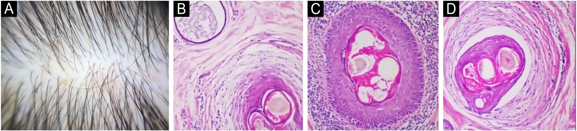
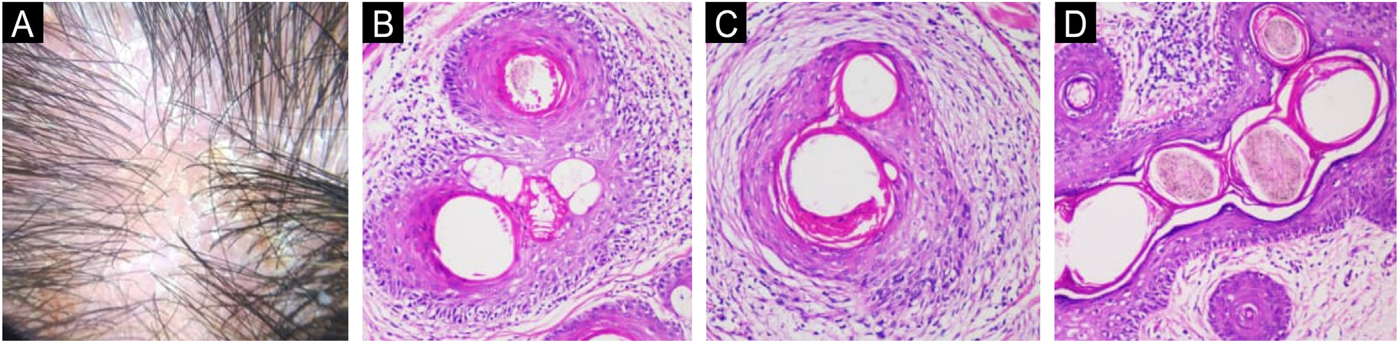
Results: AGA showed higher vellus follicle counts (p = 0.029) and lower terminal follicle and anagen hair counts (p = 0.002; p = 0.045), confirming follicular miniaturization. Perifollicular fibrosis (infundibular and/or isthmic) was frequent in both conditions, limiting its specificity for scarring alopecias. Mild basal vacuolization (14.3%; p = 0.048) and eccrine duct dilatation (23.8%; p = 0.006) occurred exclusively in AGA, whereas polytrichia was observed only in SD (17.1%; p = 0.050). Perifollicular inflammation was common and non-specific in both groups, occasionally mimicking lichenoid patterns. Demodex spp. was more prevalent in AGA (p = 0.007). Necrotic keratinocytes were rare, with no significant differences. Study limitations: The study included only male patients and had a limited sample size, a cross-sectional design, and no FAPD comparison. Nonetheless, substantial histopathological overlap exists between AGA, SD, and features attributed to FAPD, particularly perifollicular fibrosis and inflammation. Careful clinicopathological correlation is essential to avoid misclassification, with prognostic and therapeutic implications.
KEYWORDS
Androgenetic alopecia; Dermatitis; seborrheic; Fibrosis; Inflammation; Cicatricial alopecia
 Thumbnail
 Thumbnail