 Echtyma contagiosum in a goat farmer
Dear Editor,
Echtyma Contagiosum (EC), or Orf, is a zoonosis with world-wide distribution caused by the Orf Virus (OrfV), of the genus Parapoxvirus.1-3 The virus is transmissible to humans from infected small ruminants, predominantly sheep and goats. In infected animals, the virus manifests as proliferative lesions frequently appearing on the lips, muzzle, ears, eyelids, and nostrils, and less commonly on the udder, genitalia, and feet.1
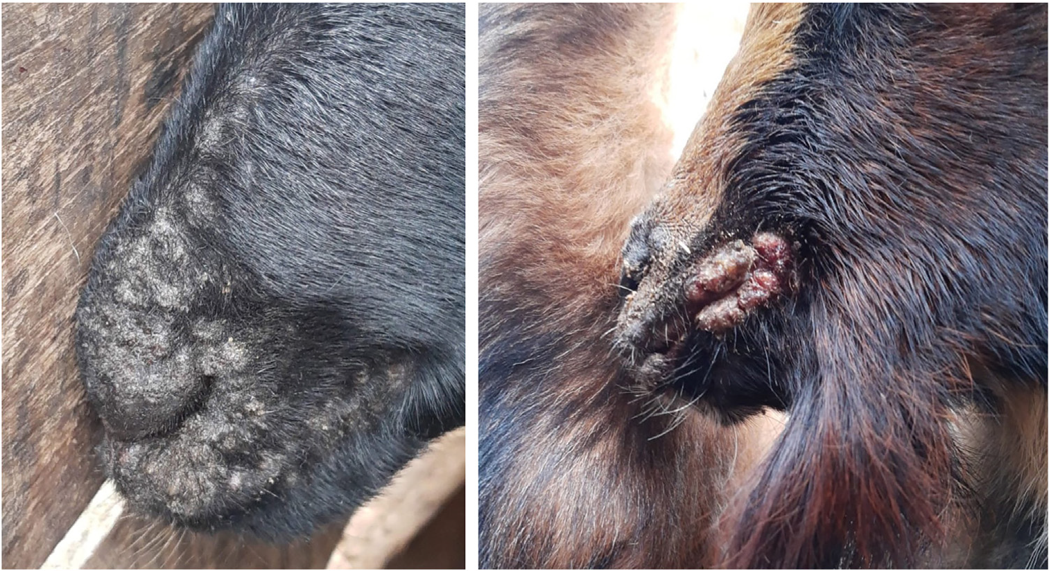
Due to its self-limited nature, treatment is not necessary in immunocompetent patients with a solitary lesion; how-ever, immunocompromised individuals have an increased risk of developing large and persistent lesions. Immunity against the virus is short-lived, and reinfection may occur.3 A 46-year-old female rural worker sought medical care due to a painful ulceration on the right thumb, measuring 1.6 × 1.0 cm, as well as a small erythematous-edematous lesion with a central crust at the base of the right index fin-ger (Fig. 1A). She reported that both lesions had appeared simultaneously seven days earlier and suspected a spider bite. When questioned about potential contact with ani-mals, the patient stated that she had cared for goats with crusted lesions on their lips (Fig. 2).

Clinical aspect. (A) Ulceration on the right thumb and a small erythematous-edematous lesion with a central crust at the base of the right index finger. (B) Partial involution after 15-days.

Laboratory tests were performed, including complete blood count, ESR, C-reactive protein, VDRL, urea, and hand radiograph - all of which were normal.
Histopathology of the affected skin revealed a central area of ulceration covered by a scale-crust with compact hyperkeratotic epidermis containing eosinophilic inclusion bodies, ballooning degeneration, and neutrophil collection. The dermis exhibited moderate edema and a dense infiltrate composed of lymphocytes, plasma cells, and occasional neu-trophils (Fig. 3). Immunohistochemistry (Fig. 4) was positive for CD2 (T-cell and NK-cell marker), CD4 (T-helper marker), and CD5 (T-cell marker), showed weaker staining for CD8 (regulatory T-cell marker), and was strongly positive for CD68 (monocyte and macrophage marker). It was negative for CD20 (B-cell marker).

Light microscopy. (A) Central area of ulceration covered by a scale-crust. (B) Neutrophils microabcess. (C) Dense dermal infiltrate (D) Eosinophilic inclusion body (arrow). (Hematoxylin & eosin, ×100, ×400, ×400, ×500).

Immunohistochemistry strongly positive for CD2, CD4, CD5 and CD68, weaker staining for CD8 and negative for CD20 (×200).
Since there was no contact with cattle, the milker’s nodule was excluded, and a diagnosis of echtyma contagiosum was established. The lesions regressed spon-taneously in three weeks (Fig. 1B), leaving mild residual erythema.
EC is an occupational zoonosis with worldwide distribution.4 The OrfV has affinity for domestic rumi-nants, such as sheep and goats; however, cases have also been reported in wild species.4 In animals, lesions are crusted and proliferative, affecting the skin and mucocutaneous junctions. The virus is highly contagious among animals. It is known to tolerate inactivation in dry environments and has been recovered from crusts after several months.1 It survives for almost one month in wool and skin after clinical improvement.1
Transmission of OrfV to humans may occur through direct contact with an infected animal or via fomites. Most cases are observed in individuals with occupational exposure to animals, such as livestock workers. However, cases have been reported among shepherds, butchers, slaughterhouse workers, handlers of animal hides and wool, veterinarians, zoo visitors, hunters, and participants in religious cere-monies involving animal sacrifice.5 The incubation period is 3-7 days,1,2 and may reach up to six weeks depending on the severity and immune status of the patient.4
The clinical diagnosis of EC depends on the character-istic appearance of the lesions, histopathological features, exposure to animals, and lesion evolution; all of which were typical in this case. Confirmation may be obtained via elec-tron microscopy, PCR, or detection of specific antibodies, which were not available in our setting. There are a few reports of human cases in Brazil,6 although many cases in animals have been published.7,8
Histopathological findings may vary depending on the stage of the lesion. Early stages are characterized by spongiform degeneration of the epidermis, with variable vesiculation of the superficial epidermis and eosinophilic cytoplasmic inclusion bodies in keratinocytes. Our immuno-histochemical findings demonstrated the involvement of T-lymphocytes and histiocytes in the inflammatory response.
In the differential diagnosis, the main entity to be con-sidered is milker’s nodule, also caused by a Parapoxvirus, with similar clinical and histopathological features.9,10 How-ever, the host animal in that condition is cattle. Neutrophilic dermatoses can also resemble Orf nodules, especially pyo-derma gangrenosum and neutrophilic dermatosis of the dorsal hands.
Infection with the OrfV does not result in long-lasting immunity, and individuals may be infected multiple times throughout life; however, subsequent infections may be less pronounced and heal more rapidly or remain unnoticed.1
Because animals are the primary reservoirs and sources of infection in humans, preventive measures should focus on vaccination of animals (there is no vaccine for humans) and hand protection during animal handling.1
Research data availability
Does not apply.
References
- 1 Kassa T. A review on human Orf: a neglected viral zoonosis. Res Rep Trop Med. 2021;12:153-72.
- 2 Estela Cubells JR, Braverman I, Kashgarian M, Lazova R. A 65-year-old female from connecticut with Orf infection. Der-matopathology (Basel). 2016;3:55-60.
- 3 Thompson HJ, Harview CL, Swick B, Powers JG. Orf virus in humans: case series and clinical review. Cutis. 2022;110:48-52.
- 4 Flores C, González E, Verna A, Peralta A, Madariaga C, Odeón A, et al. Virus orf en humanos, confirmación molecular de un caso clínico en Chile [Orf virus in human, confirmation in case report from Chile]. Rev Chilena Infectol. 2017;34:607-9.
- 5 Caravaglio JV, Khachemoune A. Orf virus infection in humans: a review with a focus on advances in diagnosis and treatment. J Drugs Dermatol. 2017;16:684-9.
- 6 Rezende ALRA, Bernardes Filho F, de Paula NA, Towersey L, Hay R, Frade MAC. Clinical manifestation, dermoscopy, and scanning electron microscopy in two cases of contagious ecthyma (Orf Nodule). Case Rep Dermatol Med. 2018;2018:2094086.
- 7 Abrahão JS, Campos RK, Trindade GS, Guedes MI, Lobato ZI, Mazur C, et al. Detection and phylogenetic analysis of Orf virus from sheep in Brazil: a case report. Virol J. 2009;6:47.
- 8 de Oliveira CH, Assis FL, Neto JD, Oliveira CM, Lopes CT, Bom-jardim Hdos A, et al. Multifocal cutaneous ORF virus infection in goats in the Amazon region. Brazil. Vector Borne Zoonotic Dis. 2012;12:336-40.
- 9 Espósito ACC, Jorge MFS, Marques MEA, Abbade LPF. Milker’s nodules: classic histological findings. An Bras Dermatol. 2017;92:838-40.
- 10 Adriano AR, Quiroz CD, Acosta ML, Jeunon T, Bonini F. Milker’s nodule - Case report. An Bras Dermatol. 2015;90:407-10.
Edited by
-
Editor
Sílvio Alencar Marques