 Donovanosis - A rare, sexually transmitted infection that the world has not yet eradicated
Dear Editor,
Donovanosis or granuloma inguinale is a chronic, indolent, and progressive disease with low infectivity, caused by the bacterium Klebsiella granulomatis.1 Donovanosis transmis-sion occurs predominantly through sexual contact,2 although cases have been reported in children and adults without sex-ual activity through contact with infected adults. There are also reports of patients with extra-genital lesions without concomitant genital lesions, making contamination through feces possible, as a bacterium similar to Klebsiella granulo-matis has been identified in the feces of a patient with the disease.1
It is considered endemic in some tropical and subtrop-ical regions, including parts of Brazil, South Africa, India, and Australia.3 In the 1990s, an increase in the number of cases was observed, mainly in South Africa, after the use of mass rapid testing,1 suggesting that the low incidence is a consequence of underdiagnosis. Currently, the number of new cases is low, being described only sporadically,1 without clear numerical data in Brazil and worldwide.
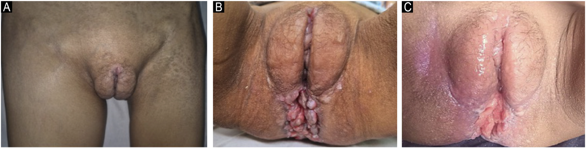
This article reports a case of donovanosis, diagnosed at a reference center for STIs in the city of Manaus, in the Brazil-ian Amazonia. The patient, a 21-year-old indigenous woman from the municipality of Tabatinga, state of Amazonas, has cognitive and motor impairment. Her mother reports that, eight months ago, she observed lesions on the vulva and anal region, associated with pain, pruritus, and bleeding. Physical examination revealed multiple coalescent vegeta-tive and ulcerated lesions in the vulvar and anal region, with areas of bleeding, as well as lymphedema of the right lower limb and bilateral adenomegaly (Figs. 1A and 1B; Fig. 1C -lesion after treatment). Family members are unaware of the patient’s any sexual exposure. Serological tests for hepati-tis B and C, syphilis, and HIV, performed in December 2023, were negative.

(A) Lymphedema of the right lower limb (June 2024). (B) Vegetative and ulcerated lesions in the perianal region and perineum (June 2024). (C) Regression of lesions after treatment (December 2024).
Donovanosis is characterized by lesions that generally begin as painless papules, developing into nodules, vege-tating lesions, or ulcers, which bleed easily and may have aspects similar to carcinoma.4 Elephantiasis, associated with ulcerated and cicatricial lesions, as observed in the present case, is relatively frequent in patients with a long course of the disease. In all these patients, serology for HIV, syphilis, hepatitis B and C is recommended. Malignant neo-plasms, American cutaneous leishmaniasis, and cutaneous tuberculosis, among other diseases, are among the main differential diagnoses.5
In the reported case, the diagnosis was attained by histopathological examination, which revealed an epider-mis with acanthosis and marked inflammatory infiltrate in the dermis, consisting of vacuolated lymphocytes and histiocytes (Fig. 2A). Warthin-Starry staining revealed a large number of Donovan bodies in the cytoplasm of his-tiocytes (Fig. 2B), characteristic of Klebsiella granulomatis infection.6

Microscopic examination showing inflammatory infiltrate of vacuolated histiocytes and lymphocytes. (A) Hematoxylin & eosin, ×400. (B) Warthin-Starry. Donovan bodies (arrows; ×1000).
For the treatment of donovanosis, azithromycin is rec-ommended as a first-line drug, given its efficacy and safety. Among the alternative drugs, doxycycline, ciprofloxacin, and sulfamethoxazole-trimethoprim are indicated,1,3,4 espe-cially in resistant cases or patients who cannot use azithromycin. In immunosuppressed patients, gentamicin or chloramphenicol is indicated.1,6 For the case of the present study, doxycycline was administered at a dose of 100 mg every 12 hours. After 21 days of treatment, partial regression of the lesions and significant regression of the lymphadenopathy were observed.
Despite the relative rarity of donovanosis in our setting, it is believed it is important to emphasize that the diagnosis of donovanosis should always be considered in patients with ulcerated lesions located in the male or female genitalia, perineum, and anorectal region. According to records from Fundação Alfredo da Matta, the last diagnosis of donovanosis occurred in 2007.
Research data availability
Does not apply.
References
- 1 Belda Junior W. Donovanosis. An Bras Dermatol. 2020;95:675-83.
- 2 Bezerra SMFMC, Jardim MML, Silva VB. Donovanose. An Bras Der-matol. 2011;86:585-6.
- 3 Workowski KA, Bachmann LH, Chan PA, Johnston CM, Muzny CA, Park I, et al. Sexually transmitted infections treatment guide-lines, 2021. MMWR Recomm Rep. 2021;70:1-187.
- 4 O’Farrell N, Moi H. 2016 European guideline on donovanosis. Int J STD AIDS. 2016;27:605-7.
-
5 Brasil. In: Ministério da Saúde. Secretaria de Vigilância em Saúde. Protocolo clínico e diretrizes terapêuticas para atenção integral às pessoas com infeçcões sexualmente transmis-síveis (IST) [Internet]. Brasília: Ministério da Saúde; 2018 [cited 2025 Oct 27]. Available from: https://www.gov.br/conitec/pt-br/midias/infeccoessexualmentetransmissiveis.pdf
» https://www.gov.br/conitec/pt-br/midias/infeccoessexualmentetransmissiveis.pdf - 6 Calonje E, Brenn T, Lazar A. McKee’s pathology of the skin. 4th ed. Philadelphia: Elsevier; 2012.
Edited by
-
Editor
Hiram Larangeira de Almeida Jr.