 Immunohistochemical analysis of MMP-1 and 2 in patients with lichen planopilaris and frontal fibrosing alopecia
Dear Editor,
Lichen planopilaris (LPP) and frontal fibrosing alopecia (FFA) are primary lymphocytic scarring alopecias. Clinically, clas-sic LPP shows an area of irregular alopecia that is more common in the vertex region, while FFA causes hair loss that occurs slowly in the frontotemporal implantation hairline, commonly associated with eyebrow alopecia.1
According to Doche et al.,2 approximately 65% of the scalp biopsy specimens from ‘‘normal-appearing’’ areas of LPP and FFA showed perifollicular inflammation around the isthmus/infundibulum region. These findings suggest that both diseases may be more generalized processes affecting the scalp. The lack of relationship between the degree of histopathological inflammation and clinical signs of inflam-mation supports the idea that some scalp areas could possibly be more prone to develop a cicatricial alopecia.
These disorders are mediated by lymphocytes and result in disruption of the basement membrane cells, destruction of the hair follicle and replacement by fibrosis. They have a chronic course, unpredictable development and, probably, autoimmune pathogenesis. Drugs, infections, genetic fac-tors and immunological abnormalities are possible triggering factors.3
Matrix metalloproteinases (MMPs) are involved in cell proliferation and differentiation, inflammation, degenera-tion, tumor metastasis, and growth. The production and activation of MMPs is rapidly induced when active tissue remodeling is needed. MMP-2, the most widely distributed of all MMPs, along with MMP-9, is said to digest type IV, V and XI collagens; laminin and aggrecan core protein, and singly digests collagens I, II and III.4
MMP-1 is capable of degrading type I and III fibrillar collagens.5
MMP-4 (or MMP-17) is involved in different pathological processes such as arthritis, cardiovascular disease, and can-cer progression.6
Our objective was to evaluate the presence of MMP-1 and MMP-2 in LPP and FFA and to assess their role in the pathogenesis of these diseases.
The patients were collected from Renata Zac Dermato-logical Clinic, Belo Horizonte, MG, Brazil, from 2019 to 2023. A university ethical committee approval was obtained prior to carrying out this study. Participants taking part gave written informed consent.
Inclusion criteria were patients with clinically and histopathologically proven disease. Patients with lesions not confirmed by biopsy or on treatment at the time of biopsy were excluded from this study.
In this study, 60 patients (48 females and 12 males) aged from 23 to 71 years were included.
To be included in the classical LPP group, the patient should have an irregular involvement of the scalp in the form of plaques presenting with hyperkeratosis, follicular plugs and perifollicular erythema or atrophic scars, mainly in the apex and in the parietal region. Trichoscopy could evi-dence perifollicular and interfollicular erythema associated with tubular perifollicular scales. Complaints of pruritus and burning sensation were common, as well as the association with the skin, nail and mucosal LP lesions.3
To be included in the FFA group, the participants should fulfill the diagnostic criteria for FFA proposed by Van˜ó-Galván et al.7 The major criteria are scarring alopecia of the scalp in the frontotemporal region (in the absence of ker-atotic follicular papules on the body) and bilateral diffuse alopecia of the eyebrows. The minor criteria include tri-choscopy with peripilar erythema, peripilar desquamation, or both; histopathological characteristics of scarring alope-cia with FFA or LPP pattern; involvement of occipital region, face, sideburns, body hair and presence of non-inflammatory facial papules. The diagnosis requires two major criteria or one major and two minor criteria.
All patients had undergone a scalp biopsy with a 4-mm punch biopsy, with one sample submitted for vertical sec-tions and the other for cross-sectional sections. Diagnosis was confirmed by one pathologist and one dermatolo-gist using hematoxylin and eosin-stained sections. The histopathological alterations found in LPP and FFA are a perifollicular lymphohistiocytic infiltrate, sometimes with a lichenoid pattern, more prominent in the isthmus and infundibulum regions; vacuolar degeneration of basal cells; necrotic keratinocytes; artifactual clefts between the fol-licle and the perifollicular fibrous band; and perifollicular fibrosis separating the inflammatory infiltrate from the fol-licle. Over time, there is a reduction and loss of sebaceous glands and destruction of the entire hair follicle.1
Twenty apparently normal scalp tissues, age and sex matched, were obtained at the time of facial plastic surgery and used as controls.
This laboratory-based study involved the use of sixty buffered, formalin-fixed, paraffin-embedded tissue blocks of histologically proven cases of LLP, FFA and controls.
The primary antibodies for immunohistochemistry stain-ing included polyclonal antibodies against MMP1 [EP1247Y] (ab52631, Abcam, Cambridge, UK) and MMP2 [6E3F8] (ab86607, Abcam, Cambridge, UK).
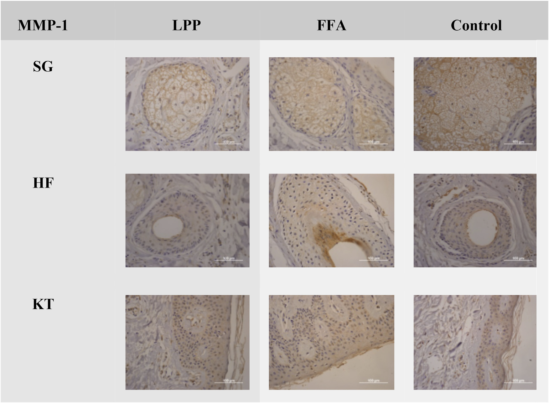
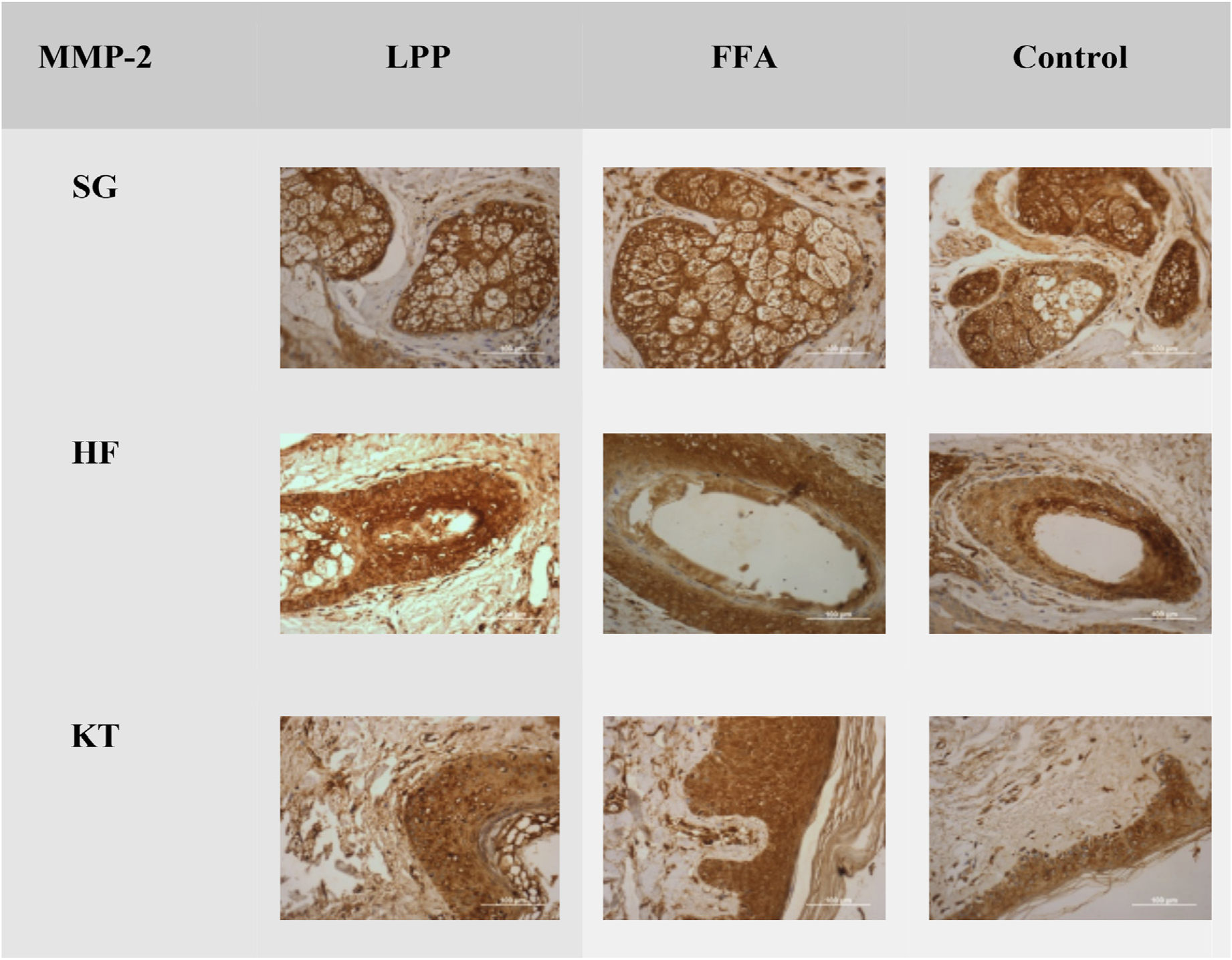
The expression of MMP-1 and MMP-2 in LPP, FFA, and con-trol groups was evaluated in the sebaceous glands (SG), all segments of the hair follicles (HF), and the epidermal ker-atinocytes (KT).
The Klein score is a semiquantitative method used to evaluate the expression of immunohistochemical markers by multiplying the percentage of positive cells by the staining intensity to obtain the final score (Klein et al., 2001).
The Klein score was obtained through the calculation described in Table 1.
Qualitative variables were presented as absolute and rel-ative frequencies, and quantitative variables as minimum, maximum, mean, standard deviation, median, first (Q1) and third (Q3) quartiles. The number of available information (valid n) was presented for each variable.
The association between qualitative variables was assessed using Fisher’s exact test. The comparison of quan-titative variables between the three comparison groups was performed using the Kruskal-Wallis test.
The analyses were carried out in the R Studio program version 2024.04.2 using the R language version 4.4.0, and p < 0.05 was considered significant.
There was no difference between the three groups evalu-ated in the MMP-1 and MMP-2 antibody markers (Figs. 1 and 2 and Tables 2 and 3).
Negative expression of MMP-1 in LPP, FFA, and control groups in the sebaceous glands (SG), hair follicles (HF) and keratinocytes (KT) ×100. Source: research original data.
Same expression of MMP-2 in LPP, FFA, and control groups in the sebaceous glands (SG), hair follicles (HF) and ker-atinocytes (KT) ×100. Source: research original data.
LPP and FFA are primary lymphocytic scarring alopecias in which the lower infundibular and isthmic areas of the hair follicle are primarily affected, the region where follicular stem cells are located.2
It has been observed that many patients being fol-lowed for LPP and FFA, without inflammatory symptoms or signs, continue with the progression of the area of alopecia.1
Wong and Goldberg,8 found statistically significant dif-ferences between the extent of the inflammatory infiltrate below the isthmus in FFA when compared with LPP (92% vs. 63%; p = 0.02) but found no differences between the inflam-matory infiltrate intensity in the two diseases.
Although the pathophysiology of both diseases remains unknown, an autoimmune aetiology is favoured. However, the dramatic increase in the incidence of FFA over the past decade has caused investigators to also postulate a strong potential environmental aetiology for this disease.2
We recommend that patients with FFA/LPP avoid daily use of chemical sunscreens and/or sunscreens containing titanium dioxide. Furthermore. General allergen avoidance is important in controlling scalp symptoms.9
In oral squamous cell carcinoma, there is an increased expression of MMPs that play an important role in tumor progression. MMP-2 and MMP-9 play an important role in the cleavage of type IV collagen, facilitating the disruption of the basement membrane and migration of dysplastic cells.4
Despite the expression of MMP-2 observed mainly in the lymphocytic band in the lamina propria in oral lichen planus,4 it was not noticed in the patients with LPP or FFA in our study.
MMP-1 positively affects the wound-healing process and reduces unwanted scar formation. Therefore, MMP-1 can potentially be used for preventing or treating hypertrophic scars.10
We believe that MMPs increase in situations where there is collagen breakdown and disruption of the basal mem-brane zone, but in cicatricial alopecias, there is mainly accumulation of this substance in the perifollicular zone and fibrosis.
A key limitation of standard immunohistochemistry when studying matrix metalloproteinases (MMPs) is that it detects the presence and location of the protein (total amount, active and inactive), but it does not provide information on its enzymatic activity, which zymography does measure.
MMP-1 and 2 aren’t probably not mediators in the patho-genesis of FFA and LPP.
-
☆
Study conducted at the Clínica Renata Zac, Belo Horizonte, MG, Brazil and Instituto de Assistência Médica do Servidor Público Estad-ual de São Paulo, São Paulo, SP, Brazil.
-
Financial supportThe study received financial support from Fundo de Apoio a Dermatologia (FUNADERM) of the Brazilian Society of Der-matology.
Acknowledgments
We thank Fundo de Apoio a Dermatologia (FUNADERM) of the Brazilian Society of Dermatology for the financial support.
Research data availability
The entire dataset supporting the results of this study was published in this article.
References
- 1 Fechine COC, Valente NYS, Romiti R. Lichen planopilaris and frontal fibrosing alopecia: review and update of diagnostic and therapeutic features. An Bras Dermatol. 2022;97:348-57.
- 2 Doche I, Romiti R, Hordinsky MK, Valente NS. ‘‘Normal-appearing’’ scalp areas are also affected in lichen planopilaris and frontal fibrosing alopecia: an observational histopathologic study of 40 patients. Exp Dermatol. 2020;29:278-81.
- 3 Andrade TCPC, Martins TY, Oliveira AMN, Santiago TM, Soares CT, Nakandakari S. Lichen planopilaris: the importance of early diagnosis. Surg Cosmet Dermatol. 2017;9:72-5.
- 4 Agarwal N, Carnelio S, Rodrigues G. Immunohistochemical and clinical significance of matrix metalloproteinase-2 and its inhibitor in oral lichen planus. J Oral Maxillofac Pathol. 2019;23:476.
- 5 Chen B, Cai S, Cui L, Yu T, Qiao K, Su Y, et al. Novel peptide inhibitor of matrix Metalloproteinases-1 from pufferfish skin col-lagen hydrolysates and its potential photoprotective activity via the MAPK/AP-1 signaling pathway. J Photochem Photobiol B. 2025;262:113088.
- 6 Yip C, Foidart P, Noël A, Sounni NE. MT4-MMP: the GPI-anchored membranetype matrix metalloprotease with multiple func-tions in diseases. Int J Mol Sci. 2019;20:354.
- 7 Van˜ó-Galván S, Saceda-Corralo D, Moreno-Arrones ÓM, Camacho-Martinez FM. Updated diagnostic criteria for frontal fibrosing alopecia. J Am Acad Dermatol. 2018;78:e21-2.
- 8 Wong D, Goldberg LJ. The depth of inflammation in frontal fibrosing alopecia and lichen planopilaris: a potential distin-guishing feature. J Am Acad Dermatol. 2017;76:1183-4.
- 9 Ezemma O, Devjani S, Kelley KJ, Senna MM. Treatment modal-ities for lymphocytic and neutrophilic scarring alopecia. J Am Acad Dermatol. 2023;89:S33-5.
- 10 Keskin ES, Keskin ER, Öztürk MB, Çakan D. The effect of MMP-1 on wound healing and scar formation. Aesthetic Plast Surg. 2021;45:2973-9.
Edited by
-
Editor
Sílvio Alencar Marques.