 Thin and in situ melanoma: an update for the dermatologist
Abstract
Background: Thin melanoma (TM, ≤1.0 mm Breslow thickness) and Melanoma In Situ (MIS) constitute the majority of melanoma diagnoses worldwide and are responsible for melanoma-related deaths in these early-stage tumors. Despite their favorable prognosis, MIS and TM represent an opportunity for improving patient outcomes through early detection, accurate risk stratification, and long-term surveillance for metastasis and new skin neoplasms.
Objective: Provide an update of current evidence regarding epidemiology, risk factors, prog-nostic indicators, genetic background, and clinical management of MIS and TM.
Methods: A comprehensive review of the literature and international guidelines was conducted, integrating epidemiologic data, clinical prognostic parameters, and molecular insights relevant to MIS and TM.
Results: MIS and TM account for over 80% of all melanomas, with increasing incidence and relatively stable mortality rates. Prognosis is primarily determined by Breslow depth and ulcer-ation, while factors such as mitotic rate, anatomic site, and age further refine risk assessment. Genetic alterations contribute to tumorigenesis but are not yet integrated into routine mana-gement. Long-term dermatological surveillance is needed, as new neoplasms, recurrence, and metastasis can develop during follow-up.
Conclusions: MIS and TM are increasingly diagnosed, and dermatologists need to be a part of early detection, multidisciplinary management, and lifelong surveillance, which remain the cornerstone of reducing melanoma-related mortality. Study limitations: The substantial heterogeneity among the included studies limits direct comparison and quantitative synthesis of the available data.
KEYWORDS
Melanoma; Neoplasm metastasis; Prognosis; Recurrence
Introduction
In recent years, advances in the genetic characterization of melanomas, early diagnosis, and new treatments have changed the perspective and knowledge of this tumor. Even in the absence of a defined model of melanoma evolution, the clinical and epidemiological importance of Melanoma In Situ (MIS) and Thin Melanoma (TM) (Breslow thickness ≤1.0 mm), which comprises the vast majority of cases, is well established.
Increased use of dermoscopy, translation of novel technologies into clinical practice, improved melanoma awareness in clinical practice and public health campaigns all contribute to earlier diagnosis of skin tumors, leading to an increasing number of patients with MIS and TM being diagnosed and managed by dermatologists.
Progression of melanoma to invasive and metastatic dis-ease has a substantial impact on public health, as these stages account for most new diagnoses and are responsible for approximately one-third of melanoma-related deaths. Moreover, MIS and TM are important risk factors for the development of subsequent primary melanomas and other cutaneous neoplasms, underscoring the necessity of long-term dermatologic surveillance.
Epidemiology
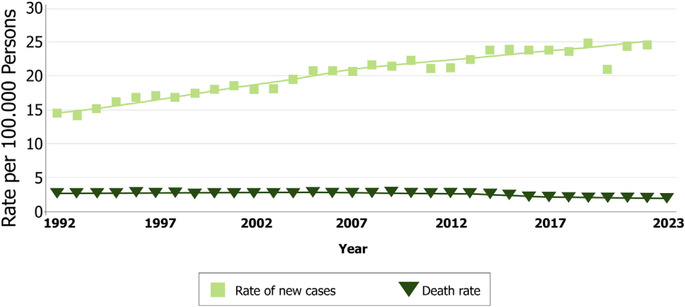
Unlike most other tumors, the overall global incidence of cutaneous melanoma has been steadily increasing in recent decades, with mortality stabilizing over the years.1 Despite being a less frequent skin tumor, its lethality is responsible for almost 73% of all deaths from skin cancers.2,3
There is debate about the cause of increased melanoma incidence and its reliability, since the increase is only observed in cutaneous melanomas and is not accompanied by increased mortality (Fig. 1).4-6 Some authors hypothe-size that the increase in incidence is primarily driven by an increased tendency for pathologists to diagnose melanoma in lesions that were previously considered to be only atypical or benign.7 Overdiagnosis of undoubted melanoma patients that died from other pathologies and were exempt from autopsy is also a confounding factor, since their mortality is often attributed to melanoma. However, it is more likely that this rise in incidence is multifactorial, including greater exposure to Ultraviolet (UV) radiation, population aging, improvement of surveillance services that record tumors, definition and standardization of histopathological criteria, early detection campaigns, and the use of diagnostic tools such as dermoscopy, which has refined the diagnostic accu-racy of melanoma.5
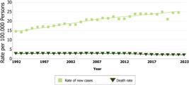
Rate of new melanoma cases and deaths per 100,000 persons in the United States of America over the years. Reprinted with permission from Cancer Stat Facts: Melanoma of the Skin. Surveillance, Epidemiology, and End Results (SEER) program.
This discrepancy between rising melanoma incidence and relatively stable or declining mortality rates should not deter individualized medical decision-making regarding patient treatment and follow-up. Similarly, patient educa-tion and early detection efforts should not be deprioritized, as they can significantly influence disease awareness, facil-itate earlier diagnosis, and ultimately improve prognosis.
The Surveillance, Epidemiology, and End Results (SEER) registry reports that in 2020 in the USA, the age-adjusted incidence rates for MIS were 18.39/100,000 and 11.32/100,000 for TM.8,9 The Instituto Nacional do Câncer (INCA) estimated 8,980 new cases of melanoma in Brazil for 2023-2025. Following a worldwide trend, the incidence of melanoma is higher in men, with 4640 new cases and 4340 in women, with a national incidence (across all stages) of 4.13 cases per 100,000 persons and a higher incidence in the south of the country. In 2020, there were 1923 melanoma-related deaths in Brazil, comprising 1120 deaths among men and 803 among women.10
Currently, most of the melanomas diagnosed are MIS and TM, globally and in the USA, accounting for 83% of all cases.9 Although they usually have a good prognosis, a small per-centage of these patients will have disease progression, and since they are very numerous, MIS and TM melanomas are responsible for 30% of all melanoma deaths.11
Recently, global and national databases have improved the registration process substantially, including the imple-mentation of automatic reporting and staging verification. Identification of late-stage melanomas is more reliable due to the use of hospital, regional pathology lab, and death records, whereas early-stage melanomas are likely to be relatively more difficult for the registry to document. This is likely responsible for some of the observed increasing incidence of melanoma and possibly contributes to the dis-parity between incidence and mortality increases. There is a significant possibility that melanoma cases may still be underreported, leading to errors in large databases, which could result in the underrepresentation of these tumors.
Risk and prognostic factors
New insights into melanoma risk and prognostic factors have been published in recent years. This is important so that patient awareness and better risk stratification can be achieved. Prognostic factors are related to disease pro-gression and are constantly being assessed and updated by the American Joint Committee on Cancer (AJCC) and the National Comprehensive Cancer Network (NCCN).
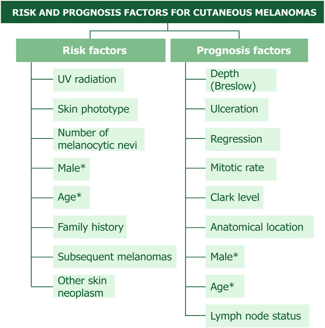
Few studies have established specific MIS and TM risk fac-tors. Fig. 2 provides a schematic view of these factors so they can be easily addressed during patient consultation and counseling.
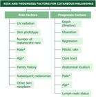
Well stablished risk and prognostic factor in patients with cutaneous melanoma. * Some features such as age and sex are mutual factors. Pigmentation phenotype includes lightly pigmented skin, hair, eyes, red hair and freckles as risk factors.
UV radiation, skin phototype and melanocytic nevi
Exposure to UV radiation is the most common environmen-tal risk factor for skin tumors. The mutations caused by UV radiation can be considered a pathogenic factor, acting from neogenesis to advanced stages of melanoma. Individuals’ phenotypes are controlled by the ratio of eumelanin and pheomelanin. Eumelanin provides protection against UV-induced DNA damage and is almost not present in red-haired and Caucasian individuals, who have more pheomelanin and have a greater tendency to develop skin cancers.12
Risk of melanoma is correlated with the number of sun-burns that a patient has suffered, which are more common in individuals with lightly pigmented phototypes. Individuals with dark skin phototypes are not exempt from melanomas, which occur preferentially in acral topography, with a worse prognosis, often with advanced disease. Recent muta-tional data have shown that acral melanomas have a low mutational burden, suggesting that it is not a UV-induced malignancy.13 The risk of individuals with lightly pigmented phototypes developing melanoma is 10 times higher when compared to individuals with dark skin.14
Evidence suggests that the number of Melanocytic Nevi (MN) is more important as an individual risk marker for the development of melanoma than as precursor lesions. About one-third of melanomas originate from pre-existing nevi, occurring most commonly on the trunk of young patients, while 70% are de novo.15 These data indicate that most melanomas do not originate from the malignant transfor-mation of nevus cells. A challenge in the MN approach is the differential diagnosis with MIS, especially in dysplastic nevi with severe atypia.
Anatomical location, sex and age
Melanomas located on the head and neck deserve particular care due to their worse prognosis. They occur in the elderly, and their frequency is considered high (up to 26.7%) for an area that corresponds to only 9% of the body surface.16-18
The trunk is the most affected site in men (41.5%) and the lower limbs (32.7%) in women.19 Previous studies reveal that a worse prognosis is expected in male patients, increased age, and sites such as the head and neck or trunk.20-22 Men generally have a higher mean age (56-years) at the time of diagnosis than women (52-years).19
Family history, subsequent melanomas and other skin neoplasms
Family History (FH) of melanoma is a well-defined risk fac-tor. Wei et al. followed 216,115 individuals, finding a 74% increased risk of melanoma (Hazard Ratio [HR = 1.74]) when compared with those without FH. Hereditary melanomas have an increased risk of cancers in other organs, such as the breast, pancreas, or central nervous system.23,24
A prior history of any melanoma should be considered a high-risk factor for cutaneous melanoma, with 1% to 8% of these patients developing multiple melanomas.14 On follow-up, 18.7% of MIS and TM patients developed a second melanoma.25 The subsequent tumor is usually thinner than the first, and its risk is higher in patients with fair skin and hair and an increased number of nevi.19
Individuals with a FH also have a 22% increased risk (HR = 1.22) of Squamous Cell Carcinoma (SCC), 27% (HR = 1.27) for Basal Cell Carcinoma (BCC), and an increased risk of melanomas on the trunk in both sexes and SCC on the extremities in women.26
In a meta-analysis, the lifetime risk of developing sec-ondary skin tumors, after a primary melanoma, was 3.8% for a new melanoma, 2.8% for BCC and 1% for SCC. The calculated 20-year cumulative risk was 5.4% for a sec-ond melanoma, 14% for BCC, and 4% for SCC. Although the analyses by subgroups and continents show substan-tial differences, the previous history of melanoma is a strong predictive factor for the development of a subsequent melanoma (approximately 10-fold increase in RR).27
As the lifespan of patients with melanoma has increased with new treatments, the likelihood of new melanomas and SCC/BCC also increases, and greater surveillance is needed in this group.25,28
Breslow
Breslow thickness (depth or index) represents the mea-surement in millimeters from the granulosa layer of the epidermis to the maximum depth of tumor invasion. This measurement is the most important prognostic factor for metastasis used by the AJCC for staging.29,30
New data indicate a ‘‘breakpoint’’ in 0.7 to 0.8 mm for the survival of T1 patients; this subgroup of TM should be assessed for their high risk of disease progression and Lymph Node Biopsy (LNB) should be considered in the multidis-ciplinary tumor board.31 Invasive melanomas with Breslow depth ≥0.8 mm have a 1.7 hazard ratio of worse survival than patients with <0.8 mm.30 TM patients with Breslow thickness between 0.8 and 1 mm also have a six-fold risk for progression to death, and the same six-fold risk for head and neck localization, when compared to tumors <0.8 mm. Melanomas with higher Breslow thickness should be mon-itored more frequently, especially if other associated risk factors are present.25,32,33
Ulceration
Besides Breslow depth, the presence of ulceration in the primary tumor is the most important pathological prognos-tic indicator in melanoma, being associated with aggressive disease and risk of Lymph Node (LN) metastasis.30,34-36 This is reflected by upstaging these patients in the AJCC when ulceration is present. MIS does not have any ulceration, and it is rare in T1 patients.
Regression
Patients with invasive tumors may present with partial regression on pathology, a phenomenon that may represent an immunological response to the melanoma. Macroscopi-cally, this may present as pink, grayish, hypopigmented or depigmented areas.37,38 The influence of regression on prog-nosis remains unknown, with some studies considering it to be a negative prognostic factor, because of the difficulty in accurately assessing Breslow thickness in regressed areas.39 This phenomenon was associated in some studies with a bet-ter prognosis, since effective activation of the host immune system against melanoma cells is likely its basis.40 Regres-sion is usually measured based on changes that are present in the dermis, which makes this factor inappropriate for MIS.38
Mitotic rate
Despite being removed from the last AJCC 8th staging edi-tion, this index remains an important prognostic factor in most studies. It is considered an independent prognostic fac-tor for LN positivity in TM, along with Breslow thickness in several studies.29,35,41,42 However, the reproducibility of this risk factor, including interobserver variability and conflict-ing data on the number of mitoses that would be considered the threshold to become a factor of worse prognosis, makes it harder to standardize.
Clark level
For decades, Clark’s levels of invasion have been used in conjunction with Breslow thickness for staging and classifi-cation in past AJCC editions. The challenge associated with reproducibility in measurements of Clark’s levels among observers has precipitated the abandonment of this param-eter in recent years.43
Although it is not used for staging in the AJCC 8th edition, the Clark level is a well-established prognostic factor and correlates with increased mortality in most studies.33,35,39 Clark level is important for TM evaluation and risk stratifi-cation and is a part of a complete pathological report.
Genetic aspects
Genetic profiling of melanomas will most likely provide miss-ing information on tumor progression, therapeutic targets and personal staging in the upcoming years. It is impor-tant for the classification and identification of mutations in different populations, stages, and anatomical sites in an academic scenario (Table 1).24,44-46
Somatic genetic mutations in early melanomas are distinct and necessary for tumorigenesis and disease pro-gression. The most common oncogenic mutations are BRAF (commonly V600E), NRAS, Kit, especially in acral and mucosal subtypes.46-48
BRAF-V600E can be found in 28% of lethal TM patients, as this could be a potential marker, probably associated with other mutations and a treatment target in the future.49 They are also found in primary, metastatic, and melanoma cell linages, suggesting that they occur before tumor pro-gression and spread and remain at a constant incidence during progression.12 BRAF mutation can occur early and be found in more than 80% of patients with common acquired MN and dysplastic nevus, and is considered a benign fea-ture of nevi formation. Since these pigmented lesions rarely progress to melanoma, it can be concluded that other muta-tions and additional genetic changes are required for tumor progression.12,49,50
Germline mutations predispose individuals to melanoma due to hereditary predisposition and syndromes. Multiple genes such as CDKN2A, CDK4, BAP1, POT1 and MITF are correlated to melanoma-dominant syndromes. Subordinate syndromes are associated with BRCA1/2, PTEN and TP53 mutations and contribute to an increased melanoma risk and other cancers (e.g., pancreatic, astrocytoma, breast, colon, ovarian, prostate, BAP1 syndrome) in an individual context.21,24,46
About 10% of melanomas are associated with germline mutations, and these can increase the risk of melanoma by four to 100-times.23 Progression to metastatic disease is probably due to a combination of mutations and the individ-ual immune system. It has been associated with mutations in the gene PTEN or TP53, there is a lack of studies in MIS and TM patients.50,51 Genetic testing in high-risk individuals with multiple primary melanomas or FH of melanoma and other cancers is available for genetic counseling.
Environmental risk factors such as UV radiation from early and intermittent sun exposure, and individual factors (lightly pigmented skin, hair, eyes, red hair and freckles) tend to result in a high mutational burden (>10 mutations per megabase), with a high number of mutations typical of UV damage.50 This environmental exposure predisposes to BRAF-driven melanomas, usually in younger patients, on non-sun damage on the skin (e.g., trunk) and melanoma of the superficial extensive type.50,52 Chronic sun exposure, on the other hand, is associated with mutations in NRAS, unrelated to the MN number.50
Gene Expression Profiling (GEP) represents an emerging adjunctive tool for the diagnostic and prognostic evaluation of cutaneous melanoma, though its integration into routine clinical practice remains under active investigation. They can aid the diagnosis of challenging melanocytic, however, GEP results should not supersede established histopatholog-ical criteria in guiding critical management decisions such as LNB or imaging surveillance strategies.45,46
Current knowledge about the genetic alterations that participate in the development of initial MIS and TM is insufficient, and genetic testing should not be performed routinely. Different combinations of mutations have been found, and new genes discovery increases the number of pos-sible genetic combinations. Mutations in high-penetrance genes, such as CDKN2A, CDK4, and BAP1, confer a 60% to 90% lifetime risk of melanoma.23 The future use of muta-tion biomarkers for risk stratification, choice of imaging, LNB, and adjuvant therapy is promising, but there is still no consensus for its use, requiring further studies.53,54
Melanoma Prevention Working Group guidelines state that genetic testing should be analyzed as continuous varia-bles to avoid lowand high-risk dichotomous interpretations that may have no biological significance. Results of these genetic profiles should always be evaluated and compared with established prognostic factors and by the risk stratifi-cation of the AJCC, and there are not yet sufficient data for their routine use.54
Currently, commercial use of genetic testing on different platforms can aid pathologists in challenging melanocytic lesions.44 There are no specific guidelines for genetic anal-ysis in MIS or TM for risk stratification, treatment, or follow-up. It is the authors’ opinion that genetic profiling of these initial tumors could contribute to the future, so treatment and follow-up can be tailored to each patient.
Diagnosis and treatment
Diagnosis should be made by clinical and dermoscopic evaluation, followed by anatomopathological examination. Dermoscopy allows the magnification of structures not vis-ible to the naked eye, in the superficial epidermis and dermis, and is mandatory for dermatologists and health pro-fessionals caring for patients with melanocytic lesions and tumors.
The ABCD rule for the clinical diagnosis of melanoma, described in the 1980s, was a milestone for its earlier detection, especially considering that until then, large and ulcerated tumors were common.55 Later, the addition of the letter ‘‘E’’ to the ABCD acronym - indicating ‘‘evolution’’ or ‘‘change’’ - further refined melanoma diagnosis.56
This dynamic behavior of the lesion may occasionally be the only indication of the tumor, facilitating even earlier diagnoses, particularly in initial melanomas that may not exhibit a striking ABCD criteria. In the 1990s, dermoscopy improved the accuracy of melanoma diagnosis by more than 30%, revolutionizing the approach to these cutaneous tumors.57 This technique has allowed the identification of increasingly early melanomas, including melanomas that do not resemble typical ones.58
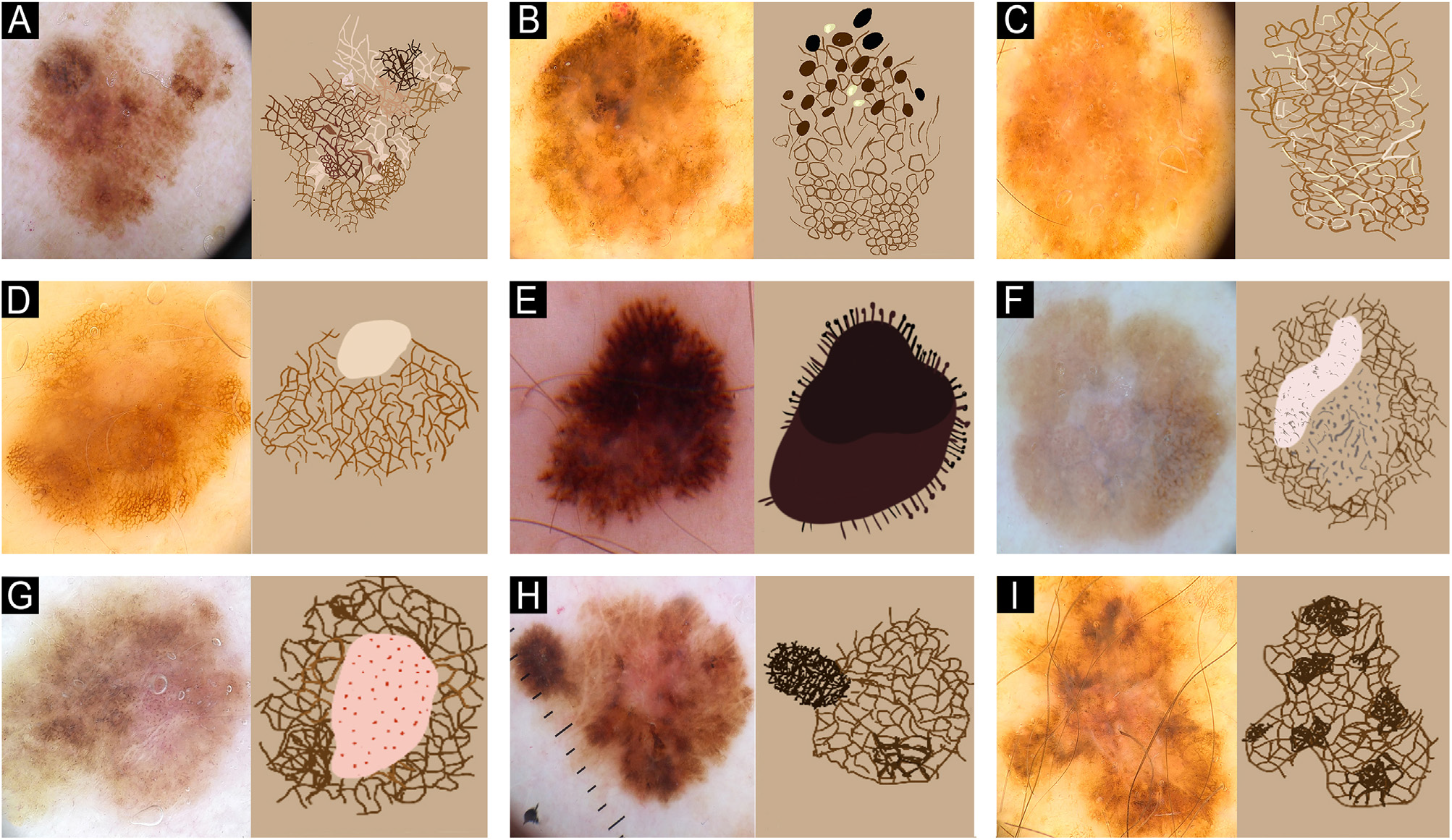
The main dermoscopic findings in MIS and TM (Fig. 3), include an irregular pigmented network, negative network, irregular globules and dots, radial streaks, irregular pig-mentation, structureless areas, and dermoscopic islands (well-circumscribed areas showing a uniform dermoscopic pattern that differs from the rest of the pigmented lesion). Photographic follow-up with total body mapping and dig-ital dermoscopy has also contributed to earlier melanoma diagnoses while avoiding unnecessary removal of MN. It is plausible that this trend toward earlier diagnoses will continue to grow, especially with the implementation of artificial intelligence in dermatology.59
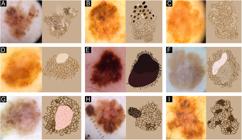
Dermoscopy features and their schematic features in MIS and TM. (A) Atypical, pigmented network. (B) Irregular globules and dots. (C) Negative pigmented network. (D) Peripheral tan and structureless areas. (E) Irregular radial streaks. (F) Regression. (G) Dotted vessels. (H) Dermoscopic islands. (I) Irregular pigmentation.
Initial biopsy should be, whenever possible, excisional, with minimal margins (1 to 3 mm), for complete patho-logical evaluation of the lesion and removing the least amount of unaffected skin, to avoid alteration in the local lymphatic drainage, with the longest axis in the same direc-tion/parallel to lymphatic vessels.46
Special sites with aesthetic or functional impact, such as the face, extremities, and genitalia, where initial com-plete resection may lead to mutilation, incisional or punch biopsy may be performed and guided by dermoscopy. Confo-cal microscopy can aid in diagnosing challenging lesions and in guiding biopsies, especially in the face.60
Diagnostic challenges in MIS may arise from het-erogeneity across histologic sections and overlap with severely atypical dysplastic nevi, which can promote inter-observer variability when dermatopathologists rely only on Hematoxylin-Eosin (HE) staining. This is rele-vant in melanocytic lesions with architectural disorder and cytologic atypia, where the differentiation between MIS and dysplastic nevi is uncertain even for experi-enced dermatopathologists.15,38 Current diagnostic guide-lines emphasize that Immunohistochemical (IHC) stains are not essential for the microscopic diagnosis of melanoma and should be reserved for selected cases in which morphology on HE is insufficient for the diagnosis; IHC should be used to support, rather than supplant, the primary histopathologic assessment.24,45,46,61,62
IHC can be helpful in challenging MIS and TM cases where standard HE examination is uncertain or when dermo-epidermal junction architecture and cytologic features overlap with benign or atypical melanocytic proliferations. IHC panels include markers such as S-100, SOX10, Melan-A/MART-1, and HMB-45 that enhance melanocytic lineage identification and can identify deeper dermal invasion that may be underestimated on HE. IHC markers can have false positives or complicate interpretation in heavily pigmented lesions.63
Molecular diagnostic platforms, such as gene-expression-based assays, DNA-based sequencing, cytogenetic analyses, and copy-number assessment, can aid in selected cases of diagnostically challenging melanocytic tumors. Gene-expression assays evaluate the transcriptional profile of tumor cells to distinguish benign from malignant melanocytic proliferations and estimate metastatic risk in specific settings. DNA-based sequencing techniques identify somatic mutations in oncogenic pathways (e.g., BRAF, NRAS, KIT), providing insight into tumor genesis and potential ther-apeutic targets. Cytogenetic and copy-number techniques detect chromosomal gains, losses, or structural rearrange-ments that are more frequently associated with melanoma than with benign nevi.45,50,54,64 These techniques also should not supersede established histopathologic criteria for diag-nosis, management, and staging. Such methods have not yet been incorporated into routine clinical practice, they require further validation before their application in risk stratification and melanoma management.24,44,45
Cutaneous melanomas are classified according to their growth pattern, clinical and histopathological characteris-tics into four subtypes of invasive melanomas: Superficial Spreading (SS), Nodular Melanoma (NM), Acral Lentiginous (AL) and Lentigo Maligna Melanoma (LMM).37,65 Lentigo Maligna (LM) is a subtype of MIS, which is slow growing and can evolve into an invasive component (LMM). These sub-types are not included as prognostic factors by the AJCC.65 After histological confirmation of melanoma, definitive surgical excision of scar tissue or residual lesion, along with adjacent tissue, should be planned and performed. NCCN 2025 guidelines for surgical margins of this definitive exci-sion should be based on Breslow thickness (Table 2), and margins greater than 2 cm had no impact on Local Recurrence (LR) and survival.46 Whenever possible, the largest margin according to the Breslow thickness of the tumor should be performed, respecting the maximum value of 2 cm.62,66
Surgery with intraoperative margin control (e.g., mod-ified Mohs) associated with the use of IHC markers has been used in some countries with similar survival rates to standard surgery.67 Regular frozen sections without IHC can undergo artifact alterations, making the correct assessment of melanocytic lesions challenging, and should not be per-formed according to NCCN guidelines.46
LNB should not be performed in MIS. T1b melanoma (Bres-low depth < 0.8 mm with ulceration or 0.8-1 mm with or without ulceration) should be assessed for LNB as a shared decision and discussed in multidisciplinary tumor boards.46,68 LNB remains a crucial factor in the staging of patients and an important prognostic factor and predictor of survival.69 It remains the most sensitive and specific test to iden-tify occult metastasis in LN, but it should not be routinely performed in TM.70 Complete LN dissection should not be performed since it does not impact patient survival.71
For greater uniformity, most studies use the AJCC system, staging tumors as in situ, according to Breslow thickness, LN involvement, and presence of metastasis (Table 3). MIS, by definition, do not exceed the basal layer and do not have Breslow depth, being staged as Tis. Accurate staging of patients by a dermatologist is mandatory so they can receive proper treatment, follow-up, and imaging when necessary. The term TM is historically used in the literature and in research and comprises tumors that have an IB ≤ 1.0 mm. Until 2002, the AJCC defined TM as lesions ≤0.76 mm, and the changes in this definition and in the staging over the years make it difficult for meta-analysis studies and often cannot be compared with present data.72
Currently, there is no recommendation for using neoadju-vant or adjuvant treatment for MIS and TM. If these patients progress to metastatic/advanced stages, they can bene-fit from anti PD-1 (pembrolizumab and nivolumab), anti CTLA-4 (ipilimumab); and/or mutation-directed therapies (dabrafenib/trametinib, vemurafenib/cobimetinib enco-rafenib/binimetinib), with well-established results, accord-ing to their staging.46
Radiotherapy remains indicated for palliative treatments or in inoperable cases, for local control of the disease. Use of topical medications should be restricted to excep-tional situations and/or palliative cases in whom resection is not feasible or desirable. Topical Imiquimod (IMQ) has been used for MIS, particularly LM, as a first-line, second-line, or adjuvant therapy, with high rates of clinical and histopatho-logical clearance. Patient response to topical medication can vary, and there is a need for long-term studies to fur-ther validate its efficacy. Therefore, the decision to use IMQ should be made collaboratively with the patient and discussed in tumor boards, in cases where surgery is not viable.46
A thorough history and clinical examination, including not only the area of the melanoma scar for the detection of LR, but also the entire body surface, with dermoscopy performed on all pigmented and non-pigmented lesions, is required. Total body digital dermoscopy can aid patient surveillance for new skin neoplasms.73
LN palpation is mandatory. Imaging should be per-formed based on specific patient signs and symptoms.46 LN Doppler ultrasound can assist dermatologists in assess-ing patients with challenging physical examinations (e.g., obesity, inguinal folds) when performed by a trained and experienced specialist. Performing a high-quality clinical examination is paramount, highlighting the dermatologist’s role in the follow-up of melanoma patients, since they are at a higher risk of developing new melanomas than metastases.46,62,65,66
There is no need for baseline/follow-up laboratory tests or imaging in MIS and TM; they should be considered in patients with a Breslow >0.8 mm. Clinical follow-up aims at early detection of recurrence, subsequent primary melanomas, and education.46
Patient education regarding SCC, BCC and new melanomas can aid early diagnosis and modify per-sonal risk factors (Fig. 2). This should be tailored to the patient’s educational level in simple language and focused on patient counseling. MIS and TM patients should not be discharged since they have an increased risk of new neoplasms and of LR and metastasis.
There are some global discrepancies on how often MIS and TM patients should be followed. Usually, a dermatologi-cal consultation every 4-months in the first year of diagnosis, followed by every 6-months in the second year and annually after is sufficient for most patients. The number of visi-tations can be modified due to patient risk and prognosis factors or due to public health-specific guidelines in each country.62,65,66,74,75
The first five years of follow-up are important because about 90% of metastases occur during this period, with almost two-thirds occurring in the first two years.65,76 The risk of late metastasis and recurrence should be kept in mind, so that if they occur, appropriate treatment is not delayed.
Despite advances in recent years, treatment is still chal-lenging in patients with metastatic disease, having a high mortality rate when diagnosed in advanced stages. Thus, the measure with the greatest impact to reduce mortal-ity is based on early detection of initial tumors, maximizing survival rates.5,60
Local recurrence and metastasis
Early diagnosis and appropriate surgical treatment for MIS and TM are the most important factors in patient survival. Despite their good prognosis, since this is the largest num-ber of melanoma patients, a significant number of patients will have LR or metastasis, and this should be promptly diag-nosed by dermatologists.
There is a lack of uniformity in the definition of LR in the literature. Most authors consider LR to be the reappearance of the tumor in the scar or adjacent to the initial surgi-cal procedure. Some studies use the nomenclature, distant recurrence, for LN involvement or metastasis.76-79
The rate of LR in MIS is variable, ranging from 0.3 to 9%, with most studies having a small number of individu-als. In TM, the LR rate ranges from 2% to 11.3%, depending on the study design and follow-up time. Table 4 summa-rizes the findings of LR and metastasis in MIS and TM in the literature.25,80-86
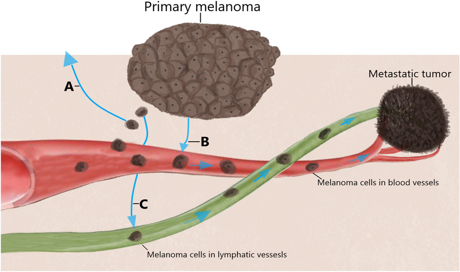
Metastases are defined as invasion of the tumor into an organ or tissue, with melanoma being a neoplasm with lymphatic and hematogenous dissemination (Fig. 4). It is estimated that in up to two-thirds of cases, they are locore-gional, affecting the skin or adjacent lymphatic system.76 In almost half of metastatic melanomas, only one organ is affected, with the skin accounting for about 20% of cases and the lungs, liver, and brain for 50%.37
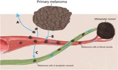
Melanoma dissemination pathways. Tumor cells can spread through adjacent skin (A), blood vessels (B) and lymphatic vessels (C). Its dissemination can occur either simultaneously or individually and lead to local recurrence, satellitosis and distant metastasis.
These regional metastases can be classified as satelli-tosis, in transit, or nodal (LN involvement), according to the distance from the primary tumor.37 Satellitoses are metastatic papules/nodules that appear within 2 cm of the primary tumor. They may be adjacent to the surgical scar, and their differential clinical diagnosis with LR may be diffi-cult, and it is necessary to highlight their dermal component on histology. In-transit metastases represent invasion of the tumor into the skin or subcutaneous tissue and are located 2 cm beyond the primary site and the LN drainage.37 Nodal metastases are more common at the nodal draining site of the primary tumor; nevertheless, they can also be found in discordant and unexpected LN drainages. In about 3% of patients with metastases, the primary site is not found.65
Presence of metastasis during follow-up of MIS patients raises the question about primary tumor depth missed by the pathologist and possible presence of another thick or unknown melanoma that may be the origin of metastatic dis-ease. Studies reporting metastasis in this group are scarce, with rates ranging from 0.24% to 1.8%.25,81,85,87
The rate of metastasis in patients with TM is rarely reported; if the authors exclude studies of LN involvement, being from 1.6% to 6.8%, according to Breslow thickness and study design.25,77,88,89
Late recurrence is generally defined by most studies as the recrudescence of the disease after 10-years (some authors consider it to be late after 5-years) and early recur-rence when it occurs before this period. Late recurrence incidence can reach up to 6.9%, varying according to the population studied.78,79 These data show that patients with melanoma have a rate of LR even after long follow-up peri-ods.
Some authors state that melanomas may possibly remain quiescent for decades in individuals until the development of LR or metastasis. In an analysis of 2,766 melanomas staged I-IV, from 1960 to 1996, Tsao et al. found a 18.1-years period for regional recurrence and 19-years for distant metastasis, showing that ultra-late recurrence (more than 15-years after diagnosis) can occur and without identifiable risk factors in the study.79
Progression rate from MIS to invasive melanoma is not known, but the rarity of LR and the exceptional deaths of these patients due to metastases suggest that not all MIS lesions would be precursors of invasive tumors and may remain without vertical or invasive growth. MIS can be considered a risk factor for the development of a sec-ond melanoma, which may present aggressive and invasive behavior.7
A limitation of long-term studies is that patients who died from causes other than melanoma may have died from occult metastasis and are not accounted. The incidence of occult metastasis can only be verified through autopsy, a procedure that is difficult to access and in some countries is subject to strict legal requirements.
Another possible bias when interpreting LR and metasta-sis is the lack of uniformity in large databases. A study of SEER data revealed that in one data center, a quarter of TM had Breslow depth errors. These tumors were reclassified as Breslow >1.0 mm, including 96% of the deaths associated with TM.90
When diagnosed in early stages (T1a), patients have a five-year survival rate of 99% and a 10-year survival rate of 98%. As these tumors progress, 5-year survival drops to 82% and 10-year survival to 75% in T4b N0 patients.
Since MIS and TM represent up to 83% of new melanoma diagnoses, even a 2% lethality rate represents a massive number of patients dying from early-stage disease, currently representing more than 30% of all melanoma deaths.9 In Aus-tralia, there are currently more deaths related to TM than to thick melanomas; these tumors comprise a substantial fraction of the overall burden of lethal melanomas in this high-incidence population.91
Careful consideration must be taken when advising MIS and TM patients on their diagnosis, follow-up and risk fac-tors. One should not state that MIS or TM patients are disease-free (‘‘cured’’) and do not require follow-up. Strong current data prove that these patients have a risk of LR and metastasis and will most likely develop a secondary melanoma, BCC, or SCC. Current prognostic tools do not allow us to stratify which patients are at higher risk for a worse outcome. Since initial melanomas are the majority of melanoma diagnoses, this gives dermatology an opportunity for patient education, screening, and facilitating secondary prevention. Aggressive behavior towards MIS and TM with expensive imaging and exams might also not be the correct approach for a vast number of patients, placing an economic and psychological burden on patients.
Conclusion
MIS and TM have a growing incidence and importance, rep-resenting a substantial part of dermatological practice. Individual risk stratification, early diagnosis, and patient information about prevention are essential to reducing incidence and mortality. Strict clinical follow-up will facili-tate timely diagnosis of recurrences, metastases, secondary melanomas, and other skin neoplasms, reinforcing the need for continuous long-term follow-up of these patients. New treatments and diagnostic tools will possibly be incorporated into the future management of these patients by dermatol-ogists, making it essential to update them.
Research data availability
The entire dataset supporting the results of this study was published in this article.
References
- 1 Long GV, Swetter SM, Menzies AM, Gershenwald JE, Scolyer RA. Cutaneous melanoma. Lancet. 2023;402:485-502. Erratum in: Lancet. 2023;402:450.
-
2 Howlader N, Noone AM, Krapcho M, Miller D, Brest A, Yu M, et al [cited 2025 Oct 10]. Available from: https://seer.cancer.gov/csr/19752016/, 2019.
» https://seer.cancer.gov/csr/19752016/ - 3 Carr S, Smith C, Wernberg J. Epidemiology and risk factors of melanoma. Surg Clin North Am. 2020;100:1-12.
- 4 Welch HG, Mazer BL, Adamson AS. The rapid rise in cutaneous melanoma diagnoses. N Engl J Med. 2021;384:72-9.
- 5 Muzumdar S, Lin G, Kerr P, Grant-Kels JM. Evidence concern-ing the accusation that melanoma is overdiagnosed. J Am Acad Dermatol. 2021;85:841-6.
-
6 National Cancer Institute. In: SEER Cancer Stat Facts: Melanoma of the Skin [Internet]. Bethesda (MD): National Insti-tutes of Health; 2025 [cited 2025 Oct 11]. Available from: https://seer.cancer.gov/statfacts/html/melan.html
» https://seer.cancer.gov/statfacts/html/melan.html - 7 Wei EX, Qureshi AA, Han J, Li TY, Cho E, Lin JY, et al. Trends in the diagnosis and clinical features of melanoma in situ (MIS) in US men and women: a prospective, observational study. J Am Acad Dermatol. 2016;75:698-705.
-
8 SEER*Stat databases: SEER november 2022 submission [Internet]. Bethesda (MD): National Cancer Institute; 2023 [cited 2025 Oct 3]. Available from: https://seer.cancer.gov/data-software/documentation/seerstat/nov2022/
» https://seer.cancer.gov/data-software/documentation/seerstat/nov2022/ -
9 SEER*Stat databases: SEER november 2021 submission [Internet]. Bethesda (MD): National Cancer Institute; 2022 [cited 2025 Oct 4]. Available from: https://seer.cancer.gov/data-software/documentation/seerstat/nov2021/
» https://seer.cancer.gov/data-software/documentation/seerstat/nov2021/ -
10 Instituto Nacional de Câncer. In: Estimativa 2023: incidência de câncer no Brasil [Internet]. Rio de Janeiro: INCA; 2022 [cited 2025 Oct 4]. Available from: https://www.inca.gov.br/publicacoes/livros/estimativa-2023-incidencia-de-cancer-no-brasil
» https://www.inca.gov.br/publicacoes/livros/estimativa-2023-incidencia-de-cancer-no-brasil - 11 Landow SM, Gjelsvik A, Weinstock MA. Mortality burden and prognosis of thin melanomas overall and by subcategory of thickness, SEER registry data, 1992-2013. J Am Acad Dermatol. 2017;76:258-63.
- 12 Hayward NK, Wilmott JS, Waddell N, Johansson PA, Field MA, Nones K, et al. Whole-genome landscapes of major melanoma subtypes. Nature. 2017;545:175-80.
- 13 Wang M, Fukushima S, Sheen YS, Ramelyte E, Pacheco NC, Shi C, et al. The genetic evolution of acral melanoma. bioRxiv [Preprint]. 2023;2023, 10.18.562802.
- 14 Rastrelli M, Tropea S, Rossi CR, Alaibac M. Melanoma: epidemi-ology, risk factors, pathogenesis, diagnosis and classification. In Vivo. 2014;28:1005-11.
- 15 Pampena R, Kyrgidis A, Lallas A, Moscarella E, Argenziano G, Longo C. A meta-analysis of nevus-associated melanoma: prevalence and practical implications. J Am Acad Dermatol. 2017;77:938-45.e4.
- 16 Dabouz F, Barbe C, Lesage C, Le Clainche A, Arnoult G, Hibon E, et al. Clinical and histological features of head and neck melanoma: a population-based study in France. Br J Dermatol. 2015;172:707-15.
- 17 Ettl T, Irga S, Müller S, Rohrmeier C, Reichert TE, Schreml S, et al. Value of anatomic site, histology and clinicopathological parameters for prediction of lymph node metastasis and overall survival in head and neck melanomas. J Craniomaxillofac Surg. 2014;42:e252-8.
- 18 Xie C, Pan Y, McLean C, Mar V, Wolfe R, Kelly J. Impact of scalp location on survival in head and neck melanoma: a retrospective cohort study. J Am Acad Dermatol. 2017;76:494-8.e2.
- 19 Bradford PT, Freedman DM, Goldstein AM, Tucker MA. Increased risk of second primary cancers after a diagnosis of melanoma. Arch Dermatol. 2010;146:265-72.
- 20 Lyth J, Falk M, Maroti M, Eriksson H, Ingvar C. Prognostic risk factors of first recurrence in patients with primary stages I-II cutaneous malignant melanoma - from the population-based swedish melanoma register. J Eur Acad Dermatol Venereol. 2017;31:1468-74.
- 21 Green AC, Baade P, Coory M, Aitken JF, Smithers M. Population-based 20-year survival among people diagnosed with thin melanomas in Queensland. Australia. J Clin Oncol. 2012;30:1462-7.
- 22 Chiaravalloti AJ, Jinna S, Kerr PE, Whalen J, Grant-Kels JM. A deep look into thin melanomas: what’s new for the clini-cian and the impact on the patient. Int J Womens Dermatol. 2018;4:119-21.
- 23 Ransohoff KJ, Jaju PD, Tang JY, Carbone M, Leachman S, Sarin KY. Familial skin cancer syndromes: increased melanoma risk. J Am Acad Dermatol. 2016;74:423-34, quiz 435-6. Erratum in: J Am Acad Dermatol. 2016;74:1290.
- 24 Leachman SA, Lucero OM, Sampson JE, Cassidy P, Bruno W, Queirolo P, et al. Identification, genetic testing, and management of hereditary melanoma. Cancer Metastasis Rev. 2017;36:77-90.
- 25 Gontijo JRV, Nelson JH, Diehl K, Korcheva VB, Bittencourt FV, Leachman SA. Thin and in situ melanomas of unfavorable prognosis: a retrospective observational analysis of local recur-rence, metastasis, and death in early-stage disease. J Am Acad Dermatol. 2025;92:325-7.
- 26 Wei EX, Li X, Nan H. Having a first-degree relative with melanoma increases lifetime risk of melanoma, squamous cell carcinoma, and basal cell carcinoma. J Am Acad Dermatol. 2019;81:489-99.
- 27 van der Leest RJ, Flohil SC, Arends LR, de Vries E, Nijsten T. Risk of subsequent cutaneous malignancy in patients with prior melanoma: a systematic review and meta-analysis. J Eur Acad Dermatol Venereol. 2015;29:1053-62.
- 28 Pastor-Tomas N, Martinez-Franco A, Banuls J, Pen˜alver JC, Traves V, García-Casado Z, et al. Risk factors for the devel-opment of a second melanoma in patients with cutaneous melanoma. J Eur Acad Dermatol Venereol. 2020;34:2295-302.
- 29 Conic RRZ, Ko J, Damiani G, Funchain P, Knackstedt T, Vij A, et al. Predictors of sentinel lymph node positivity in thin melanoma using the national cancer database. J Am Acad Der-matol. 2019;80:441-7.
- 30 Gershenwald JE, Scolyer RA, Hess KR, Sondak VK, Long GV, Ross MI, et al. Melanoma staging: evidence-based changes in the American joint committee on cancer eighth edition cancer staging manual. CA Cancer J Clin. 2017;67:472-92.
- 31 Varey AH, Lo SN, Williams GJ, Cust AE, Ollila D, Scolyer RA, et al. Analysis of the long-term survival of patients with thin melanomas using comprehensive, population-based australian data. EJC Skin Cancer. 2024;2:100073.
- 32 Claeson M, Baade P, Brown S, Soyer HP, Smithers BM, Green AC, et al. Clinicopathological factors associated with death from thin (</= 1.00 mm) melanoma. Br J Dermatol. 2020;182:927-31.
- 33 Marghoob AA, Koenig K, Bittencourt FV, Kopf AW, Bart RS. Bres-low thickness and clark level in melanoma: support for including level in pathology reports and in american joint committee on cancer staging. Cancer. 2000;88:589-95.
- 34 Wilkinson MJ, Gyorki DE. Extent of ulceration in cutaneous melanoma: is this biomarker ready for primetime? Br J Derma-tol. 2021;184:192-3.
- 35 Elder DE. Thin melanoma. Arch Pathol Lab Med. 2011;135:342-6.
- 36 Portelli F, Galli F, Cattaneo L, Cossa M, De Giorgi V, Forte G, et al. The prognostic impact of the extent of ulceration in patients with clinical stage I-II melanoma: a multicentre study of the Italian Melanoma Intergroup (IMI). Br J Dermatol. 2021;184:281-8.
- 37 Kibbi N, Kluger H, Choi JN. Melanoma: clinical presentations. Cancer Treat Res. 2016;167:107-29.
- 38 Cartron AM, Aldana PC, Khachemoune A. Reporting regression with melanoma in situ: reappraisal of a potential paradox. Arch Dermatol Res. 2021;313:65-9.
- 39 Mansson-Brahme E, Carstensen J, Erhardt K, Lagerlöf B, Ring-borg U, Rutqvist LE. Prognostic factors in thin cutaneous malignant melanoma. Cancer. 1994;73:2324-32.
- 40 El Sharouni MA, Aivazian K, Witkamp AJ, Sigurdsson V, van Gils CH, Scolyer RA, et al. Association of histologic regression with a favorable outcome in patients with stage 1 and stage 2 cuta-neous melanoma. JAMA Dermatol. 2021;157:166-73.
- 41 Kruper LL, Spitz FR, Czerniecki BJ, Fraker DL, Blackwood-Chirchir A, Ming ME, et al. Predicting sentinel node status in AJCC stage I/II primary cutaneous melanoma. Cancer. 2006;107:2436-45.
- 42 Sinnamon AJ, Neuwirth MG, Yalamanchi P, Gimotty P, Elder DE, Xu X, et al. Association between patient age and lymph node positivity in thin melanoma. JAMA Dermatol. 2017;153:866-73.
- 43 Garbe C, Ellwanger U, Tronnier M, Brocker EB, Orfanos CE. The new american joint committee on cancer staging system for cutaneous melanoma: a critical analysis based on data of the German central malignant melanoma registry. Cancer. 2002;94:2305-7.
- 44 Leachman SA, Mengden Koon S, Korcheva VB, White KP. Assess-ing genetic expression profiles in melanoma diagnosis. Dermatol Clin. 2017;35:537-44.
- 45 Kashani-Sabet M, Leachman SA, Stein JA, Arbiser JL, Berry EG, Celebi JT, et al. Early detection and prognostic assessment of cutaneous melanoma: consensus on optimal practice and the role of gene expression profile testing. JAMA Dermatol. 2023;159:545-53.
-
46 National Comprehensive Cancer Network. In: NCCN clinical practice guidelines in oncology: Melanoma: cutaneous. version 2.2025 [Internet]; 2025 [cited 2025 Oct 4]. Available from: https://www.nccn.org/guidelines/category1
» https://www.nccn.org/guidelines/category1 - 47 van den Hurk K, Niessen HE, Veeck J, van den Oord JJ, van Steensel MA, Zur Hausen A, et al. Genetics and epigenetics of cutaneous malignant melanoma: a concert out of tune. Biochim Biophys Acta. 2012;1826:89-102.
- 48 Gu F, Chen TH, Pfeiffer RM, Fargnoli MC, Calista D, Ghiorzo P, et al. Combining common genetic variants and non-genetic risk factors to predict risk of cutaneous melanoma. Hum Mol Genet. 2018;27:4145-56.
- 49 Claeson M, Tan SX, Lambie D, Brown S, Walsh MD, Baade PD, et al. The association between BRAF-V600E mutations and death from thin (≤ 1.00 mm) melanomas: a nested case-case study from Queensland. Australia. J Eur Acad Dermatol Venereol. 2023;37:e1168-72.
- 50 Schadendorf D, van Akkooi ACJ, Berking C, Griewank KG, Gutzmer R, Hauschild A, et al. Melanoma. Lancet. 2018;392:971-84.
- 51 Shain AH, Yeh I, Kovalyshyn I, Sriharan A, Talevich E, Gagnon A, et al. The genetic evolution of melanoma from precursor lesions. N Engl J Med. 2015;373:1926-36.
- 52 Spathis A, Katoulis AC, Damaskou V, Liakou AI, Kottaridi C, Leventakou D, et al. BRAF mutation status in primary, recurrent, and metastatic malignant melanoma and its rela-tion to histopathological parameters. Dermatol Pract Concept. 2019;9:54-62.
- 53 Leong SP, Aktipis A, Maley C. Cancer initiation and progres-sion within the cancer microenvironment. Clin Exp Metastasis. 2018;35:361-7.
- 54 Grossman D, Okwundu N, Bartlett EK, Marchetti MA, Othus M, Coit DG, et al. Prognostic gene expression profiling in cutaneous melanoma: identifying the knowledge gaps and assessing the clinical benefit. JAMA Dermatol. 2020;156:1004-11.
- 55 Friedman RJ, Rigel DS, Kopf AW. Early detection of malig-nant melanoma: the role of physician examination and self-examination of the skin. CA Cancer J Clin. 1985;35: 130-51.
- 56 Abbasi NR, Shaw HM, Rigel DS, Friedman RJ, McCarthy WH, Osman I, et al. Early diagnosis of cutaneous melanoma: revisit-ing the ABCD criteria. JAMA. 2004;292:2771-6.
- 57 American Academy of Dermatology Ad Hoc Task Force for the ABCDEs of MelanomaTsao H, Olazagasti JM, Cordoro KM, Brewer JD, Taylor SC, Bordeaux JS, et al. Early detection of melanoma: reviewing the ABCDEs. J Am Acad Dermatol. 2015;72:717-23.
- 58 Menzies SW. Evidence-based dermoscopy. Dermatol Clin. 2013;31:521-4, vii.
- 59 von Schuckmann L, Banney L, Soyer HP. Melanoma imaging and diagnosis: what does the future hold? Aust J Gen Pract. 2024;53:633-4.
- 60 Blundo A, Cignoni A, Banfi T, Ciuti G. Comparative analysis of diagnostic techniques for melanoma detection: a systematic review of diagnostic test accuracy studies and meta-analysis. Front Med (Lausanne). 2021;8:637069.
- 61 Xavier-Júnior JCC, KMPA Coelho, Macedo MP, Lellis RF, Pin-heiro Junior NF, Rocha RF, Dermatopathology Committee of the Brazilian Society of Pathology. Pre and post-analytical guidelines for the microscopic diagnosis of melanoma: recommendations from the Brazilian Society of Pathology. An Bras Dermatol. 2025;100:501139.
- 62 Swetter SM, Tsao H, Bichakjian CK, Curiel-Lewandrowski C, Elder DE, Gershenwald JE, et al. Guidelines of care for the management of primary cutaneous melanoma. J Am Acad Der-matol. 2019;80:208-50.
- 63 Voiculescu VM, Popescu AI, Costache M. Immunohistochemistry for skin cancers: new insights into diagnosis and treatment of melanoma. Cancers. 2025;17:1769.
- 64 Testa U, Castelli G, Pelosi E. Melanoma: genetic abnormalities, tumor progression, clonal evolution and tumor initiating cells. Med Sci (Basel). 2017;5:28.
- 65 Garbe C, Amaral T, Peris K, Hauschild A, Arenberger P, Basset-Seguin N, et al. European consensus-based interdisciplinary guideline for melanoma. part 1: diagnostics: update 2022. Eur J Cancer. 2022;170:236-55.
-
66 Grupo Brasileiro de Melanoma. Recomendação para o tratamento do melanoma cutâneo [Internet]. 2023 [cited 2025 Oct 4]. Available from: https://gbm.org.br/wp-content/uploads/2023/05/CartilhaRecomendacoesGBMmaio23.pdf
» https://gbm.org.br/wp-content/uploads/2023/05/CartilhaRecomendacoesGBMmaio23.pdf - 67 Heath M, Woody M, Leitenberger J, Latour E, Bar A. Inva-sive melanoma and melanoma in situ treated with modified mohs micrographic surgery with en face permanent section-ing: a 10-year retrospective review. Dermatol Surg. 2020;46: 1004-13.
- 68 Shannon AB, Sharon CE, Straker RJ 3rd, Carr MJ, Sinnamon AJ, Bogatch K, et al. Sentinel lymph node biopsy in patients with T1a cutaneous malignant melanoma: a multicenter cohort study. J Am Acad Dermatol. 2023;88:52-9.
- 69 Quintanilla-Dieck MJ, Bichakjian CK. Management of early-stage melanoma. Facial Plast Surg Clin North Am. 2019;27:35-42.
- 70 Rosko AJ, Vankoevering KK, McLean SA, Johnson TM, Moyer JS. Contemporary management of early-stage melanoma: a system-atic review. JAMA Facial Plast Surg. 2017;19:232-8.
- 71 Faries MB, Thompson JF, Cochran AJ, Andtbacka RH, Mozzillo N, Zager JS, et al. Completion dissection or observation for sentinel-node metastasis in melanoma. N Engl J Med. 2017;376:2211-22.
- 72 Balch CM, Buzaid AC, Soong SJ, Atkins MB, Cascinelli N, Coit DG, et al. Final version of the American Joint Committee on Cancer staging system for cutaneous melanoma. J Clin Oncol. 2001;19:3635-48.
- 73 Barcaui C, Bakos RM, Paschoal FM, Bittencourt FV, Sá BCS, Miot HA. Total body mapping in the follow-up of melanocytic lesions: recommendations of the Brazilian Society of Dermatology. An Bras Dermatol. 2021;96:472-6.
- 74 Castro LG, Bakos RM, Duprat Neto JP, Bittencourt FV, Di Giacomo TH, Serpa SS, et al. Brazilian guidelines for diagnosis, treatment and follow-up of primary cutaneous melanoma - part II. An Bras Dermatol. 2016;91:49-58.
- 75 Swetter SM, Johnson D, Albertini MR, Barker CA, Bateni S, Baumgartner J, et al. NCCN Guidelines(R) insights: melanoma: cutaneous, version 2.2024. J Natl Compr Canc Netw. 2024;22:290-8.
- 76 Tas F, Erturk K. Recurrence behavior in early-stage cutaneous melanoma: pattern, timing, survival, and influencing factors. Melanoma Res. 2017;27:134-9.
- 77 Richetta AG, Bottoni U, Paolino G, Clerico R, Cantisani C, Ambrifi M, et al. Thin melanoma and late recurrences: it is never too thin and never too late. Med Oncol. 2014;31:909.
- 78 Faries MB, Steen S, Ye X, Sim M, Morton DL. Late recurrence in melanoma: clinical implications of lost dormancy. J Am Coll Surg. 2013;217:27-34, discussion 34-36.
- 79 Tsao H, Cosimi AB, Sober AJ. Ultra-late recurrence (15-years or longer) of cutaneous melanoma. Cancer. 1997;79:2361-70.
- 80 Huilgol SC, Selva D, Chen C, Hill DC, James CL, Gramp A, et al. Surgical margins for lentigo maligna and lentigo maligna melanoma: the technique of mapped serial excision. Arch Der-matol. 2004;140:1087-92.
- 81 Joyce KM, Joyce CW, Jones DM, Donnellan P, Hussey AJ, Regan PJ, et al. An assessment of histological margins and recur-rence of melanoma in situ. Plast Reconstr Surg Glob Open. 2015;3:e301.20150306.
- 82 Kunishige JH, Brodland DG, Zitelli JA. Surgical margins for melanoma in situ. J Am Acad Dermatol. 2012;66:438-44.
- 83 Bene NI, Healy C, Coldiron BM. Mohs micrographic surgery is accurate 95.1% of the time for melanoma in situ: a prospective study of 167 cases. Dermatol Surg. 2008;34:660-4.
- 84 Akhtar S, Bhat W, Magdum A, Stanley PR. Surgical excision margins for melanoma in situ. J Plast Reconstr Aesthet Surg. 2014;67:320-3.
- 85 Moura FS, Homer LE, McKirdy SW. Histological peripheral mar-gins and recurrence of melanoma in situ treated with wide local excision. J Skin Cancer. 2020;2020:8813050.
- 86 Nosrati A, Berliner JG, Goel S, McGuire J, Morhenn V, de Souza JR, et al. Outcomes of melanoma in situ treated with mohs micrographic surgery compared with wide local excision. JAMA Dermatol. 2017;153:436-41.
- 87 Bricca GM, Brodland DG, Ren D, Zitelli JA. Cutaneous head and neck melanoma treated with Mohs micrographic surgery. J Am Acad Dermatol. 2005;52:92-100.
- 88 Gimotty PA, Elder DE, Fraker DL, Botbyl J, Sellers K, Elenitsas R, et al. Identification of high-risk patients among those diagnosed with thin cutaneous melanomas. J Clin Oncol. 2007;25:1129-34.
- 89 Durham AB, Schwartz JL, Lowe L, Zhao L, Johnson AG, Harms KL, et al. The natural history of thin melanoma and the utility of sentinel lymph node biopsy. J Surg Oncol. 2017;116:1185-92.
- 90 Gimotty PA, Shore R, Lozon NL, Whitlock J, He S, Vigneau FD, et al. Miscoding of Melanoma thickness in SEER: research and clinical implications. J Invest Dermatol. 2016;136:2168-72.
- 91 Whiteman DC, Baade PD, Olsen CM. More people die from thin melanomas (≤1 mm) than from thick melanomas (>4 mm) in Queensland, Australia. J Invest Dermatol. 2015;135:1190-3.
- 92 Leiter U, Buettner PG, Eigentler TK, Bröcker EB, Voit C, Gollnick H, et al. Hazard rates for recurrent and secondary cuta-neous melanoma: an analysis of 33,384 patients in the German Central Malignant Melanoma Registry. J Am Acad Dermatol. 2012;66:37-45.
- 93 Cordeiro E, Gervais MK, Shah PS, Look Hong NJ, Wright FC. Sentinel lymph node biopsy in thin cutaneous Melanoma: a systematic review and meta-analysis. Ann Surg Oncol. 2016;23:4178-88.
- 94 Hou JL, Reed KB, Knudson RM, Mirzoyev SA, Lohse CM, Frohm ML, et al. Five-year outcomes of wide excision and Mohs micro-graphic surgery for primary lentigo maligna in an academic practice cohort. Dermatol Surg. 2015;41:211-8.
- 95 Murali R, Haydu LE, Long GV, Quinn MJ, Saw RP, Shannon K, et al. Clinical and pathologic factors associated with distant metastasis and survival in patients with thin primary cutaneous melanoma. Ann Surg Oncol. 2012;19:1782-9.
Edited by
-
Editor
Sílvio Alencar Marques.