
 The intergluteal cleft involvement in hidradenitis suppurativa: clinical characteristics and potential clinical implications
Dear Editor,
Hidradenitis suppurativa (HS) is a chronic inflammatory disease characterized by painful nodules, abscesses, and draining tunnels affecting apocrine gland-bearing regions, often resulting in scarring and substantial impairment in quality of life.1,2 Although genotype-based stratification could theoretically inform individualized prognosis, the lack of validated biomarkers and inconsistent genotype-phenotype correlations limit its applicability in daily practice. Clinically defined phenotype-based assessment, therefore, remains the most pragmatic strategy for evalu-ating severity and guiding treatment.3 Several phenotypic frameworks have been proposed, including anatomical clustering and lesion-based or comorbidity-driven sub-types; however, consensus is lacking, and underrecognized topographic patterns may provide clinically meaningful insights.3-5
Among these, the intergluteal cleft lesion (ICL), a mid-line fissure or ulceration with fibrosis, scarring, and drainage (Fig. 1), is one such pattern, infrequently described in the literature and often misclassified as pilonidal sinus disease (PSD). Given the established association between hidradenitis suppurativa and inflammatory bowel disease (IBD), particularly Crohn’s disease (CD),6 we sought to explore whether the presence of ICL has clinical relevance in this context.
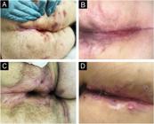
(A and B) Linear ulceration with surrounding inflammation in a patient with Hurley stage III HS and extensive gluteal involvement. (C) Fibrotic scarring and fissuring along the midline, suggestive of chronic disease and structural remodeling. (D) Superficial ulceration and papules confined to the intergluteal fold, in a patient without other follicular occlusion disorders.
We conducted a cross-sectional observational study to evaluate whether ICL represents a distinct clinical pattern associated with more severe HS or systemic involvement. We assessed 119 consecutive patients with confirmed HS at a tertiary dermatology center in Brazil. Standardized evaluation included demographic and clinical data, comor-bidities, disease duration, number of affected regions, history of HS-related hospitalizations and surgeries, and sys-temic therapies. Disease activity was evaluated using the Hurley system and the International HS Severity Score Sys-tem (IHS4). In 10 ICL cases, punch biopsies were performed for histopathological characterization. Comparative analy-ses were conducted between patients with and without ICL, and logistic regression identified independent predictors of ICL and of poor outcomes, defined as the presence of ≥ 2 of the following: Hurley III, IHS4 ≥ 11, hospitalization due to HS, or need for multiple systemic therapies.
ICL was observed in 18 of 119 patients (15.1%). Patients with ICL were more frequently male (72.2% vs. 30.7%; p = 0.002) and exhibited a markedly higher prevalence of CD (16.7% vs. 2.0%; p = 0.024) (Table 1). Disease duration and BMI did not differ significantly between groups. How-ever, ICL was associated with substantially higher disease burden: mean IHS4 scores were more than double those of patients without ICL (16.0 vs. 7.03; p < 0.001), draining tunnels were more common (88.9% vs. 50.5%; p = 0.006), and the number of affected anatomical regions was greater (3.39 vs 2.34; p < 0.001) (Table 2). Patients with ICL also required biologic therapy more frequently and had signifi-cantly more HS-related hospitalizations in the preceding five years (55.6% vs. 12.0%; p < 0.001). Although Hurley stage III was numerically more prevalent, the lack of statistical sig-nificance suggests that ICL identifies severity dimensions not fully captured by traditional staging.
Clinical and demographic characteristics of patients with and without intergluteal cleft lesions.
Histopathological examination demonstrated chronic inflammation, granulation tissue, and fibrosis, without epithelialized tracts or embedded hairs typical of PSD, and without granulomatous inflammation suggestive of CD. Anaerobic or fungal growths were occasionally observed and likely reflected secondary colonization. Clinically, most ICLs were refractory: 66.7% exhibited only partial improvement, and 27.8% no improvement despite systemic therapy, pre-dominantly adalimumab.
Multivariate analysis identified male sex (OR = 11.09; p < 0.001), CD (OR = 66.41; p = 0.004), older age (OR = 1.04 per year; p = 0.032), and HS-related surgery (OR = 14.52; p = 0.031) as independent predictors of ICL. Conversely, the presence of follicular occlusion syndromes was associated with lower odds (OR = 0.23; p = 0.041), suggesting partial divergence from follicular-occlusion-dominant phenotypes (Table 3). Importantly, ICL conferred an eightfold higher likelihood of poor clinical outcomes (OR = 8.37; p = 0.043), even after adjustment for demographic and clinical varia-bles.
These findings identify ICL as a potential clinical marker of disease severity in HS, associated with higher inflamma-tory burden, adverse outcomes, and systemic involvement. HS is a heterogeneous disorder with diverse clinical endo-types, and clinically defined phenotype-based stratification has gained emphasis as a tool to identify patients at risk for refractory disease or complications.4,5 Our results align with existing gluteal-focused clinical phenotypes described in the literature, such as the ‘‘LC3’’ subtype described by Canoui-Poitrine et al.7 and the ‘‘fistulous’’ variant described by Riera-Martí,8 both characterized by draining tunnels and aggressive behavior. The association between ICL and the absence of follicular occlusion syndromes suggests a more inflammatory pattern, less responsive to keratinization-directed therapies.
The consistently higher severity markers observed in ICL - elevated IHS4, tunnel predominance, increased healthcare utilization - support its role as a visible marker of advanced and treatment-resistant HS. Tunneling, a frequent feature of ICL, has previously been linked to chronicity and refrac-toriness, reinforcing the need for early recognition before irreversible fibrosis develops.9
A striking observation in this study was the strong and independent association between ICL and CD. Given the well-established coexistence of HS and IBD,6 this study aimed to evaluate whether the presence of ICL is clini-cally associated with CD. The disproportionate prevalence of CD among patients with ICL suggests that this lesion may function as a clinical marker associated with systemic inflammatory burden in HS. Given the clinical challenges in distinguishing perianal CD from gluteal HS,10 recognition of ICL may support earlier gastrointestinal evaluation.
Therapeutic refractoriness further underscores the need for targeted and timely interventions, potentially beyond current anti-TNF strategies, as suggested by isolated reports of IL-17 pathway efficacy in gluteal disease.11
This study has limitations, including a small sample size, single-center recruitment, and a cross-sectional design that precludes determining whether ICL precedes or results from severe HS. Nonetheless, the magnitude and coherence of the associations observed support the clinical relevance of this understudied lesion.
In conclusion, ICL appears to be a readily identifi-able, high-yield clinical marker associated with a severe and potentially systemic form of HS. Its recognition may prompt earlier therapeutic escalation, multidisciplinary management, and targeted screening for IBD. As precision dermatology advances in HS, incorporating underrecognized clinical clues such as ICL may improve prognostication and optimize patient care.
Ethics statement
The patients in this manuscript have given written informed consent to the publication of their case details. This study was reviewed and approved by the Research Ethics Commit-tee of the State University of Campinas (UNICAMP) on March 26 (2024), under approval number 77223323.1.0000.5404.
Research data availability
The entire dataset supporting the results of this study was published in this article.
References
- 1 Sabat R, Alavi A, Wolk K, Wortsman X, McGrath B, Garg A, et al. Hidradenitis suppurativa. Lancet. 2025;405:420-38.
- 2 Zouboulis CC, Bechara FG, Benhadou F, Bettoli V, Bukvi´c Mokos Z, Del Marmol V, et al. European S2k guidelines for hidradenitis suppurativa/acne inversa part 2: treatment. J Eur Acad Derma-tol Venereol. 2025;39:899-941.
- 3 Frew JW, Hawkes JE, Sullivan-Whalen M, Gilleaudeau P, Krueger JG. Inter-rater reliability of phenotypes and exploratory genotype-phenotype analysis in inherited hidradenitis suppura-tiva. Br J Dermatol. 2019;181:566-71.
- 4 Vişan MA, Ca˘runtu C, Costache RS, Tiplica G, Costache DO. Hidradenitis suppurativa: detangling phenotypes and identi-fying common denominators. J Eur Acad Dermatol Venereol. 2024;38:62-76.
- 5 Jørgensen AHR, Nicolai SA, Yao Y, Holm JG, Nielsen VW, Ring HC, et al. Cluster analysis identifies six clinical sub-types of hidradenitis suppurativa characterized by distinct comorbidities, inflammatory and metabolic profiles, patient-reported outcomes and treatment patterns. JEADV Clin Pract. 2023;2:80-91.
- 6 Egeberg A, Jemec GBE, Kimball AB, Bachelez H, Gislason GH, Thyssen JP, et al. Prevalence and risk of inflammatory bowel disease in patients with hidradenitis suppurativa. J Invest Der-matol. 2017;137:1060-4.
- 7 Canoui-Poitrine F, Revuz JE, Wolkenstein P, Viallette C, Gabison G, Pouget F, et al. Clinical characteristics of a series of 302 French patients with hidradenitis suppurativa, with an analysis of factors associated with disease severity. J Am Acad Dermatol. 2009;61:51-7.
- 8 Riera-Martí N, Vilarrasa E, López-Llunell C, Gamissans M, Sin M, Romaní J. Gluteal hidradenitis suppurativa: analysis of 83 patients. Actas Dermosifiliogr. 2024;115:137-42.
- 9 Ingram JR, Marzano AV, Prens E, Schneider-Burrus S, Warren RB, Keal A, et al. Hidradenitis suppurativa with and without draining tunnels: a real-world study characterizing differences in treatment and disease burden. J Eur Acad Dermatol Venereol. 2025;39:1431-41.
- 10 Yamanaka-Takaichi M, Yalon M, Loftus EV, Grimaldo AB, Todd A, Wetter DA, et al. Magnetic resonance imaging for distinguishing perianal hidradenitis suppurativa from fistulizing Crohn disease. Dermatology. 2025;241:437-46.
- 11 Arenbergerova M, Arenberger P, Marques E, Gkalpakiotis S. Successful treatment of recalcitrant gluteal hidradenitis suppurativa with brodalumab after anti-TNF failure. Int J Dermatol. 2020;59:733-5.
Edited by
-
Editor
Hiram Larangeira de Almeida Jr.

