
Abstract
Netherton syndrome is a rare autosomal recessive disease characterized by erythroderma, ichthyosis linearis circumflexa, atopy, failure to thrive and a specific hair shaft abnormality called trichorrhexis invaginata or bamboo hair, considered pathognomonic. We report the case of a 4-year-old boy with erythroderma since birth, growth deficit and chronic diarrhea. Trichoscopy was used to visualize typical bamboo and "golf tee" hair and of key importance to diagnose Netherton syndrome. We suggest the use of this procedure in all children diagnosed with erythroderma.
Dermoscopy; Hair; Netherton syndrome
INTRODUCTION
Netherton syndrome (NS) is a rare recessive autosomal disease characterized by
erythroderma, ichthyosis linearis circumflexa, atopy, growth retardation and a
specific hair shaft alteration, identified as trichorrhexis
invaginata (TI) or bamboo hair. 11 Sun JD, Linden KG. Netherton syndrome: a case report and review of
the literature. Int J Dermatol. 2006;45:693-7.,22 Duquia RP, Almeida Jr HL, Souza PRM, Vettorato G. Netherton’s
Syndrome with a 20-year follow-up. An Bras Dermatol.
2006;81:559-62. TI is
pathognomonic of NS and presents itself microscopically as an invagination of the
shaft's distal portion to its proximal portion, giving it an appearance of a “ball
in a hoop”.22 Duquia RP, Almeida Jr HL, Souza PRM, Vettorato G. Netherton’s
Syndrome with a 20-year follow-up. An Bras Dermatol.
2006;81:559-62.,33 Powell J, Dawber RP, Ferguson DJ, Griffiths WA. Netherton's
syndrome: increased likelihood of diagnosis by examining eyebrows hairs. Br J
Dermatol. 1999;141:544-6. When there is fracture of hair at the site of the
invagination, its end becomes similar to a shell and this type of fractured hair is
called “golf tee hair”.11 Sun JD, Linden KG. Netherton syndrome: a case report and review of
the literature. Int J Dermatol. 2006;45:693-7.,44 Rakowska A, Kowalska-Oledzka E, Slowinska M, Rosinska D, Rudnicka L.
Hair shaft videodermoscopy in Netherton syndrome. Pediatr Dermatol.
2009;26:320-2. In recent years the application of
trichoscopy has increased for diagnosing cicatricial and noncicatricial alopecias,
inflammatory diseases and scalp hair shaft disorders such as monilethrix,
trichorrhexis nodosa, pili torti, pili annulati and TI.44 Rakowska A, Kowalska-Oledzka E, Slowinska M, Rosinska D, Rudnicka L.
Hair shaft videodermoscopy in Netherton syndrome. Pediatr Dermatol.
2009;26:320-2.
5 Rakowska A, Slowinska M, Kowalska-Oledzka E, Rudnicka L. Trichoscopy
in genetic hair shaft abnormalities. J Dermatol Case Rep.
2008;2:14-20.
6 Burk C, Hu S, Lee C, Connelly EA. Netherton syndrome and
trichorrhexis invaginata - a novel diagnostic approach. Pediatr Dermatol.
2008;25:287-8.
7 Liu CI, Hsu CH. Rapid diagnosis of monilethrix using dermoscopy. Br
J Dermatol. 2008;159:741-3.-88 Kharkar V, Gutte R, Thakkar V, Khopkar U. Trichorrhexis nodosa with
nail dystrophy: diagnosis by dermoscopy. Int J Trichology.
2011;3:105-6.
CASE REPORT
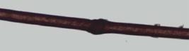
A four-year-old boy presented history of erythroderma after birth, chronic diarrhea and growth deficit. He was brought for dermatological evaluation presenting eczematous plaques disseminated in the tegument. He had been using topical corticoids, oral antihistamine and zinc regularly, with persistence of lesions and recurrent episodes of exacerbation. His mother also reported that he presented brittle and fragile hair. At the dermatological examination we observed short hair, diffuse erythema and scaling (especially on the face) and erythematous scaly plaques on the trunk, abdomen and back of hands (Figure 1). Trichoscopy showed trichorrhexis invaginata and golf tee hair (Figure 2). Optical microscopy analysis confirmed characteristic TI changes (Figures 3, 4 and 5). Laboratory examinations revealed eosinophilia (14%), thrombocytosis, and elevated IgE serum levels. Serology for HIV 1 and 2, HTLV 1 and 2 were negative.

Clinical aspects: A) Short hair, erythema and desquamation of face; B), C) and D) Erythematous and scaly plaques disseminated on the tegument


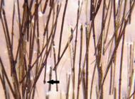
Aspects of trichoscopy in detail, showing trichorrhexis invaginata (*) and golf tee hair (arrow)
DISCUSSION
NS is a recessive autosomal disorder, described for the first time in 1958. Cutaneous changes are usually visible right after birth. Patients present different degrees of erythroderma, which many times cause diagnostic difficulties. Despite advancements in molecular diagnostics and increasing knowledge about NS, diagnosing it correctly remains difficult. Concomitant atopy may lead to errors in diagnosing it as atopic dermatitis or severe eczema. Among NS manifestations, the most specific is hair follicle alterations, which must always be looked for in erythrodermic children. The basis for the diagnosis remains the visualization of TI in the optical microscope.33 Powell J, Dawber RP, Ferguson DJ, Griffiths WA. Netherton's syndrome: increased likelihood of diagnosis by examining eyebrows hairs. Br J Dermatol. 1999;141:544-6.,44 Rakowska A, Kowalska-Oledzka E, Slowinska M, Rosinska D, Rudnicka L. Hair shaft videodermoscopy in Netherton syndrome. Pediatr Dermatol. 2009;26:320-2.,77 Liu CI, Hsu CH. Rapid diagnosis of monilethrix using dermoscopy. Br J Dermatol. 2008;159:741-3. A single hair strand with the characteristic invagination is enough to establish the diagnosis. However, it is not uncommon for hundreds of hair samples to be examined before TI is found. This way, the diagnosis is many times confirmed after several months or even years after follow-up. Eyebrows are a good place to visualize this abnormality.33 Powell J, Dawber RP, Ferguson DJ, Griffiths WA. Netherton's syndrome: increased likelihood of diagnosis by examining eyebrows hairs. Br J Dermatol. 1999;141:544-6.
Trichoscopy is a non-invasive and quick diagnostic method that shows the typical
changes of TI. By this method it is possible to visualize hairs that invaginate in
several spots and nodular structures along its axis, typical of bamboo hair as well
as golf tee hair.44 Rakowska A, Kowalska-Oledzka E, Slowinska M, Rosinska D, Rudnicka L.
Hair shaft videodermoscopy in Netherton syndrome. Pediatr Dermatol.
2009;26:320-2.
5 Rakowska A, Slowinska M, Kowalska-Oledzka E, Rudnicka L. Trichoscopy
in genetic hair shaft abnormalities. J Dermatol Case Rep.
2008;2:14-20.-66 Burk C, Hu S, Lee C, Connelly EA. Netherton syndrome and
trichorrhexis invaginata - a novel diagnostic approach. Pediatr Dermatol.
2008;25:287-8. This method also allows diagnosing other hair
shaft genetic dystrophies, such as monilethrix, trichorrhexis nodosa, pili
torti, pili annulati .55 Rakowska A, Slowinska M, Kowalska-Oledzka E, Rudnicka L. Trichoscopy
in genetic hair shaft abnormalities. J Dermatol Case Rep.
2008;2:14-20.
6 Burk C, Hu S, Lee C, Connelly EA. Netherton syndrome and
trichorrhexis invaginata - a novel diagnostic approach. Pediatr Dermatol.
2008;25:287-8.
7 Liu CI, Hsu CH. Rapid diagnosis of monilethrix using dermoscopy. Br
J Dermatol. 2008;159:741-3.-88 Kharkar V, Gutte R, Thakkar V, Khopkar U. Trichorrhexis nodosa with
nail dystrophy: diagnosis by dermoscopy. Int J Trichology.
2011;3:105-6. Electronic
microscopy studies suggest that TI occurs due to a transient defect in the
keratinization in the inner root sheath, which is keratinized but the hair shaft is
not. It is suggested that the weakness in the hair cortical region arises from the
incomplete sulfhydryl conversion into sulfide bridges in the cortical region.22 Duquia RP, Almeida Jr HL, Souza PRM, Vettorato G. Netherton’s
Syndrome with a 20-year follow-up. An Bras Dermatol.
2006;81:559-62.
Genetic studies identified several mutations in the SPINK5 gene located in chromosome 5q31-32 which codifies serine protease inhibitor LEKTI.99 Komatsu N, Saijoh K, Jayakumar A, Clayman GL, Tohyama M, Suga Y, et al. Correlation between SPINK5 gene mutations and clinical manifestations in Netherton syndrome patients. J Invest Dermatol. 2008;128:1148-59. In NS, mutations of loss of function in LEKTI lead to increased skin proteolytic activity, affecting scaling and barrier function. Patients may also present angioedema, urticaria, high levels of serum immunoglobulin E and hypereosinophilia, which was also observed in our case.22 Duquia RP, Almeida Jr HL, Souza PRM, Vettorato G. Netherton’s Syndrome with a 20-year follow-up. An Bras Dermatol. 2006;81:559-62. Short hair due to follicle alterations are the norm in NS in the first years of life.22 Duquia RP, Almeida Jr HL, Souza PRM, Vettorato G. Netherton’s Syndrome with a 20-year follow-up. An Bras Dermatol. 2006;81:559-62.
Differential diagnosis of small dark nodules in hair axis also includes trichorrhexis
nodosa, monilethrix and black piedra. NS is a rare and complex disease, which
frequently presents serious complications in the neonatal period due to dehydration,
hypothermia, weight loss, respiratory infection and sepsis. The differential
diagnosis includes Omenn syndrome, generalized seborrheic dermatitis, erythrodermic
psoriasis, staphylococcal scalded skin syndrome and non-bullous ichthyosiform
erythroderma. 2 In our case, trichoscopy visualized TI (bamboo hair) and golf tee
hair and proved itself useful in establishing the correct diagnosis of NS.44 Rakowska A, Kowalska-Oledzka E, Slowinska M, Rosinska D, Rudnicka L.
Hair shaft videodermoscopy in Netherton syndrome. Pediatr Dermatol.
2009;26:320-2.
5 Rakowska A, Slowinska M, Kowalska-Oledzka E, Rudnicka L. Trichoscopy
in genetic hair shaft abnormalities. J Dermatol Case Rep.
2008;2:14-20.-66 Burk C, Hu S, Lee C, Connelly EA. Netherton syndrome and
trichorrhexis invaginata - a novel diagnostic approach. Pediatr Dermatol.
2008;25:287-8. Thus, we believe that trichoscopy may be a painless, non-invasive
diagnostic tool, accessible and precise in evaluating erythroderma and ichthyosis in
infants and children.
-
Financial Support: none
-
How to cite this article: Bittencourt MJS, Mendes AD, Moure ERD, Deprá MM, Pies OTC, Mello ALP. Trichoscopy as a diagnostic tool in trichorrhexis invaginata and Netherton syndrome. An Bras Dermatol. 2015;90(1):114-6.
-
*
Work performed at Dermatology Service, Centro Universitário do Estado do Pará (CESUPA) – Belém (PA), Brazil.
REFERENCES
-
1Sun JD, Linden KG. Netherton syndrome: a case report and review of the literature. Int J Dermatol. 2006;45:693-7.
-
2Duquia RP, Almeida Jr HL, Souza PRM, Vettorato G. Netherton’s Syndrome with a 20-year follow-up. An Bras Dermatol. 2006;81:559-62.
-
3Powell J, Dawber RP, Ferguson DJ, Griffiths WA. Netherton's syndrome: increased likelihood of diagnosis by examining eyebrows hairs. Br J Dermatol. 1999;141:544-6.
-
4Rakowska A, Kowalska-Oledzka E, Slowinska M, Rosinska D, Rudnicka L. Hair shaft videodermoscopy in Netherton syndrome. Pediatr Dermatol. 2009;26:320-2.
-
5Rakowska A, Slowinska M, Kowalska-Oledzka E, Rudnicka L. Trichoscopy in genetic hair shaft abnormalities. J Dermatol Case Rep. 2008;2:14-20.
-
6Burk C, Hu S, Lee C, Connelly EA. Netherton syndrome and trichorrhexis invaginata - a novel diagnostic approach. Pediatr Dermatol. 2008;25:287-8.
-
7Liu CI, Hsu CH. Rapid diagnosis of monilethrix using dermoscopy. Br J Dermatol. 2008;159:741-3.
-
8Kharkar V, Gutte R, Thakkar V, Khopkar U. Trichorrhexis nodosa with nail dystrophy: diagnosis by dermoscopy. Int J Trichology. 2011;3:105-6.
-
9Komatsu N, Saijoh K, Jayakumar A, Clayman GL, Tohyama M, Suga Y, et al. Correlation between SPINK5 gene mutations and clinical manifestations in Netherton syndrome patients. J Invest Dermatol. 2008;128:1148-59.
Publication Dates
-
Publication in this collection
Jan-Feb 2015
History
-
Received
27 July 2013 -
Accepted
30 Oct 2013


