Tetanus in feline - case report
ABSTRACT
The article describes a case of tetanus in a feline, a neuromuscular disease caused by the toxin tetanospasmin, produced by the anaerobic bacterium Clostridium tetani. This toxin interferes with the transmission of nerve impulses, resulting in progressive muscle rigidity, spasms and motor difficulties. Although tetanus is uncommon in cats, when it does occur, it usually manifests as a localized form of the disease. The case report presents a 13-year old male cat with severe neurological clinical signs, including spastic tetraplegia and cranial nerve involvement. After extensive diagnostic investigation, including magnetic resonance imaging and cerebrospinal fluid analysis, other potential causes were ruled out, and the diagnosis of tetanus was established by exclusion. Treatment involved antibiotic therapy, symptomatic support and gradual withdrawal of phenobarbital, which resulted in the patient's complete recovery after two months of follow-up. The report highlights the importance of clinical diagnosis with differential screening of motor neuropathies in cats.
Keywords:
Clostridiosis; cat; motor neuropathy; toxin
RESUMO
O presente artigo descreve um caso de tétano em felino, doença neuromuscular causada pela toxina tetanospasmina, produzida pela bactéria anaeróbica Clostridium tetani. Essa toxina interfere na transmissão dos impulsos nervosos, resultando em rigidez muscular progressiva, espasmos e dificuldades motoras. Embora o tétano seja incomum em gatos, quando ocorre, manifesta-se geralmente na forma localizada da doença. Este relato de caso apresenta um felino macho, de 13 anos, com sinais clínicos neurológicos graves, incluindo tetraplegia espástica e comprometimento dos nervos cranianos. Após investigação diagnóstica extensiva, incluindo ressonância magnética e análise do líquor, descartaram-se outras causas potenciais, sendo o diagnóstico de tétano estabelecido por exclusão. O tratamento envolveu antibioticoterapia, suporte sintomático e retirada gradual de fenobarbital, o qual resultou em recuperação completa do paciente após dois meses de acompanhamento. O relato destaca a importância do diagnóstico clínico com triagem diferencial das neuropatias motoras em gatos.
Palavras-chave:
clostridiose; gato; neuropatia motora
INTRODUCTION
Tetanus is a neuromuscular disease caused by the toxin produced by the anaerobic gram-negative bacterium Clostridium tetani, a microorganism found in soil, feces and contaminated objects. Tetanospasmin interferes with the transmission of nerve impulses, resulting in progressive muscle rigidity, spasms and motor difficulties. Under anaerobic conditions, it binds to the presynaptic membrane of nerve endings and migrates retrogradely to the neuronal body of inhibitory interneurons in the central nervous system (Propoff, 2020).
In cats, the occurrence of tetanus is rare, and when it does occur, it manifests itself predominantly in the localized form of the disease (Popoff, 2020). This predominance is due to cats' resistance to tetanus toxin, which hinders its spread through the central nervous system and keeps the infection restricted to the muscles near the infectious focus (Zilli et al., 2023). Clinical signs include muscle rigidity, difficulty walking, lip retraction and exacerbated reflex spasms, which can lead to respiratory impairment and dysphagia in severe cases. Diagnosis is based on the typical clinical signs of the disease associated with a history of wounds, as specific laboratory tests to detect the toxin are not widely available. Treatment involves the use of anti-tetanus serum, antibiotics, pain control and hospital support, and recovery can take weeks or even months, depending on the severity of the condition (Jones and Bennett, 2004).
This case report describes an episode of generalized tetanus in a feline. The study highlights the importance of differential diagnosis in neuromuscular disorders in cats.
ETHICAL ASPECTS
This study was not submitted to the Ethics Committee on Animal Use, but the person responsible signed a free and informed consent form authorizing the publication of the data.
CASE REPORT
A 13-year-old neutered male cat was referred for neurological assessment after being hospitalized for paraparesis. The animal had a history of a non-infected lesion on the cushion of the left thoracic limb (LTL), which healed properly. Six weeks later, with the wound completely healed, he began to show slight lameness in the LTL, followed by a head turn to the left side. This was followed by muscle stiffness and difficulty supporting the limbs on the left. Within five days, the condition progressed to loss of mobility, with spasticity in all limbs. Previous hematological tests indicated the presence of echinocytes (+/+++) and acanthocytes (++/+++) in the erythrogram, a leukogram within the reference values, alanine aminotransferase (ALT) 35, 0 IU/L (6.0 - 83.0 IU/L), alkaline phosphatase (AP) 94.0 IU/L (28.0 - 93.0 IU/L), urea 52.0 mg/dL (10.0 - 54.0 mg/dL) and creatinine 1.69 mg/dL (0.50 - 1.80 mg/dL). The guardian reported that the animal had undergone PCR testing for Feline Immunodeficiency Virus (FIV) and Feline Leukemia Virus (FeLV), the results of which were negative. The patient had urinary retention. The general physical examination revealed serous nasal discharge and abnormal pulmonary sounds with adventitious lung sounds compatible with crackles in the cranio-ventral fields.
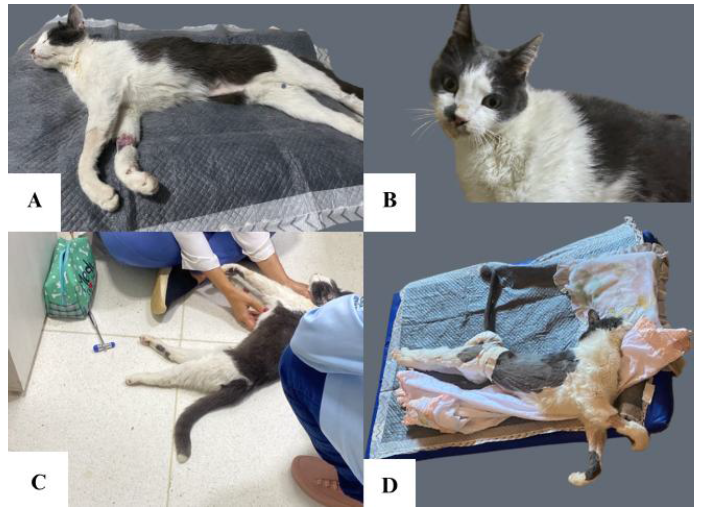
The neurological examination revealed a reduced level of consciousness (depression), altered posture due to head and body curvature (head turn and pleurothotonus), bilateral reduction of the photopupillary reflex, the threat test and nasal sensitivity. In addition, there was spastic tetraplegia with increased muscle tone in all limbs and preserved nociception (Fig.1). In view of the history and clinical signs, neurolocation was attributed to the brainstem region or neuromuscular disease. To rule out possible brain diseases, the decision was made to perform magnetic resonance imaging of the region and cytological, biochemical and molecular analysis of the cerebrospinal fluid.
The magnetic resonance imaging showed that the brain was within normal standards for the species, with preserved proportion between gray and white matter, absence of alterations in the ventricular system, cranial nerves and middle/inner ear. No lymph node enlargement or brain masses were observed (Fig. 2). Due to the patient's general clinical condition, symptomatic supportive treatment was performed with clindamycin (Clinbacter®, Agener União, São Paulo, São Paulo, Brazil) at a dose of 10 mg/kg every 12 hours, orally, for 21 days, and prednisone (Prednisona®, Aché, São Paulo, São Paulo, Brazil) at a dose of 1 mg/kg, orally, every 24 hours for 7 days.
1mL of cerebrospinal fluid was collected, the results of which are shown in Table 1. The cytological analysis showed 90% neutrophils and 10% lymphocytes; there were no fungi, bacteria or protozoa, or any other noteworthy alterations.

Neurological presentation of a 13-year-old neutered male cat with tetanus. Patient presenting spastic tetraparesis. A) Spastic tetraparesis in thoracic and pelvic limbs preventing head and neck movements at the time of hospitalization. B) Patient presenting head turn. C) Neurological evaluation of the patient. D) Spastic tetraparesis with limb abduction after worsening of the clinical condition.

Clinical recovery of a 13-year-old neutered male cat diagnosed with tetanus after supportive treatment. After two months of therapy, the patient regained motor function, being able to stand, walk, and move both thoracic and pelvic limbs normally.
The day after the magnetic resonance imaging, the patient presented with generalized cluster epileptic seizures, for which the use of phenobarbital (Gardenal pediátrico®, Sanofi, Suzano, São Paulo, Brazil) was indicated at a dose of 2.5mg/kg, orally, every 12 hours. The patient's clinical condition progressed to spastic tetraplegia with opisthotonus, as well as urinary and fecal retention. A n° 4 urethral tube was used for bladder emptying, which remained in place for 5 days. For constipation, lactulose (Lactuliv®, Legrand Pharma, Campinas, São Paulo, Brazil) was prescribed at a dose of 1ml, orally, every 12 hours for 7 days and then this medication was replaced with a pediatric commercial rectal laxative (Minilax®, Momenta, São Paulo, São Paulo, Brazil) in a single dose rectal tube.
Electrolyte analysis and urinalysis were requested. The results of the electrolyte profile showed hyponatremia and hypochloremia, shown in Table 2. The results of the urinalysis are shown in Table 3.
Molecular analysis of the cerebrospinal fluid tested the sample for polymerase chain reaction (PCR) for Bartonella spp., Cryptococcus spp., Toxoplasma gondii, FIV, FeLV and Feline Panleukopenia virus. The results were available 7 days after collection and were all negative. In view of these results, the antibiotic and anti-inflammatory drugs were discontinued.
Given the absence of brain lesions and the ruling out of inflammatory and infectious diseases, the conditions were ruled out, with tetanus being the presumptive diagnosis. The hypothesis was reinforced by the improvement in neurological signs over the days without specific treatment.
As the patient still had urinary retention, it was decided to carry out a new urinalysis, the results of which are shown in Tab. 3. Uroculture was requested and Enterobacter spp. was isolated, sensitive to doxycycline, amikacin, ceftriaxone, ciprofloxacin, cephalexin and gentamicin. Following the results, doxycycline (Doxy suspension®, CEPAV, Água Branca São Paulo, São Paulo, Brazil) was prescribed at a dose of 5mg/kg, every 12 hours, orally, for 14 days. The results of the laboratory tests showed normal blood count values, and the biochemistry values are shown in Tab. 4. Given the favorable evolution of the neurological condition and the hematological findings, phenobarbital was gradually withdrawn over 15 days. The patient remained stable with no neurological signs and was discharged after two months of follow-up.
DISCUSSION
Clostridium tetani is a non-invasive bacterium and therefore has no ability to penetrate healthy cells. Because it does not have the ability to cross the skin barrier in intact epithelial tissue, it requires a gateway, which is usually a deep wound with intense injury and tissue necrosis, which contributes to the formation of a medium conducive to the growth of the microorganism, through an anaerobic microenvironment (Malinovská et al., 2020). However, this case involves a feline patient in whom, during the physical examination, no skin wounds were evident. In addition, the patient's history of recent surgeries was not verified in the anamnesis, although he did have a previous history of a healed lesion on the LTL. As the diagnosis of tetanus considers the patient's clinical history (Langner et al., 2011; Farrar and Liebel, 2019), based on factors that may increase clinical suspicion, it is concluded that the form of contagion was atypical.
The disease occurs due to the release of the toxin tetanospasmin, which is responsible for the clinical manifestation (Farrar and Liebel, 2019). Tetanus is classified as generalized and localized, with the generalized form being considered a rare presentation in cats, due to the few reports described in the literature (Popoff, 2020; Zilli et al., 2023). However, the feline in this case presented clinical symptoms compatible with the systemic form of the disease, which is manifested by muscle hyperactivity, resulting from tonic contractions and reflex spasms to sensory stimuli, characterized by progressive spastic paresis or plegia of the trunk and limb muscles (Popoff, 2020). The presence of hypertonic myotatic reflexes with preserved proprioception, in addition to the involvement of cranial nerves, is in line with the report by Linnenbrink and McMichael (2006), alterations that result from hypertonia of the motor nuclei.
The diagnosis of feline tetanus is challenging, since the initial infection resembles other myopathies and paralytic neuropathies (Langner et al., 2011; Popoff, 2020). Laboratory tests, including hematology and serum biochemistry, can show normal values or only nonspecific alterations, but these can help with supportive therapy, such as the creatine kinase (CK) enzyme, evaluated in this report, whose result was 340 U/L and which shows the degree of muscle damage generated by myotonicity (Costa et al., 2002). However, for this case, the measurement of other enzymes, such as aspartate aminotransferase (AST) and lactate dehydrogenase (LDH), could have been used in order to identify the kinetics of muscle damage and contribute to the diagnostic approach, since these can show results above the reference values for the species in these cases (Oikonomidis and Milne, 2023).
Another factor refers to the effect exerted by tetanospasmin on the muscles of the urinary tract, which was observed in this patient. This is a particular feature of tetanus in cats, which is related to urinary obstruction secondary to spasticity of the striated muscles of the external urethral ostium, which can induce post-renal azotemia because of urinary retention or even acute kidney injury, justifying the use of a urethral tube, as in this case (Costa et al., 2002). Despite this, we found no renal impairment in the laboratory tests of the animal presented here.
For definitive diagnosis, the technique of inoculating mice with material extracted from a culture with isolation of the microorganism is recommended, and the disease is confirmed when clinical symptoms develop within a period of one to four days (Silva et al., 2023). However, the bioassay was not used in this case because there was no apparent wound from which it was possible to obtain a sample for isolation and because of the unavailability of laboratory analysis. In addition, the uroculture and urinary antibiogram in this case did not imply a diagnosis of tetanus, but it did allow the identification and targeting of the drug therapy to be used for bacterial cystitis.
It should be noted that the differential diagnosis should consider intoxications (strychnine, organophosphates, metaldehyde and others), immune-mediated polymyositis, spinal cord trauma, meningoencephalitis (infectious and idiopathic), subarachnoid hemorrhage or cyst, acute cerebellar alterations and electrolyte disorders, mainly linked to calcium homeostasis (Malinovská et al., 2020). In this case, to rule out infectious and metabolic alterations with neurological repercussions, a molecular technique (PCR) was carried out on cerebrospinal fluid (CSF) for the agents Bartonella spp., Cryptococcus spp., Toxoplasma gondii, Feline Immunodeficiency Virus (FIV), Feline Leukemia Virus (FeLV) and Feline Panleukopenia Virus, as well as electrolyte dosage, respectively. For this same purpose, magnetic resonance imaging (MRI) was recommended in this case, as it is a highly sensitive imaging technique, to rule out orthopedic, spinal or meningeal alterations that could affect the final diagnosis (Dussaux et al., 2024).
This patient's treatment was aimed at preventing the progression of the disease and alleviating the clinical signs, based mainly on supportive therapy with intensive care (Langner et al., 2011). Due to the poor prognosis, prolonged hospitalization was necessary to carry out essential supportive management, including the administration of anticonvulsants and muscle relaxants to reduce the patient's discomfort (Saravanan et al., 2024; Jardim et al., 2023). With appropriate treatment and management, the clinical symptoms of generalized tetanus tend to resolve within weeks, a situation observed in this case (Risio and Gelati, 2003).
CONCLUSIONS
The exclusion of diseases that could lead to similar clinical signs made it possible to make a diagnosis of exclusion in this case. This reinforces the importance of the clinician associating the clinical manifestation of tetanus even in the absence of a classic history of skin continuity solutions.
REFERENCES
- COSTA, F.S.; AGUIAR, D.M.D.; GIUFFRIDA, R. et al. Tétano em um gato. Braz. J. Vet. Res. Anim. Sci., v.39, p.160-162, 2002.
- DUSSAUX, A.; FUHRER, L.; DORNER, M.B. et al. Clinical findings and outcome in feline tetanus: a multicentric retrospective study of 27 cases and review of the literature. Front. Vet. Sci., v.11, p.1425917, 2024.
- FARRAR, B.; LIEBEL, F.X. Generalised tetanus in a cat. Vet. Rec. Case Rep., v.7, p.e000907, 2019.
- JARDIM, G.D.C.; SOUZA L.D.P.D.; BONATO, K.S. et al. Tétano canino-relato de caso. Braz. J. Dev., v.9, p.6563-6572, 2023.
- JONES, B.R.; BENNETT, D. The nervous and musculoskeletal systems. In: CHANDLER, E.A.; GASKELL, R.M.; GASKELL, C.J. (Eds.). Feline medicine and therapeutics. 3.ed. Oxford: Blackwell Publishing, 2004. p.125-235.
- LANGNER, K.F.A.; SCHENK, H.C.; LEITHAEUSER, C. et al. Localised tetanus in a cat. Vet. Rec., v.169, p.126, 2011.
- LINNENBRINK, T.; MCMICHAEL, M. Tetanus: pathophysiology, clinical signs, diagnosis, and update on new treatment modalities. J. Vet. Emerg. Crit. Care, v.16, p.199-207, 2006.
- MAGALHÃES, L.; MIZIARA, R.; TEIXEIRA, B. et al. Tétano equino - relato de cinco casos. Med. Vet. Zoo., n.1, p.8-13, 2016.
- MALINOVSKÁ, Z.; ČONKOVÁ, E.; VÁCZI, P. Tetanus in animals-summary of knowledge. Folia Vet., v.64, p.54-60, 2020.
- OIKONOMIDIS, I.L.; MILNE, E. Clinical enzymology of the dog and cat. Austr. Vet. J., v.101, n.12, 2023.
- POPOFF, M.R. Tetanus in animals. J. Vet. Diag. Inv., v.32, p.184-91, 2020.
- RISIO, L.; GELATI, A. Tetanus in the cat-an unusual presentation. J. Feline Med. Surg., v.5, p.237-240, 2003.
- SARAVANAN, S.; RAMPRABHU, R.; THANGADURAI, R. Generalized and chronic form of tetanus in a cat-Its diagnosis and ethno-veterinary management. Indian J. Anim. Health, v.63, p.136-138, 2024.
- SILVA, G.P.D.; GARCIA, C.; SILVA, R.S.D. et al. Tétano em um canino: aspectos clínicos e terapêuticos. Ciênc. Anim. Bras., v.24, p.e-73825, 2023.
- ZILLI, J.; HÄUßLER, T.C.; IVES, E.J.; SCHMIDT, M.J. Case report: A severe case of generalized tetanus in a young cat. Front. Vet. Sci., v.10, p.1328331, 2023.
The research data are available within the article itself.