
 Antioxidant, antimicrobial, anti-inflammatory, and wound healing potency of moon jellyfish Aurelia aurita: in vitro study
ABSTRACT
Jellyfish bioactive compounds are a promising, sustainable resource for biomedical applications, including impaired wound healing. This study aimed to assess the antioxidant, antimicrobial, anti-inflammatory, and wound-healing properties of Aurelia aurita in vitro. The bioactive compounds in A. aurita powder were identified using high-performance liquid chromatography (HPLC). The total phenolic content, flavonoids, and saponins were measured with spectrophotometric methods. Also, Total antioxidant capacity and DPPH radical-scavenging assays of A. aurita powder were used to evaluate antioxidant activity, and the disk diffusion method was used to determine its antimicrobial activity. A wound scratch assay evaluates the migration potential of human melanoma cells A375 after treatment with A. aurita powder. Results showed that A. aurita powder contains active phenolic and flavonoid compounds, such as gallic and chlorogenic acids, hesperidin, and daidzein. The powder exhibited significant antioxidant capacity, DPPH scavenging activity, antibacterial properties, and anti-inflammatory effects by stabilizing the RBC membrane. The wound scratch assay also showed significant differences in width, area, closure rate, and cell migration between treated and control cells. In conclusion, A. aurita can enhance wound healing in vitro through its antimicrobial, anti-inflammatory, and antioxidant properties, making it a promising candidate for wound treatment.
Keywords:
Aurelia aurita; antioxidant; anti-inflammatory; antimicrobial; wound healing
RESUMO
Os compostos bioativos das águas-vivas representam um recurso promissor e sustentável para aplicações biomédicas, incluindo o tratamento de feridas com cicatrização comprometida. Este estudo teve como objetivo avaliar as propriedades antioxidantes, antimicrobianas, anti-inflamatórias e cicatrizantes da Aurelia aurita in vitro. Os compostos bioativos presentes no pó de A. aurita foram identificados por cromatografia líquida de alta eficiência (CLAE). O teor total de fenólicos, flavonoides e saponinas foi determinado por métodos espectrofotométricos. Além disso, a capacidade antioxidante total e o ensaio de sequestro do radical DPPH foram utilizados para avaliar a atividade antioxidante do pó de A. aurita, e o método de difusão em disco foi empregado para determinar sua atividade antimicrobiana. Um ensaio de migração celular (ensaio de arranhão em ferida) avaliou o potencial de migração de células de melanoma humano A375 após o tratamento com o pó de A. aurita. Os resultados demonstraram que o pó de A. aurita contém compostos fenólicos e flavonoides ativos, como os ácidos gálico e clorogênico, hesperidina e daidzeína. O pó apresentou capacidade antioxidante significativa, atividade sequestradora de DPPH, propriedades antibacterianas e efeitos anti-inflamatórios, estabilizando a membrana dos eritrócitos. O ensaio de migração celular em ferida também mostrou diferenças significativas na largura, área, taxa de fechamento e migração celular entre as células tratadas e as do grupo controle. Em conclusão, A. aurita pode promover a cicatrização de feridas in vitro por meio de suas propriedades antimicrobianas, anti-inflamatórias e antioxidantes, tornando-se um candidato promissor para o tratamento de feridas.
Palavras-chave:
Aurelia aurita; antioxidante; anti-inflamatória; antimicrobiana; cicatrizante
INTRODUCTION
Wound healing is a complex, highly coordinated biological process that involves four overlapping phases: hemostasis, inflammation, proliferation, and tissue remodeling (Elmotazbellah et al., 2025a). Immediately after injury, the hemostasis phase is initiated by platelet aggregation and fibrin clot formation, which prevent blood loss and provide a temporary scaffold for cell migration. This is followed by the inflammatory phase in which recruited immune cells, such as neutrophils and macrophages, remove debris, eliminate pathogens, and release cytokines and growth factors (Barroso et al., 2020). The proliferative phase is characterized by fibroblast proliferation, collagen synthesis, angiogenesis, and keratinocyte migration, leading to granulation tissue formation and re-epithelialization of the wound surface (Chandra et al., 2026). During the remodeling phase, collagen fibers reorganize and cross-link to restore tissue strength and skin integrity. Disruptions in any of these phases can result in chronic wounds, posing a major clinical challenge and a leading cause of morbidity globally.
Chronic wounds can lead to serious complications due to poor public hygiene, which may result in secondary bacterial infections requiring urgent antimicrobial therapy (Ramirez-Acuña et al., 2019). Unfortunately, the widespread use of antibiotics has led to increased microbial drug resistance, resulting in decreased treatment effectiveness and significant economic losses (Huang et al., 2017a). The rise in wound complications can be attributed to factors such as limited access to proper management, drug resistance, and high costs. Additionally, persistent oxidative stress and inflammation at the wound site further delay healing, highlighting the need for multifunctional therapeutic agents that promote tissue repair while preventing infection (Lopes et al., 2024). This issue requires urgent attention and strategic actions to mitigate its impact on public health. Consequently, researchers worldwide are exploring natural, biocompatible, and affordable options, especially those derived from marine and plant sources, as promising candidates for complementary wound-healing therapies that can overcome antibiotic resistance and support tissue regeneration (Kumar et al., 2023; Hatem et al., 2026).
Recent studies have emphasized the importance of natural bioactive compounds, particularly antioxidants, anti-inflammatory agents, and collagen-stimulating molecules, in enhancing the different stages of wound healing (Sabra, 2025). These compounds help reduce oxidative stress caused by excessive reactive oxygen species (ROS), regulate inflammatory signaling pathways, stimulate fibroblast proliferation and migration, and promote collagen synthesis and extracellular matrix formation, ultimately accelerating wound closure and tissue regeneration (Alberts et al., 2025). Among these biomolecules, collagen has received particular attention as a biomaterial for wound-healing applications due to its structural similarity to the extracellular matrix, excellent biocompatibility, and ability to support cell adhesion, proliferation, and tissue regeneration (Wosicka-Frąckowiak et al., 2024; Tassara et al., 2026).
In recent years, marine-derived collagen has emerged as a promising alternative to mammalian collagen sources because it is biodegradable, biocompatible, and associated with lower risks of pathogen transmission and fewer ethical concerns (Saeed et al., 2025). Marine invertebrates represent an abundant and renewable source of collagen with vast pharmacological potential (Fol et al., 2024; Hamdi et al., 2024; Abdel-Khalek et al., 2025). Among these organisms, cnidarian jellyfish found in the Mediterranean Sea contain diverse bioactive compounds, including diterpenes, flavonoids, phenolic compounds, terpenoids, and monoterpenoids, that have demonstrated promising biological activities (Rocha et al., 2011; Leone et al., 2019). Jellyfish have attracted particular attention because their bodies consist largely of collagen, and climate-driven seasonal blooms have increased their abundance, providing an easily accessible and renewable biological resource (Ahmed et al., 2021). Several studies have demonstrated that jellyfish extracts possess antimicrobial and antioxidant properties, including bioactive peptides that can enhance the natural antioxidant capacity of biological systems and food products (Zhang et al., 2018; Jeyachandran and Aman, 2026). The wound-healing potential of these natural compounds is closely related to their antioxidant activity, which enables the scavenging of reactive oxygen species and improves the local cellular environment required for tissue repair (Atiyah and Al-Falahi, 2021; Bardaa et al., 2021). Moreover, jellyfish-derived biomolecules, particularly collagen peptides and polysaccharides, have shown promising therapeutic potential by promoting cell adhesion, proliferation, and extracellular matrix remodeling during tissue regeneration (Sumiyoshi et al., 2021). Notably, jellyfish collagen exhibits a high degree of structural and biochemical similarity to human collagen, making it suitable for biomedical applications, such as tissue-engineering scaffolds and cell-culture substrates (Mearns-Spragg et al., 2020; Hu et al., 2025).
This study uniquely investigates Aurelia aurita (the moon jellyfish) as a sustainable marine source of collagen with wound-healing potential. A. aurita exhibits a distinctive biochemical composition, rich in collagen peptides, polysaccharides, and antioxidant molecules, that synergistically promotes tissue repair and regeneration (Barzkar et al., 2024). Its collagen demonstrates high biocompatibility, a structure like human collagen, low immunogenicity, and minimal zoonotic risk, making it a safer alternative to mammalian biomaterials (Davison-Kotler et al., 2019; Ballesteros et al., 2025). Additionally, utilizing A. aurita, an abundant yet underutilized marine species, supports sustainable bioproduct development and helps address environmental issues associated with jellyfish blooms. Furthermore, Stabili et al. (2021) reported that jellyfish of the Aurelia genus contain collagenous compounds and oligosaccharides, underscoring their valuable biochemical and therapeutic potential. Despite limited research, mainly focused on biochemical characterization and collagen extraction, the full wound-healing potential of A. aurita, including its antioxidant, antimicrobial, and anti-inflammatory effects, remains largely unexplored. Therefore, the present study aims to provide a thorough in vitro assessment of the antioxidant, antimicrobial, anti-inflammatory, and wound-healing properties of A. aurita powder. By exploring its bioactive potential, this research seeks to establish A. aurita as an innovative, eco-friendly candidate for future biomedical and pharmaceutical uses.
ETHICS ASPECTS
All laboratory animal use procedures in this study were agreed upon according to the Ethics of Research Committee regulations at the Faculty of Science, Cairo University, and received the approval number (No. CUIF 2724).
MATERIALS AND METHODS
In August 2024, healthy Aurelia aurita jellyfish were collected from the North Coast of Alexandria, Egypt, during peak bloom season. The specimens were transported to the lab in an icebox and washed thoroughly with running tap water to remove debris and salt residues. Following Doerr and Stoskopf's protocol (Doerr and Stoskopf, 2019), magnesium chloride (MgCl₂) was used to euthanize and fully immobilize the jellyfish before further processing. A marine taxonomist at the Department of Marine Biology, Faculty of Science, Alexandria University, confirmed the species’ identification through morphological features, such as bell transparency, four horseshoe-shaped gonads, bell diameters ranging from 10 to 25 cm, and the pattern of marginal tentacles, which align with established taxonomic descriptions of Aurelia species
Jellyfish samples were cut into small pieces (3-5 cm), soaked in distilled water for 36 hours to remove excess salt, and washed three times. Then, the samples were stored at -80°C and subjected to vacuum pressure for 72 hours to achieve freeze-drying. All the dried samples were finely ground into a powder and stored at 4°C until further use.
The bioactive compounds present in Aurelia aurita powder were analyzed using high-performance liquid chromatography (HPLC) according to the method of Aroua et al. (2025). The analysis was conducted on an Agilent 1260 HPLC system equipped with a reverse-phase C18 column (250 × 4.6mm, 5µm particle size). The mobile phase consisted of phosphate buffer (pH 6.5) as solvent A and acetonitrile as solvent B, with a gradient elution applied at a flow rate of 0.5mL/min. Detection was carried out at 280nm. A 20µL aliquot of A. aurita powder solution (dissolved in methanol) was injected into the system, with a total run time of 30 minutes. The secondary metabolites were identified by comparing their retention times with those of authentic reference standards, and their concentrations were quantified by integrating the corresponding peak areas.
According to Singleton et al. (1965), the Folin-Ciocalteu method was used to determine total phenolic content. 200 μL of A. aurita powder at 1 mg/mL in ethanol was diluted with 2.8mL of distilled water. The suspension was thoroughly mixed with 0.5 mL of the Folin-Ciocalteu reagent, which contains phosphotungstic acid and phosphomolybdic acid, for 3 minutes, followed by the addition of 2mL of 20% (w/v) sodium carbonate. The mixture was left in the dark for 60 minutes, and the absorbance was measured using a UV-Visible spectrophotometer (U-2001, model 121 0032 Hitachi, Tokyo, Japan) at 650nm. The results are expressed as mg of gallic acid equivalent (GAEs) per gram of dry weight.
The total flavonoid content of the A. aurita powder was determined using an aluminum chloride colorimetric method (Chang et al., 2002). Briefly, 50μL of A. aurita powder (1mg/mL in ethanol) was mixed with 4 mL of distilled water and 1mL of methanol, then incubated for 5 minutes. Next, 0.3mL of a 5% NaNO2 solution was added, followed by a 10% AlCl3 solution after 5 minutes of incubation. The mixture was left to stand for 6 minutes, then 2mL of NaOH solution (1 M) was added. After 15 minutes, the absorbance was measured using a UV-Visible spectrophotometer (U-2001, model 121 0032 Hitachi, Tokyo, Japan) at 510 nm, and the results were expressed as mg rutin equivalents per gram of dry weight.
Spectrophotometry was used to quantify the saponin content in A. aurita powder, as previously described by Uematsu et al. (2000). The following coloring reagent solutions were prepared: Reagent (A) was made by mixing p-anisaldehyde with ethyl acetate in a 0.5:99.5 ratio, and Reagent (B) was prepared by combining 50 mL of H2SO4 with 50 mL of ethyl acetate. The A. aurita powder was dissolved in 2 mL of ethyl acetate, and 1 mL of each reagent was added. The mixture was stirred and incubated for 10 minutes in a water bath at 60°C. After cooling, the absorbance was measured using a UV-Visible spectrophotometer (Jenway, England) at 430nm. Ethyl acetate served as a control for absorbance measurement. A solution containing 20-45µg of standard saponin in 2mL of ethyl acetate was used.
The total antioxidant capacity of A. aurita powder was measured using a phospho-molybdenum assay, following the method of Prieto et al. (1999). 0.1mL of A. aurita powder at different concentrations (100-400µg/mL in DMSO) was combined with 1 mL of molybdate reagent solution containing 0.6 M H2SO4, 28 mM sodium phosphate, and 4 mM ammonium molybdate. The mixture was incubated at 95°C for 90 minutes and then cooled. Absorbance was read using a UV-Visible spectrophotometer (U-2001, model 121 0032 Hitachi, Tokyo, Japan) at 695 nm. All tests were performed in triplicate, and the overall antioxidant capacity was expressed as ascorbic acid equivalents (AAE).
A. The free radical scavenging ability of A. aurita powder was evaluated using a DPPH radical assay with some modifications (Villaño et al., 2007). Specifically, 200µL of A. aurita powder and ascorbic acid (100-500 µg/ml in methanol) were added to a 2mL solution of 0.1 mM DPPH and incubated in the dark at 37°C for 30 minutes. After incubation, the absorbance was measured using a UV-Visible spectrophotometer (U-2001, model 121 0032 Hitachi, Tokyo, Japan) at 517 nm against a methanol blank. The control tube (DPPH only) was prepared identically, and all experiments were performed in triplicate. Radical scavenging activity (%) was calculated using the following equation
Then, the half-maximal effective concentration (EC50) was calculated using an equation derived from the plot of percentage DPPH radical scavenging activity versus the various sample concentrations.
The anti-inflammatory activity of A. aurita powder was assessed through an in vitro red blood cell (RBC) membrane stabilization assay and compared to aspirin at various concentrations (Gandhidasan et al., 1991). A rat blood sample was taken, and a 10% RBC suspension was prepared. A. aurita powder or aspirin (50-400μg/ml) was mixed with the RBC suspension and incubated at 56ºC. Afterward, the tubes were centrifuged at 2500 rpm for 5 minutes. Hemoglobin content was measured using a UV-Vis spectrophotometer (U-2001, model 121 0032 Hitachi, Tokyo, Japan) at 560 nm. The control tube containing RBC suspension and standard saline solution was processed similarly. The percentage of RBC membrane stabilization was calculated using the following equation:
Then, the half-maximal effective concentration (EC50) was calculated from the plot of percentage HRBC membrane stabilization versus the various sample concentrations.
The antimicrobial potential of Aurelia aurita powder (AAP) was evaluated against a range of pathogenic microorganisms, including Gram-positive bacteria (Streptococcus faecalis, Staphylococcus aureus, and Bacillus subtilis), Gram-negative bacteria (Escherichia coli, Pseudomonas aeruginosa, and Neisseria gonorrhoeae), and fungal strains (Aspergillus flavus and Candida albicans), using a modified Kirby-Bauer disc diffusion method (Bauer et al., 1966). The bacterial and fungal strains were obtained from the Microbiology Laboratory at the Micro-Analytical Center, Faculty of Science, Cairo University (Egypt). All cultures were sub-cultured and maintained under sterile conditions before testing. The microorganisms were inoculated into Mueller-Hinton Broth supplemented with 5% horse blood and incubated overnight at 37°C to achieve a turbidity equivalent to the 0.5 McFarland standard, corresponding to a final inoculum of 1.5 × 10^8 CFU/mL. A 50mg/mL stock solution of AAP was prepared in 5% dimethyl sulfoxide (DMSO), filtered through a sintered glass filter, and stored at 4°C until use. Sterile Petri plates containing solidified Mueller-Hinton agar were inoculated evenly with the test microorganisms, and six wells (6 mm in diameter) were aseptically bored using a sterile cork borer. Each well was filled with 50µL of AAP solution, the positive control (Ampicillin, 0.1 mg/mL for bacteria; Amphotericin B, 1mg/mL for fungi), and the negative control (DMSO). Plates were left at room temperature for 30 minutes to allow diffusion and then incubated at 37 °C for 18-24 hours. After incubation, the zones of inhibition were examined and measured in millimeters (mm) to assess antimicrobial activity. All assays were performed in triplicate, and mean values were calculated for accuracy.
The effectiveness of A. aurita powder in healing wounds was assessed using a cell migration assay (Main et al., 2020). Human melanoma cells (A375) were obtained from Nawah Scientific Inc. (Mokatam, Cairo, Egypt). They were seeded at a density of 3 × 10^5 cells per well in a coated 6-well plate and cultured overnight until 80% confluence was reached. The next day, the complete medium was replaced with serum-free medium containing 1μg/mL mitomycin C, and the cells were incubated at 37°C for 2 hours to stop cell proliferation. A sterile pipette tip was used to create a vertical scrape on the well, and cell debris was removed by washing with phosphate-buffered saline. In serum-free medium, the cells were treated with a control solvent or A. aurita powder at an IC50 concentration, determined from a dose-response curve of cell viability (%) versus A. aurita powder concentration using nonlinear regression analysis. Images of the wounded cells were captured at specific intervals using an inverted fluorescence microscope (Nikon Eclipse TS-100, Nikon Corporation, Tokyo, Japan). The plate was kept at 37°C with 5% CO2 between time points. The acquired images were analyzed using MII Image View version 3.7 to calculate the wound width, migration rate, wound area, and percentage of wound closure.
The current study was conducted in accordance with the relevant ARRIVE guidelines. The Cairo University Institutional Animal Care and Use Committee (CU-IACUC) approved experimental procedures using animals in this study, as outlined in the relevant document (No. CUIF 2724). All methods were performed in accordance with the relevant guidelines and regulations.
SPSS software (Version 22, SPSS Inc., Chicago, IL, USA) was used to analyze the data. All data were expressed as mean ± standard error (SEM) for three measurements. All data were analyzed using Student’s T-test at a 95% confidence level. P<0.05 was considered statistically significant.
RESULTS
A chromatogram was obtained at various retention times (3.56, 4.26, 5.59, 16.15, 21.32), which revealed a total of five active ingredients (Fig. 1). As shown in Table 1, A. aurita powder contains several bioactive constituents, mainly phenolic compounds, which make up 92.60% of the total bioactive components. Gallic and chlorogenic acids, types of phenolic compounds, were the most prevalent active components, accounting for 48.29% and 42.34%, respectively. Conversely, methyl gallate accounted for only 1.97% of the phenolic compounds. Additionally, some flavonoids were present, with hesperidin being the most abundant at 6.63%, followed by daidzein at 0.75%.

HPLC chromatogram of phenolic and flavonoid constituents of A. aurita powder detected at 280nm.
Table 2 shows that the phenolic compound content exceeds the flavonoid content. The phenolic content is 183.69±0.35mg GAEs/g, while the flavonoid content is 26.86±0.008mg rutin equivalent/g. Additionally, A. aurita saponin content is reported as 3263.97±0.04.
As shown in Fig. 2a, the crude powder of A. aurita exhibits significant antioxidant activity, with a total antioxidant capacity of 737.91±12.21mg/g and 829.43±12.84mg/g ascorbic acid at low and high concentrations, respectively.
Figure 2b shows that the capacity of A. aurita (an antioxidant) to scavenge free radicals compared to the standard ascorbic acid increases with its concentration. At a concentration of 100µg/mL, the crude powder could scavenge more than 80% of the DPPH radical. The standard ascorbic acid could scavenge 89.02 ± 0.34% of the DPPH at the same concentration. Additionally, at a high concentration, AAP exhibited a highly effective free radical scavenging ability, with a percentage of 89.82±1.10%, which is comparable to that of ascorbic acid (94.71±0.23%) at the same concentration. Furthermore, the EC50 value of A. aurita was 83.88±0.22μg/mL, indicating higher antioxidant activity compared to ascorbic acid (71.62±0.40μg/mL), as illustrated in Table 3.
As shown in Fig. 3, A. aurita powder exhibits concentration-dependent anti-inflammatory potency by stabilizing the RBC membrane, compared with aspirin. At a concentration of 50µg/mL, the crude powder has a noticeable effect on RBC membrane stabilization, with a value of 60.28±0.15%, which is almost the same as that of aspirin (61.15±0.12%) at the same concentration. Furthermore, it exhibited a maximum stabilization of 83.14±0.06% at 400µg/mL, comparable to that of aspirin (86.14±0.07%) at the same concentration. Table 3 demonstrates that the EC50value of the anti-inflammatory effect of A. aurita powder (33.11±0.57μg/mL) was approximately the same as that of aspirin (33.98±0.28μg/mL).
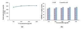
Antioxidant potency of A. aurita powder. a: Total antioxidant capacity; b: DPPH radical scavenging activity. Values are expressed as the mean of three replicate determinations ± SEM. AAE: Ascorbic acid equivalents.

Stabilization percentage of red blood cell (RBC) membrane by A. aurita powder and Aspirin, demonstrating anti-inflammatory potency. Values expressed as mean ± SEM of three determinants.
Table 4 shows that the crude powder of A. aurita effectively suppresses the growth of pathogenic microorganisms with variable potency comparable to antibacterial and antifungal standards. Regarding antibacterial potency, the powder exhibited the highest inhibition zone diameter against Pseudomonas aeruginosa (19.82 ± 0.17mm), followed by Escherichia coli (19.66 ± 0.30mm). On the other hand, the lowest inhibition zone diameter was against Neisseria gonorrhoeae (16.83±0.17mm). The growth-inhibition potency against fungi was lower than that observed against the bacterial strains. This is evidenced by the powder producing a zone diameter of 14.20±0.18 mm for Aspergillus flavus, whereas it produces a zone diameter of 14.88±0.17 mm for Candida albicans.
Microscopic images showed that A. aurita powder at 100 µg/ml significantly enhanced cell migration and promoted wound healing, confirming its potential for wound repair (Fig. 4a). Specifically, the scratch wound width was significantly reduced in cells treated at 24-, 48-, and 96-hours post-wounding compared to the untreated cells (Fig. 4b). A. aurita powder also significantly (P < 0.05) decreased the wound area after 48 hours compared to the control (Fig. 4c). Additionally, it notably increased the cells' ability to migrate toward the wound across all time points compared to control cells (Fig. 4d). After 96 hours post-wounding, A. aurita powder increased the wound closure rate by approximately 70% relative to untreated cells (Fig. 4e).
DISCUSSION
Conventional wound-healing treatments are often costly, which may trigger allergic reactions and contribute to the growing problem of antimicrobial resistance (D’Ercole, 2025). In recent years, natural-based therapies have gained significant attention as safer and more affordable alternatives. Marine invertebrates represent a promising source of bioactive compounds with potent pharmacological properties (Nurkolis, 2025). In this study, the bioactive components of the jellyfish Aurelia aurita were characterized, and their antioxidant, anti-inflammatory, antimicrobial, and wound-healing potentials were comprehensively evaluated in vitro to assess their suitability as natural therapeutic agents for enhancing skin regeneration.
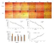
In vitro wound healing potency of A. aurita powder. (a) Inverted light microscopic images demonstrate the effect of A. aurita powder on the migration of Human Melanoma (A375). (b) Wound width of untreated and treated A375 cells at different intervals. (c) Wound area of untreated and treated A375 cells at different intervals. (d) The migration rate of untreated and treated A375 cells at different intervals. (e) Wound closure percentage of untreated and treated A375 cells at different intervals. Values are expressed as mean ± SEM (n=3); * indicates significance at P<0.05 at each time interval.
Identification of bioactive compounds in natural products is essential for drug discovery. After conducting HPLC analysis, it was found that A. aurita powder contains various phenolic and flavonoid compounds, including gallic acid, chlorogenic acid, methyl gallate, hesperidin, and daidzein. The high level of phenolic compounds measured 183.69±0.35mg GAE/g. The flavonoid content was 26.86±0.008mg rutin equivalent/g. The functional role of secondary metabolites, such as phenolics and flavonoids, in mediating biological activities has been extensively studied in plants, where their composition is influenced by multiple biotic and abiotic factors (Özay and Pehlivan, 2024; Kumar et al., 2025). Similar principles may underline the variability in metabolite profiles and bioactivity in marine organisms, such as Aurelia aurita. Therefore, the rich phenolic and flavonoid profile of A. aurita significantly contributes to its antioxidant defense and wound-healing potential, supporting the in vitro results and emphasizing its potential as a natural biomaterial for skin regeneration applications. Previous research has demonstrated that polyphenolic compounds, particularly gallic acid, exhibit potent antioxidant and anti-inflammatory effects (Karatas and Gevrek, 2021). These compounds accelerate the healing process by reducing wound size, increasing fibroblast proliferation, decreasing inflammatory cell infiltration, and stimulating TGF-β expression. They also enhance collagen type I synthesis, extracellular matrix production, and osteoblast proliferation (Huang et al., 2017b; Alharbi et al., 2026). Similarly, Thanikachalam et al. (2019) reported that gallic acid promoted cell migration from wound edges and improved both in vitro and in vivo wound healing. Chlorogenic acid, an ester of caffeic and quinic acids, is another primary bioactive compound identified in A. aurita. It possesses vigorous radical-scavenging activity, prevents DNA damage, and exhibits antihypertensive, anticancer, and anti-inflammatory properties (He et al., 2025). Likewise, flavonoids such as hesperidin facilitate collagen synthesis and cross-linking. At the same time, daidzein, an isoflavone, enhances skin collagen production and prevents its degradation in both in vitro and in vivo models (Zhao et al., 2014; Ahmad et al., 2017; Duarte et al., 2026).
Oxidative stress significantly delays wound healing by producing excess reactive oxygen species (ROS), which can damage cells, inhibit collagen production, and prolong inflammation (Ukaegbu et al., 2025). Therefore, compounds with potent antioxidant properties are crucial for protecting cells from oxidative damage, promoting fibroblast growth, and facilitating tissue repair. The DPPH radical scavenging assay results showed significant antioxidant capacity and DPPH scavenging activity of the A. aurita powder compared to ascorbic acid. The EC50 value of A. aurita powder (83.88±0.22µg/mL) was slightly higher than that of ascorbic acid (71.62±0.40µg/mL), confirming its strong antioxidant potency. These results suggest that the phenolic and flavonoid compounds in A. aurita effectively scavenge ROS, thereby supporting its potential role in enhancing wound healing. Similar findings were reported by Khalil et al. (2022), who demonstrated that extracts from sea urchins (Diadema savignyi) and jellyfish (Aurelia aurita) exhibited comparable antioxidants and DPPH scavenging activities, reaching approximately 75% of the ascorbic acid activity.
Inflammation is a crucial stage of wound healing that involves attracting immune cells and releasing cytokines to eliminate debris and prevent infection (Raziyeva et al., 2021; Xie et al., 2026). However, if inflammation persists or becomes excessive, it can lead to oxidative stress (OXS), characterized by the overproduction of reactive oxygen species (ROS). This imbalance can impair cellular homeostasis and slow tissue repair (Gao et al., 2024). Therefore, agents with both antioxidant and anti-inflammatory activities can play a crucial role in accelerating wound healing by maintaining redox balance and modulating inflammatory responses. In this study, A. aurita powder demonstrated promising anti-inflammatory activity by stabilizing red blood cell (RBC) membranes, with an EC50 of 33.11 ± 0.57 µg/mL, comparable to that of aspirin (33.98 ± 0.28 µg/mL). This membrane stabilization suggests that A. aurita may prevent lysosomal membrane lysis, thereby inhibiting the release of pro-inflammatory mediators. These findings align with previous studies indicating that jellyfish-derived extracts, including polysaccharides and collagen hydrolysates, possess notable anti-inflammatory properties and could serve as natural therapeutic agents against inflammation (Cao et al., 2021; Lv et al., 2022; Summat et al., 2023). The combined antioxidant and anti-inflammatory activities of A. aurita thus support its potential as a bioactive material for promoting wound healing and protecting tissues from oxidative and inflammatory damage.
Microbial infection is one of the most serious complications that hinders wound healing. Pathogenic bacteria, especially Staphylococcus aureus, Pseudomonas aeruginosa, and Escherichia coli, can invade damaged tissue, form biofilms, and produce toxins that exacerbate inflammation, prolong epithelialization, and disrupt collagen synthesis (Cavallo et al., 2024; Elmotazbellah et al., 2025b). Effective antimicrobial agents are therefore essential for preventing infection, promoting tissue repair, and ensuring faster wound closure. In this study, A. aurita powder exhibited vigorous antimicrobial activity against common wound-associated pathogens, particularly Pseudomonas aeruginosa and Escherichia coli. This antimicrobial effect may be attributed to its major phenolic constituents, gallic acid and chlorogenic acid, which are abundant in A. aurita powder. Yue et al. (2015) reported that these compounds exhibit potent bactericidal activity against various bacterial strains (Liu et al., 2025; Haddada et al., 2026). Similarly, Umamageswari et al. (2016) demonstrated that extracts from the jellyfish Porpita porpita showed antibacterial effects against Staphylococcus aureus, Bacillus sp., Pseudomonas sp., Klebsiella pneumoniae, and Escherichia coli, as well as antifungal activity against Aspergillus niger and Candida albicans. Furthermore, Chen et al. (2022) found that chlorogenic acid exerted strong antibacterial effects on Yersinia enterocolitica by disrupting cell membrane integrity, increasing permeability, and leading to cell death. Likewise, gallic acid exhibited bactericidal activity against E. coli and inhibited bacterial biofilm formation (Tian et al., 2022). These findings suggest that the antimicrobial potential of A. aurita powder contributes to its overall wound-healing efficacy by reducing microbial burden, preventing infection, and supporting tissue regeneration.
The wound-healing process is difficult to simulate in vitro due to the lack of cell debris and the complex interactions among different cell types. Indeed, cell migration is a rate-limiting step in wound healing and understanding the factors that affect cell migration will help target therapies to enhance wound healing (Alonso-Matilla et al., 2025). In the present study, microscopic examination revealed that treatment with Aurelia aurita powder markedly enhanced cell migration and promoted wound closure compared to the untreated control. The wound width and area were significantly reduced (P < 0.05), while the rate of cell migration and wound closure increased notably after treatment. These findings are consistent with those of Felician et al. (2019), who demonstrated that collagen peptides derived from the jellyfish Rhopilema esculentum stimulate fibroblast migration, likely through chemotactic signaling mechanisms. The abundance of amino acid residues in jellyfish collagen peptides may also provide essential nutrients that support cell proliferation and the synthesis of the extracellular matrix (ECM) (Ballesteros et al., 2025).
The improved wound-healing activity of A. aurita powder observed in this study can be attributed to the synergistic effects of its bioactive compounds, including collagen peptides, polysaccharides, phenolics, and flavonoids. Collagen peptides provide structural support, promote fibroblast adhesion and migration, and enhance ECM deposition, all of which are important for tissue regeneration (Ahmed et al., 2021; Thuy Diep et al., 2026). Polysaccharides from jellyfish help maintain moisture, stimulate keratinocyte migration, and modulate inflammatory responses, thereby creating a favorable microenvironment for wound repair (Migone et al., 2022). Additionally, the phenolic and flavonoid antioxidants in A. aurita effectively neutralize reactive oxygen species (ROS), reducing oxidative stress and preventing cellular damage. This antioxidant system maintains cell integrity, supports collagen deposition, and accelerates re-epithelialization, thereby improving wound-healing outcomes (Zulkefli et al., 2023; Liu et al., 2026).
CONCLUSION
This study shows that the moon jellyfish Aurelia aurita is a promising source of bioactive compounds, particularly gallic acid and chlorogenic acid, which may aid wound healing in vitro. Its antioxidants, antimicrobial, and anti-inflammatory effects support its potential to promote tissue repair and prevent infection and oxidative damage during healing. However, this study has some limitations due to the use of crude A. aurita powder, which encompasses numerous bioactive compounds that may interact synergistically or antagonistically, thereby complicating the attribution of biological activities and impacting reproducibility. Future research should therefore focus on fractionating and purifying the bioactive components of Aurelia aurita, followed by molecular characterization to identify the specific compounds responsible for the observed biological activities. In addition, in vivo studies using appropriate wound models are necessary to confirm their therapeutic efficacy. Furthermore, the development of standardized biomedical formulations, such as gels, films, or nanocomposite systems containing purified A. aurita compounds, may facilitate their practical application as effective wound-healing therapies.
ACKNOWLEDGMENT
The Faculty of Science, Cairo University, Egypt, supports this work. The authors thank members of the Zoology Department for their assistance.
REFERENCES
- ABDEL-KHALEK, R.R.; ABDEL-GHAFFAR, F.; HAMDI, S.AH. et al Orobanche aegyptiaca-chitosan nanocomposite efficacy against the freshwater snail Biomphalaria Alexandrina Sci. Rep, v.15, p.1-17, 2025.
- AHMAD, M.; SULTANA, M.; RAINA, R. et al Hypoglycemic, hypolipidemic, and wound healing potential of Quercetin in streptozotocin-induced diabetic rats. Pharmacogn. Mag., v.13, p.S633-S639, 2017.
- AHMED, Z.; POWELL, L.C.; MATIN, N. et al Jellyfish Collagen: A Biocompatible Collagen Source for 3D Scaffold Fabrication and Enhanced Chondrogenicity. Mar Drugs., v.19, p.405, 2021.
- ALBERTS, A.; LUNGESCU, I.A.; NICULESCU, A.G.; GRUMEZESCU, A.M. Natural products for improving soft tissue healing: mechanisms, innovations, and clinical potential. Pharmaceutics, v.17, p.758, 2025.
- ALHARBI, H.O.A.; SARWAR, T.; RAHMANI, A.H. Unveiling the therapeutic potential of gallic acid: mechanistic insights into the management of pathogenesis: a narrative review. Int. J. Mol. Sci., v.27, p.1536, 2026.
- ALONSO-MATILLA, R.; PROVENZANO, P.P.; ODDE, D.J. Physical principles and mechanisms of cell migration. NPJ Biol. Phys. Mech., v.2, p.2, 2025.
- AROUA, N.; BOUKHRIS, M.; AYOUNI, W. et al High-performance liquid chromatography analysis of phenolics inCasuarina cunninghamiana: extraction, quantification and antioxidant evaluation. Nat. Prod. Res., v.9, p.1-11, 2025.
- ATIYAH, A.; AL-FALAHI, N. The role of Helianthus tuberosus powder in the healing of full-thickness wounds in mice. Vet. World, v.14, p.1290-1298, 2021.
- BALLESTEROS, A.; TORRES, R.; PASCUAL-TORNER, M. et al Jellyfish Collagen in the Mediterranean Spotlight: Transforming Challenges into Opportunities. Mar. Drugs, v.23, p.200, 2025.
- BARDAA, S.; MAKNI, K.; BOUDAOUARA, O.; BARDAA, T. et al Development and evaluation of the wound healing effect of a novel topical cream formula based on ginkgo biloba extract on wounds in diabetic rats. Biomed. Res. Int, v.2021, p.6474706, 2021.
- BARROSO, A.; MESTRE, H.; ASCENSO, A. et al Nanomaterials in wound healing: from material sciences to wound healing applications. Nano Select., v.1, p.443-460, 2020.
- BARZKAR, N.; SUKHIKH, S.; ZHIKHREVA, A. et al Aurelia aurita jellyfish collagen: recovery properties. Foods Raw Mater., v.13. 296-305, 2024.
- BAUER, A.W.; KIRBY, W.; SHERRIS, J.; TURCK, M. Antibiotic susceptibility testing by a standardized single disk method. Am. J. Clin. Pathol, v. 45, p. 493-496, 1966.
- CAO, Y.; GAO, J.; ZHANG, L. et al Jellyfish skin polysaccharides enhance intestinal barrier function and modulate the gut microbiota in mice with DSS-induced colitis. Food Funct, v.12, p.10121-10135, 2021.
- CAVALLO, I.; SIVORI, F.; MASTROFRANCESCO, A. et al Bacterial biofilm in chronic wounds and possible therapeutic approaches. Biology, v.13, p.109, 2024.
- CHANDRA, P.; FAIZAN, M.; PORWAL, M. et al An Overview and review of growth factors in wound healing: emerging trends and innovations. Curr. Diabetes Rev., v.22, p.1-27, 2026.
- CHANG, C.C.; YANG, M.H.; WEN, H.M.; CHERN, J.C. Estimation of total flavonoid content in propolis by two complementary colorimetric methods. J. Food Drug Anal, v.10, p.178-182., 2002.
- CHEN, K.; PENG, C.; CHI, F. et al Antibacterial and antibiofilm activities of chlorogenic acid against Yersinia enterocolitica Front. Microbiol, v.13, p.885092 2022.
- DAVISON-KOTLER, E.; MARSHALL, W.S.; GARCÍA-GARETA, E. Sources of collagen for biomaterials in skin wound healing. Bioengineering, v.6, p.56, 2019.
- D’ERCOLE, S. Eco-friendly antimicrobial strategies to fight chronic wound infections associated with multidrug-resistant pathogens for the development of innovative medical systems (SCIAMI). Med. Sci. Forum, v.29, p.1, 2025.
- DOERR, M.; STOSKOPF, M.K. Evaluation of euthanasia of moon jellyfish (Aurelia aurita) using simple salt solutions. J. Zoo Wildl. Med, v.50, p.123-126, 2019.
- DUARTE, M.; PEDROSA, S.S.; KHUSIAL, P.R.; MADUREIRA, A.R. Bridging psychological stress and skin cellular aging: flavonoids as a dual-action therapeutic strategy. Phytother. Res, v.40, p.1970-2002, 2026.
- ELMOTAZBELLAH, M.; SOLIMAN, S.M.A.; EL-GHANY, M.N.A. et al Nanoparticles in wound healing: classification, recent advances, and limitations. Regen. Eng. Transl. Med., 2025a.
- ELMOTAZBELLAH, M.; SOLIMAN, S.M.A.; EL-GHANY, M.N.A. et al. Fabrication of epidermal growth factor (EGF)- and vancomycin-loaded chitosan nanoparticles to enhance wound healing. Naunyn Schmiedebergs Arch. Pharmacol., v.399, p.4973-4985, 2025b.
- FELICIAN, F.F.; YU, R.H.; LI, MZ. et al The wound healing potential of collagen peptides derived from the jellyfish Rhopilema esculentum Chin. Traumatol, v.22, p.12-20, 2019.
- FOL, M.F.; HAMDI, S.A.H.; ABDEL RAHMAN, H.A. et al The protective effect of crawfish chitosan on the reproductive performance of obese male rats. Food Biosci., v.57, p.103463, 2024.
- GANDHIDASAN, R.; THAMARAICHELVAN, A.; BABURAJ, S. Anti-inflammatory action of Lannea coromandelica by HRBC membrane stabilization. Fitoterapia, v. 62, p. 81-83, 1991.
- GAO, M.; GUO, H.; DONG, X. et al Regulation of inflammation during wound healing: the function of mesenchymal stem cells and strategies for therapeutic enhancement. Front. Pharmacol, v.15, p.1345779, 2024.
- HADDADA, A.; LAOUANI, A.; TEKFA, M.I.B. et al Polyphenolic profile, antioxidant, antibacterial and antidiabetic activities of acorns from Three Tunisian Quercus L. Species. Plants, v.15, p.762, 2026.
- HAMDI, S.A.H.; EL-SHAZLY, M.A.M.; FOL, M.F. et al Octopus vulgaris ink chemical profiling and validation of its potential as antioxidant, antimicrobial, anti-cancer, as well as anti-Schistosomal drug in vitro. Arq. Bras. Med. Vet. Zootec, v.76, n.5, 2024.
- HATEM, S.; ABDEL-GAWAD, R.; HUSSEIN, D.K. et al Biological modulation and repair using plant-derived bioactives: advancements in tissue engineering and regenerative medicine. Fut. J. Pharm. Sci., v.12, p.14, 2026.
- HE, Y.; MAO, S.; ZHAO, Y.; YANG, J. Research advances in the synthesis, metabolism, and function of chlorogenic acid. Foods, v.14, p.1914, 2025.
- HU, B.; ZONG, Z.; HAN, L. et al Jellyfish collagen: A promising and sustainable marine biomaterial with emerging applications in food, cosmetics, and biomedical- a review. Appl. Food Res., v.5. p.101165. 2025.
- HUANG, X.; BAO, X.; LIU, Y. et al Catechol-functional chitosan/silver nanoparticle composite as a highly effective antibacterial agent with species-specific mechanisms. Sci. Rep., v.7, p.1860, 2017a.
- HUANG, L.; JIN, P.; LIN, C. et al Beneficial effects of sulfonamide-based gallates on osteoblasts in vitro. Mol. Med. Rep., 15, p.1149-1156, 2017b.
- JEYACHANDRAN, S.; AMAN, M. Jellyfish-derived bioplastics: properties, degradation, and marine applications. Front. Mar. Sci., v.12, p.1666791, 2026.
- KARATAS, O.; GEVREK, F. Gallic acid liposome and powder gels improved wound healing in Wistar rats. Ann. Med. Res, v.26, p.2720-2727, 2021.
- KHALIL, E.A.; SWELIM, H.; EL-TANTAWI, H. et al Characterization, cytotoxicity and antioxidant activity of sea urchins (Diadema savignyi) and jellyfish (Aurelia aurita) extracts. Egy. J. Aquat. Res, v.48, p.343-348, 2022.
- KUMAR, S.; BHARALI, A.; SARMA, H. et al Traditional complementary and alternative medicine (TCAM) for diabetic foot ulcer management: a systematic review. J. Ayurveda. Integr. Med., v.14, p.100745, 2023.
- KUMAR, P.; KUMAR, D.; PAL, S.; SINGH, S. Plant secondary metabolites in defense against phytopathogens: mechanisms, biosynthesis, and applications. Physiol. Mol. Plant Pathol, v.138, p.102639, 2025.
- LEONE, A.; LECCI, R.M.; MILISENDA, G.; PIRAINO, S. Mediterranean jellyfish as novel food: effects of thermal processing on antioxidant, phenolic, and protein contents. Eur. Food Res. Technol, v.245, p.1611-1627, 2019.
- LIU, Y.; GUAN, L.; YANG, D. et al Investigating the synergistic antibacterial effects of chlorogenic and p-coumaric acids on Shigella dysenteriae Food Chem, v.1, p.462, 2025.
- LIU, H.; LIU, Z.; REN, K. et al An in situ-forming hydrogel with a sol-spray system promotes diabetic wound healing via synergistic anti-inflammatory and antioxidant effects. Biomater. Adv, v.184, p.214812, 2026.
- LOPES, F.B.; SARANDY, M.M.; NOVAES, R.D. et al OxInflammatory responses in the wound healing process: a systematic review. Antioxidants, v.13, p.823, 2024.
- LV, Z.; ZHANG, C.; SONG, W. et al Jellyfish collagen hydrolysate alleviates inflammation and oxidative stress and improves gut microbe composition in high-fat diet-fed mice. Mediators Inflamm, v.2022, p.5628702, 2022.
- MAIN, K.A.; MIKELIS, C.M.; DOÇI, C.L. In Vitro wound healing assays to investigate epidermal migration BT - epidermal cells: methods and protocols. In: TURKSEN, K. (Ed.). Epidermal cells: methods and protocols. New York, NY: Springer, 2020. p.147-154.
- MEARNS-SPRAGG, A.; TILMAN, J.; TAMS, D.; BARNES, A. The biological evaluation of jellyfish collagen as a new research tool for the growth and culture of iPSC-derived microglia. Front. Mar. Sci, v.7, p.689, 2020.
- MIGONE, C.; SCACCIATI, N.; GRASSIRI, B. et al Jellyfish polysaccharides for wound healing applications. Int. J. Mol. Sci, v.23, p.11491, 2022.
- NURKOLIS, F. Marine bioactives: pioneering sustainable solutions for advanced cosmetics and therapeutics. Pharmacol. Res, v.218, p.107868, 2025.
- ÖZAY, C.; PEHLIVAN, E. Factors affecting the biosynthesis and accumulation of plant secondary metabolites. J. Fac. Pharm. Ankara, v.48, p.1248-1263, 2024.
- PRIETO, P.; PINEDA, M.; AGUILAR, M. Spectrophotometric quantitation of antioxidant capacity through the formation of a phosphomolybdenum complex: specific application to the determination of vitamin E. Anal. Biochem., v. 269, p. 337-341, 1999.
- RAMIREZ-ACUÑA, J.M.; CARDENAS-CADENA, S.A.; MARQUEZ-SALAS, P.A. et al Diabetic foot ulcers: current advances in antimicrobial therapies and emerging treatments. Antibiotics., p.8, p.193, 2019.
- RAZIYEVA, K.; KIM, Y.; ZHARKINBEKOV, Z.; et al A. Immunology of acute and chronic wound healing. Biomolecules, v.11, p.700, 2021.
- ROCHA, J.; PEIXE, L.; GOMES, N.C.M.; CALADO, R. Cnidarians as a source of new marine bioactive compounds-an overview of the last decade and future steps for bioprospecting. Mar. Drugs, v.9, p.1860-1886, 2011.
- SABRA, S.A. Natural compounds for wound healing: an integrated bioactive prospective. Trends Pharmacol. Drug Deliv, v.1, p.1-20, 2025.
- SAEED, M.; ANJUM, S.; ZHANG, Y. Harnessing marine-derived materials for therapeutics innovations:a in biomaterials from the ocean. Mater. Today Bio, v.35, p.102375, 2025.
- SINGLETON, V.L.; JOSEPH JR.; ROSSI, A. Colorimetry of total phenolics with phosphomolybdic-phosphotungstic acid reagents. Am. J. Enol. Viticult, v.16, p.144-158, 1965.
- STABILI, L.; RIZZO, L.; CAPRIOLI, R. et al Jellyfish Bioprospecting in the mediterranean sea: antioxidant and lysozyme-like activities from Aurelia coerulea (Cnidaria, Scyphozoa) extracts. Mar. Drugs, v.19, p.619, 2021.
- SUMIYOSHI, H.; OKAMURA, Y.; KAWAGUCHI, A.T. et al External administration of moon jellyfish collagen solution accelerates physiological wound healing and improves delayed wound closure in diabetic model mice. Regen. Ther, v.18, p.223-230, 2021.
- SUMMAT, T.; WANGTUEAI, S.; YOU, S. et al In Vitro anti-inflammatory activity and structural characteristics of polysaccharides extracted from Lobonema smithii Jellyfish. Mar. Drugs, v.21, p.559, 2023.
- TASSARA, E.; GIOVINE, M.; POZZOLINI, M. Collagen from marine sources for potential application in wound treatment. In: MÜLLER, W.E.G.; SCHRÖDER, H.C.; WANG, X. (Eds.). Wound healing: advanced strategies in energy-dependent tissue regeneration. Cham: Springer Nature Switzerland, 2026. p.47-82. (Serie: Progress in Molecular and Subcellular Biology, v.63).
- THANIKACHALAM, T.; SELVARAJ, T.K.R.; AYYAPPAN, M.; ARUMUGAM, G. Gap closure of different shape wounds: In vitro and in vivo experimental models in the presence of engineered protein adhesive hydrogel. J. Tissue Eng. Regen. Med, v.13, p.174-178, 2019.
- THUY DIEP, T.T.; TAKAHASHI, N.; TSUZUNO, T. et al The favorable role of recombinant collagen peptide in periosteal cell-derived osteoregeneration. Regen. Ther., v.31, p.101064, 2026.
- TIAN, Q.; WEI, S.; SU, H. et al Bactericidal activity of gallic acid against multi-drug resistance Escherichia coli Microb. Pathog, v.173, pt.A, p.105824, 2022.
- UEMATSU, Y.; HIRATA, K.; SAITO, K.; KUDO, I. Spectrophotometric determination of saponin in yucca extract used as food additive. J. AOAC Int, v.83, p.1451-1454, 2000.
- UKAEGBU, K.; ALLEN, E.; SVOBODA, K.K.H. Reactive oxygen species and antioxidants in wound healing: mechanisms and therapeutic potential. Int. Wound J v.22, p.e70330, 2025.
- UMAMAGESWARI, P.; DINESHKUMAR, R.; JAYASINGAM, P. et al Antimicrobial activities of jellyfish, Porpita porpita from Southeast Coast of India. Pharm. Biotechnol. Microbiol, v.2016, p.73-76, 2016.
- VILLAÑO, D.; FERNÁNDEZ-PACHÓN, M.S.; MOYÁ, M.L. et al Radical scavenging ability of polyphenolic compounds towards DPPH free radical. Talanta, v.71 p.230-235, 2007.
- WOSICKA-FRĄCKOWIAK, H.; PONIEDZIAŁEK, K.; WOŹNY, S. et al Collagen and its derivatives serving biomedical purposes: a review. Polymers, v.16, p.2668, 2024.
- YUE, Y.; YU, H.; LI, R. et al Exploring the antibacterial and antifungal potential of jellyfish-associated marine fungi by cultivation-dependent approaches. PLoS One, v.10, p.e0144394, 2015.
- XIE, C.; HUANG, H.; JIANG, L. et al The application of inflammation-modulating dermal scaffolds in chronic wound repair. Mater. Today Commun, v.51, p.114757, 2026.
- ZHANG, Q.; SONG, C.; ZHAO, J. et al Separation and characterization of antioxidative and angiotensin converting enzyme inhibitory peptide from jellyfish gonad hydrolysate. Molecules, v.23, p.94, 2018.
- ZHAO, D.; SHI, Y.; DANG, Y. et al Daidzein stimulates collagen synthesis by activating the TGF-β/smad signal pathway. Aust. J. Dermatol, v.56, p.e7-e14, 2014.
- ZULKEFLI, N.; CHE ZAHARI, C.N.; SAYUTI, N.H. et al Flavonoids as potential wound-healing molecules: emphasis on pathways perspective. Int. J. Mol. Sci, v.24, n.5, 2023.
All data during this study are available upon request from the corresponding author.


