ABSTRACT
The influence of the proximal tibia conformation in the rupture of the cranial cruciate ligament (CCL) in dogs is still controversial, especially in Labrador Retrievers. The aim of this study was to compare the angles of the proximal tibia between Labrador Retrievers and other large breeds of dogs, both groups with and without CCL rupture. Radiographic images of 64 stifle joints were obtained and divided into four groups of 16 images. Group 1 consisted of Labrador dogs without orthopedic disorders, group 2 consisted of Labrador dogs with CCL rupture, group 3 consisted of dogs of various large breeds without orthopedic disorders, and group 4 consisted of dogs of various large breeds with CCL rupture. The radiographs were performed in mediolateral projection with the stifle joint positioned at an approximate angle of 135°. The tibial plateau angle showed an overall average of 22.17°±4.20°, and there was no statistically significant difference between the groups. The patellar ligament angle in relation to the tibial plateau had a mean of 103°±4.20°, and there was a significant difference between groups 1 and 4. The patellar ligament angle in relation to the common tangent at the tibiofemoral contact point showed an average of 99.06°±6.08°, and there was no difference between the groups. The patellar ligament insertion angle had an overall average of 51.45°±5.06°, and there was a significant difference between the two groups of normal dogs and two groups of ruptured dogs. In conclusion, the tibial plateau angle, the patellar ligament angles and the patellar ligament insertion angle do not seem to be predisposing factors for rupture of the CCL in Labrador Retriever dogs. In general, there seems to be no relationship between the angles of the proximal tibia and the CCL rupture in dogs.
tibial plateau; patellar ligament; tibiofemoral tangent; stifle joint
RESUMO
A influência da conformação da porção proximal da tíbia na ruptura do ligamento cruzado cranial (LCC) em cães ainda é controversa, principalmente na raça Labrador Retriever. O objetivo deste estudo foi comparar os ângulos da tíbia proximal entre cães da raça Labrador Retriever e cães de outras raças grandes com e sem ruptura do LCC. Foram selecionadas 64 imagens radiográficas da articulação do joelho de cães, que foram divididas em quatro grupos com 16 imagens cada. O grupo 1 foi formado por cães da raça Labrador sem alterações ortopédicas, o grupo 2 por cães da raça Labrador com ruptura do LCC, o grupo 3 por cães de várias raças grandes sem alterações ortopédicas, e o grupo 4 por cães de diversas raças grandes com ruptura do LCC. Foram realizadas radiografias na projeção mediolateral com a articulação do joelho posicionada em angulação média de 135°. O ângulo do platô tibial apresentou média geral de 22,17°±4,20°, não sendo encontrada diferença estatisticamente significativa entre os grupos. O ângulo do ligamento patelar em relação ao platô tibial apresentou média geral de 103,00°±4,20°, havendo diferença significativa entre os grupos 1 e 4. O ângulo do ligamento patelar em relação à tangente comum no ponto de contato tibiofemoral apresentou média geral de 99.06°±6.08°, não havendo diferença estatística entre os grupos. O ângulo de inserção do ligamento patelar teve média geral de 51.45°±5.06°, com diferença significativa entre os grupos dos cães normais e dos cães com ruptura. Conclui-se que o ângulo do platô tibial, os ângulos do ligamento patelar e o ângulo de inserção do ligamento patelar não parecem ser fatores predisponentes para a ruptura do LCC em cães da raça Labrador Retriever. Em geral, não há relação entre os ângulos da tíbia proximal e a ruptura do LCC em cães.
platô tibial; ligamento patelar; tangente tibiofemoral; articulação do joelho
INTRODUCTION
The rupture of the cranial cruciate ligament (CCL) is one of the most common orthopedic conditions in dogs (Shahar and Milgram, 2006SHAHAR, R.; MILGRAM, J. Biomechanics of tibial plateau leveling of the canine cruciate-deficient stifle joint: a theoretical model. Vet. Surg., v.35, p.144-149, 2006.; Duer et al., 2008), leading to instability of the stifle joint and degenerative joint disease (Fettig et al., 2003FETTIG, A.A.; RAND, W.M.; SATO, A.F. et al. Observer variability of tibial plateau slope measurement in 40 dogs with cranial cruciate ligament-deficient stifle joints. Vet. Surg., v.32, p.471-478, 2003.; Robinson et al., 2006ROBINSON, D.A.; MASON, D.R.; EVANS, R.; CONZEMIUS, M.G. The effect of tibial plateau angle on ground reaction forces 4-17 months after tibial plateau leveling osteotomy in Labrador Retrievers. Vet. Surg., v.35, p.294-299, 2006.; Canapp, 2007CANAPP, S.O. The canine stifle. Clin. Tech. Small Anim. Pract., v.22, p.195-205, 2007.; Kim et al., 2008KIM, S.E.; POZZI, A.; KOWALESKI, M.P.; LEWIS, D.D. Tibial osteotomies for cranial cruciate ligament insufficiency in dogs. Vet. Surg., v.37, p.111-125, 2008.). There are some factors that can predispose dogs to rupture of the CCL and the tibial plateau angle (TPA) can be one of them (Zeltzman et al., 2005ZELTZMAN, P.A.; PARÉ, B.; JOHNSON, G.M. et al. Relationship between age and tibial plateau angle in dogs with cranial cruciate rupture. J. Am. Anim. Hosp. Assoc., v.41, p.117-120, 2005.; Osmond et al., 2006OSMOND, C.S.; MARCELLIN-LITTLE, D.J.; HARRYSSON, O.L.; KIDD, L.B. Morphometric assessment of the proximal portion of the tibia in dogs with and without cranial cruciate ligament rupture. Vet. Radiol. Ultrasound, v.47, p.136-141, 2006.; Kim et al., 2008).
The cranial tibial thrust is an active force that is created when the animal supports body weight against the ground, propelling the tibia cranially by the action of the active joint components. In healthy animals, the cranial tibial thrust should not occur and it is antagonized mainly by CCL (Harasen, 2005HARASEN, G.L. Feline cranial cruciate rupture: 17 cases and a review of the literature. Vet. Comp. Orthop. Traumatol., v.18, p.254-257, 2005.; Canapp, 2007; kim et al., 2008). The magnitude of the cranial tibial thrust depends not only on the compressive strength but also the inclination of the tibial plateau. It is believed that excessive TPA can lead to large tension on the ligament predisposing it for rupture (Duer et al., 2008; Kim et al., 2008; Griffon, 2010GRIFFON, D.J. A review of the pathogenesis of canine cranial cruciate ligament disease as a basis for future preventive strategies. Vet. Surg., v.39, p.399-409, 2010.).
There is a biomechanical theory that the force created by the weight bearing on the hindlimb is parallel to the patellar ligament (Renwick et al., 2009RENWICK, A.I.; MCKEE, W.M.; EMMERSON, T.D.; HOUSE, A.K. Preliminary experiences of the triple tibial osteotomy procedure: tibial morphology and complications. J. Small Anim. Pract., v.50, p.212-221, 2009.; Cadmus et al., 2014CADMUS, J.; PALMER, R.H.; DUNCAN, C. The effect of preoperative planning method on recommended tibial tuberosity advancement cage size. Vet. Surg., v.43, p.995-1000, 2014.). This theory was developed from studies in humans where the shear forces in the knee joint moved from anterior to posterior at different angles of flexion and extension. At the meeting point of forces, the shear force would be neutralized, which occurs in dogs when the patellar ligament is perpendicular to the tibial plateau (Dennler et al., 2006DENNLER, R.; KIPFER, N.M.; TEPIC, S. et al. Inclination of the patellar ligament in relation to flexion angle in stifle joints of dogs without degenerative joint disease. Am. J. Vet. Res., v.67, p.1849-1854, 2006.; Griffon, 2010; Cadmus et al., 2014).
The CCL rupture is often observed in Labrador dogs and the slope of the tibial plateau can be considered a predisposing factor for ligament rupture in large breed dogs (Morris and Lipowitz, 2001MORRIS, E.; LIPOWITZ, A.J. Comparison of tibial plateau angles in dogs with and without cranial cruciate ligament injuries. J. Am. Vet. Med. Assoc., v.218, p.363-366, 2001.; Griffon, 2010). In a comparative study of TPA in Labrador and Greyhound dogs, it was showed that the tibial plateau slope is an important factor in CCL rupture in Labradors (Wilke et al., 2002WILKE, V.L.; CONZEMIUS, M.G.; BESANCON, M.F. et al. Comparison of tibial plateau angle between clinically normal Greyhounds and Labrador Retrievers with and without rupture of the cranial cruciate ligament. J. Am. Vet. Med. Assoc., v.221, p.1426-1429, 2002.).
The aim of this study was to evaluate and compare the angles of the proximal portion of the tibia in Labrador Retrievers and other large breeds of dogs, both groups with and without spontaneous CCL rupture. We hypothesized that the angles of the proximal tibia in Labrador dogs would be different from the angles found in other large dog breeds and this anatomical difference could predispose the Labradors to CCL rupture.
MATERIAL AND METHODS
This study was approved by the Ethics Committee on Animal Use from the Institution (CEUA-UFLA 068/11). A total of 64 radiographic images of the stifle joints (60 dogs) were selected and divided into four groups of 16 images. Group 1 consisted of 16 Labrador dogs without orthopedic disorders, group 2 consisted of 12 Labrador dogs with CCL rupture (4 dogs with bilateral rupture), group 3 consisted of 16 dogs of various large breeds without orthopedic disorders, and group 4 consisted of 16 dogs of various large breeds with CCL rupture. In groups 1 and 3, the selected dogs were adults and did not show any orthopedic disorders in the clinical, radiographic or ultrasonographic examinations. In groups 2 and 4, the selected dogs had complete spontaneous CCL rupture that was confirmed surgically.
Radiographs were obtained by standardizing the mediolateral projection of the stifle joint positioned at an approximate angle of 135° such that the condyles of the femur overlapped and included the tibiotarsal joint. In animals with CCL rupture, the tibia was kept as close as possible to the anatomical position, without promoting the tibial compression to prevent cranial displacement of the tibia relative to the femur.
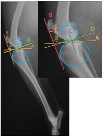
The tibial plateau was determined by a line connecting the cranial limit (insertion of CCL) to the caudal limit (insertion of the caudal cruciate ligament) of the medial condyle of the tibia. The TPA was established according to the conventional methodology recommended (Morris and Lipowitz, 2001; Osmond et al., 2006). The patellar ligament angle in relation to the tibial plateau and the patellar ligament angle in relation to the common tangent at the tibiofemoral contact point were measured based on different papers (Dennler et al., 2006; Schwandt et al., 2006SCHWANDT, C.S.; BOHORQUEZ-VANELLI, A.; TEPIC, S. et al. Angle between the patellar ligament and tibial plateau in dogs with partial rupture of the cranial cruciate ligament. Am. J. Vet. Res., v.67, p.1855-1860, 2006.). The methods of obtaining the patellar ligament angles are demonstrated in Fig. 1.
Measurement of the patellar ligament insertion angle (PLIA) was also performed. A straight line was drawn from the site of insertion of the patellar ligament at the tibial tuberosity to the tibiofemoral contact point (line X), and a second line was drawn to the most cranial aspect of the patella (line Y). The angle formed at the meeting point of these two lines is the PLIA (Fig. 2). Furthermore, the ratio between the lengths of the two lines (X/Y) was calculated.
Radiographic image of the stifle joint in the mediolateral projection of a healthy dog showing the measurement of the patellar ligament angles: the angle of the patellar ligament in relation to the tibial plateau (γ) and the angle of the patellar ligament in relation to the common tangent at the tibiofemoral contact point (α).
All measurements were performed blinded, by a single observer with experience in orthopedics and radiographic interpretation in small animals. The intraobserver analysis was performed using 25% of the random sample; three measurements were performed with a three-day interval between them. All data obtained in each of the variables were tested for normality (Kolmogorov-Smirnov) and analyzed by analysis of variance (ANOVA). When the data were significant with ANOVA, the variables were compared among the four different groups by the Tukey test or F test. The variables that were not normal were presented descriptively. Differences were considered statistically significant when P <0.05. The evaluation of the intraobserver variability was determined based on the evaluation of the coefficient of variation of repeated measurements. All statistical analyses were performed with statistical package software (IBM, 2011IBM. SPSS Statistics for Windows. Version 20.0, Release 0.0.0. Armonk, .New York: IBM Corp., 2011.).
Radiographic image of the stifle joint in the mediolateral projection of a healthy dog showing the measurement of the patellar ligament insertion angle (PLIA).
RESULTS AND DISCUSSION
In group 1, healthy Labrador Retrievers were selected, eight males and eight females, with a mean body weight of 34.31 kg and average age of 5.2 years. Group 2 was composed of Labradors with a CCL rupture, consisting of five males and seven females, with a mean body weight of 33.25 kg and average age of 5.4 years. Group 3 contained healthy dogs of different large mixed breeds (4), Golden Retriever (3), Neapolitan Mastiff (2), Rottweiler (2), Australian Cattle Dog (1), Brazilian Mastiff (1), German Shepherd (1), Belgian Shepherd (1) and American Pit Bull Terrier (1). This group included eight males and eight females with a mean body weight of 33.18 kg and average age of 5.6 years. In group 4, we included different breeds of dogs with CCL rupture, including Golden Retrievers (5), Boxers (4), Dalmatians (2), large mixed breeds (2), American Bulldog (1), Doberman Pinscher (1) and Weimaraner (1). This group included 11 males and five females with a mean body weight of 35.62 kg and an average age of 6.3 years. In general, the dogs had a mean body weight of 34.1 kg and an average age of 5.6 years, and 51.6% were males and 48.4% females. There was no statistically significant difference among the groups in relation to body weight and age.
The variables measured in the study had low levels of variability, with a coefficient of variation of 17.16% for the measurements of the TPA, 5.3% for the measurements of the patellar ligament angle in relation to the tibial plateau, 18.02% for the measurements of the patellar ligament angle in relation to the common tangent at the tibiofemoral contact point, 5.07% for the PLIA and 7.65% for the X/Y ratio. The intra-observer variability between days was considered low and the coefficient of variation was less than 3% for all measurements.
In this study, radiographs were performed with the stifle joint positioned at an average angle of 135.56°±5.73°. The measures specifically related to the slope of the tibial plateau are not influenced by the angle of the stifle joint, but the angles related to the patellar ligament are directly influenced by the stifle joint angle. With the stifle flexed, the angles related to the patellar ligament are reduced, while stifle extensions increase these angles. Therefore, it is ideal to perform the measurement of the angles of the proximal tibia with the stifle joint positioned around 135°, which corresponds to the normal articular angle during the support phase on the ground in dogs (Boudrieau, 2009BOUDRIEAU, R.J. Tibial plateau leveling osteotomy or tibial tuberosity advancement? Vet. Surg., v.38, p.1-22, 2009.; Cadmus et al., 2014).
The measured values of the TPA are shown in Tab. 1. The general average was 22.17°±4.20°, and this value was considered normal for dogs that had physiological variation of the TPA between 18° and 24° (Watt, 2000WATT, P. Tibial plateau leveling. Aust. Vet. J., v.78, p.385-386, 2000.; Kim et al., 2008). An average angle of 23.76° in dogs with CCL rupture and 18.10° in dogs without rupture was described (Morris and Lipowitz, 2001). The authors stated that high TPA might have been responsible for the CCL rupture. However, in the current study, there was no statistically significant difference among groups and the TPA alone does not seem to be a predisposing factor for CCL rupture in dogs.
The average angle of the tibial plateau observed in the Labrador Retriever dogs with CCL rupture was 24.25°, which is similar to the values described in a study that found an average of 25.30° for the TPA in 32 Labradors Retrievers with CCL rupture (Robinson et al., 2006). A similar result was reported in a retrospective study of TPA in dogs with CCL rupture. Of the 200 animals studied, 28% were Labrador Retrievers and the average angle of the tibial plateau was 24.90° (Zeltzman et al., 2005).
In a study with 68 dogs, of which 38.8% were Labrador Retrievers, the mean reported TPA in healthy and CCL ruptured dogs were 23.6° and 31.8°, respectively (Osmond et al., 2006). The values are higher than those reported in this current research, which were 20.75° and 24.25°, respectively. In one study, the mean TPA of 27.97° in healthy Labrador dogs was significantly higher than those with CCL rupture, which was 25.55° (Wilke et al., 2002). However, these authors stated that the results should be evaluated carefully because the group of healthy Labradors had a mean age of 51.3 months, and these young dogs could still develop the disease. Meanwhile, another study that selected only Labrador Retrievers over 8 years of age found a major average slope of the tibial plateau in dogs without CCL rupture but that was not statistically significant (Reif and Probst, 2003REIF, U.; PROBST, C.W. Comparison of tibial plateau angles in normal and cranial cruciate deficient stifles of Labrador retrievers. Vet. Surg., v.32, p.385-389, 2003.).
In this current study, the group of Labrador Retrievers with CCL rupture had a higher average for TPA than the two groups of healthy dogs; however, the measure was more similar to the group of dogs of various breeds with CCL rupture. There was no statistically significant difference between the groups and it is not possible to assert that the higher average slope of the tibial plateau found in both groups of dogs with CCL rupture may contribute to the pathogenesis of the disease in dogs.
The Labrador Retriever has a higher frequency of CCL rupture; however, the mean of the TPA for German Shepherds is superior to other dog breeds (Guastella et al., 2008GUASTELLA, D.B.; FOX, D.B.; COOK, J.L. Tibial plateau angle in four common canine breeds with cranial cruciate ligament rupture, and its relationship to meniscal tears. Vet. Comp. Orthop. Traumatol., v.21, p.125-128, 2008.). The TPA in dogs with CCL disorders ranges from 23.50° to 28.30°, and when it exceeds 34°, it is considered excessive (Duer et al., 2008). In the current study, none of the dogs showed an excessive angle of the tibial plateau, and the largest angle was 30° in one Labrador Retriever with CCL rupture and in one German Shepherd without an orthopedic disorder.
The two groups of dogs with CCL rupture had the highest averages for TPA, and the mean difference between the two groups of dogs with rupture and two groups of healthy dogs was 5.69°, which is similar to a reported difference of 5.66° (Morris and Lipowitz, 2001). In the current study, the mean difference between Labrador Retrievers with CCL rupture in relation to the group of the same breed of healthy dogs was 3.5°, which is close to the mean difference of 2.19° observed between the groups of dogs of various breeds with and without CCL rupture. Therefore, Labrador dogs with CCL rupture have a TPA similar to the other large dogs with rupture, and it implies that Labrador Retriever predisposition to the disease is not justified by the tibial plateau slope.
The values for the patellar ligament angle in relation to the tibial plateau are shown in Tab. 2. Only the group of healthy Labrador Retrievers was significantly higher than the group of various breeds with CCL rupture; however, this difference may be due to the angle of the stifle joint and the tibial position at the time of radiographic exposure in ruptured dogs. The average for all groups was 103°±4.20°, which is similar to the described patellar ligament angle in relation to the tibial plateau that was approximately 105° when the stifle joint is positioned at 135° (Kim et al., 2008; Sathya et al., 2014SATHYA, S.; GILBERT, P.; SHARMA, A.; HENDRICK, S. Effect of tibial plateau levelling osteotomy on patellar tendon angle: a prospective clinical study. Vet. Comp. Orthop. Traumatol., v.27, p.346-350, 2014.). However, the values in these studies were lower than those described for joints without CCL rupture evaluated in cadavers of different dog breeds that had an average patellar ligament angle in relation to the tibial plateau of 119.9° (Drygas et al., 2010DRYGAS, K.A.; POZZI, A.; GORING, R.L. et al. Effect of tibial plateau leveling osteotomy on patellar tendon angle: a radiographic cadaveric study. Vet. Surg., v.39, p.418-424, 2010.).
The values for the patellar ligament angle in relation to the common tangent at the tibiofemoral contact point are shown in Tab. 3. The general average was 99.06°±6.08° and there was no statistically significant difference between groups. In a previous study in dogs no statistically significant difference was found between the two methods of measurement of the patellar ligament angle (Schwandt et al., 2006). Conversely, in the current study, the mean value obtained by the tangent method was significantly lower than the average obtained by the method of the tibial plateau, although the measurement performed by the tangent method had greater variability. It seems there is a tendency to overestimate the patellar ligament angles when using the method of the tibial plateau (Drygas et al., 2010). Both methods can be used to measure the angles related to the patellar ligament, but there is a preference for the common tangent at the tibiofemoral contact point (Schwandt et al., 2006).
In the theory of tibial tuberosity advancement for treating CCL rupture, neutralizing the cranial tibial impulse is necessary for reducing the patellar ligament angle approximately 90° with the stifle joint maintained in the stance position (Kim et al., 2008; Cadmus et al., 2014). Therefore, the angles obtained in the current study did not approach the ideal for neutralizing the forces on the CCL, even when they were measured with the common tangent method. Although the group of healthy Labradors had the highest mean, there was no significant difference between the groups evaluated, and the predisposition of the Labrador Retriever to CCL rupture is not justified by the angle of the patellar ligament in relation to the tibial plateau or common tangent.
Values of the patellar ligament angle obtained by common tangent at the tibiofemoral contact point method in dogs
The lever arm of the extensor mechanism is represented by the distance between the tibiofemoral contact point and the point of insertion of the patellar ligament in the tibia (Boudrieau, 2009). In the current study, with the intention of developing a more comparative method for evaluating the lever arm, the ratio of the lengths of the lines (X/Y) was calculated and the PLIA was measured to compare values between animals with different sizes. The values calculated by the ratio between the lengths of the lines (X/Y) are shown in Tab. 4. There was no significant difference between the groups, and the average X/Y ratio in all groups was 0.69±0.05. Theoretically, the greater the extent of the lever arm, the lower the strength of the quadriceps mechanism for moving the tibia and promoting extension of the stifle joint (Boudrieau, 2009). In the present study, it was found that the length of the lever arm did not differ between the groups and this does not seem to be an important factor in the etiology of CCL rupture. However, the variable had a low coefficient of variation and can be considered a promising measure for morphometric studies of the proximal tibia.
An overall average of 51.45°±5.06° was found for the PLIA and the data are shown in Tab. 5. Regarding the PLIA, a smaller angle is believed to be associated with a greater force required to extend the knee joint; this increased force could overload the patellar ligament and CCL. In the present study, the mean of this angle was significantly lower in both groups of dogs with CCL rupture, which suggest that a reduced PLIA could be a cause of CCL rupture. However, these data need to be analyzed with caution because the dogs with CCL rupture may present slight cranial displacement of the proximal tibia at the time of radiographic exposure, which could influence the reduction of the PLIA.
CONCLUSIONS
In conclusion, the tibial plateau angle, the patellar ligament angles and the patellar ligament insertion angle do not seem to be predisposing factors for rupture of the CCL in Labrador Retriever dogs. In general, there seems to be no relationship between the angles of the proximal tibia and the CCL rupture in dogs.
ACKNOWLEDGEMENTS
We would like to acknowledge FAPEMIG and CNPq for partial financial support.
REFERENCES
- BOUDRIEAU, R.J. Tibial plateau leveling osteotomy or tibial tuberosity advancement? Vet. Surg., v.38, p.1-22, 2009.
- CADMUS, J.; PALMER, R.H.; DUNCAN, C. The effect of preoperative planning method on recommended tibial tuberosity advancement cage size. Vet. Surg., v.43, p.995-1000, 2014.
- CANAPP, S.O. The canine stifle. Clin. Tech. Small Anim. Pract., v.22, p.195-205, 2007.
- DENNLER, R.; KIPFER, N.M.; TEPIC, S. et al. Inclination of the patellar ligament in relation to flexion angle in stifle joints of dogs without degenerative joint disease. Am. J. Vet. Res., v.67, p.1849-1854, 2006.
- DRYGAS, K.A.; POZZI, A.; GORING, R.L. et al. Effect of tibial plateau leveling osteotomy on patellar tendon angle: a radiographic cadaveric study. Vet. Surg., v.39, p.418-424, 2010.
- DUERR, F.M.; DUNCAN, C.G.; SAVICKY, R.S. et al. Comparison of surgical treatment options for cranial cruciate ligament disease in large-breed dogs with excessive tibial plateau angle. Vet. Surg., v.37, p.49-62, 2008.
- FETTIG, A.A.; RAND, W.M.; SATO, A.F. et al. Observer variability of tibial plateau slope measurement in 40 dogs with cranial cruciate ligament-deficient stifle joints. Vet. Surg., v.32, p.471-478, 2003.
- GRIFFON, D.J. A review of the pathogenesis of canine cranial cruciate ligament disease as a basis for future preventive strategies. Vet. Surg., v.39, p.399-409, 2010.
- GUASTELLA, D.B.; FOX, D.B.; COOK, J.L. Tibial plateau angle in four common canine breeds with cranial cruciate ligament rupture, and its relationship to meniscal tears. Vet. Comp. Orthop. Traumatol., v.21, p.125-128, 2008.
- HARASEN, G.L. Feline cranial cruciate rupture: 17 cases and a review of the literature. Vet. Comp. Orthop. Traumatol., v.18, p.254-257, 2005.
- IBM. SPSS Statistics for Windows. Version 20.0, Release 0.0.0. Armonk, .New York: IBM Corp., 2011.
- KIM, S.E.; POZZI, A.; KOWALESKI, M.P.; LEWIS, D.D. Tibial osteotomies for cranial cruciate ligament insufficiency in dogs. Vet. Surg., v.37, p.111-125, 2008.
- MORRIS, E.; LIPOWITZ, A.J. Comparison of tibial plateau angles in dogs with and without cranial cruciate ligament injuries. J. Am. Vet. Med. Assoc., v.218, p.363-366, 2001.
- OSMOND, C.S.; MARCELLIN-LITTLE, D.J.; HARRYSSON, O.L.; KIDD, L.B. Morphometric assessment of the proximal portion of the tibia in dogs with and without cranial cruciate ligament rupture. Vet. Radiol. Ultrasound, v.47, p.136-141, 2006.
- REIF, U.; PROBST, C.W. Comparison of tibial plateau angles in normal and cranial cruciate deficient stifles of Labrador retrievers. Vet. Surg., v.32, p.385-389, 2003.
- RENWICK, A.I.; MCKEE, W.M.; EMMERSON, T.D.; HOUSE, A.K. Preliminary experiences of the triple tibial osteotomy procedure: tibial morphology and complications. J. Small Anim. Pract., v.50, p.212-221, 2009.
- ROBINSON, D.A.; MASON, D.R.; EVANS, R.; CONZEMIUS, M.G. The effect of tibial plateau angle on ground reaction forces 4-17 months after tibial plateau leveling osteotomy in Labrador Retrievers. Vet. Surg., v.35, p.294-299, 2006.
- SATHYA, S.; GILBERT, P.; SHARMA, A.; HENDRICK, S. Effect of tibial plateau levelling osteotomy on patellar tendon angle: a prospective clinical study. Vet. Comp. Orthop. Traumatol., v.27, p.346-350, 2014.
- SCHWANDT, C.S.; BOHORQUEZ-VANELLI, A.; TEPIC, S. et al. Angle between the patellar ligament and tibial plateau in dogs with partial rupture of the cranial cruciate ligament. Am. J. Vet. Res., v.67, p.1855-1860, 2006.
- SHAHAR, R.; MILGRAM, J. Biomechanics of tibial plateau leveling of the canine cruciate-deficient stifle joint: a theoretical model. Vet. Surg., v.35, p.144-149, 2006.
- WATT, P. Tibial plateau leveling. Aust. Vet. J., v.78, p.385-386, 2000.
- WILKE, V.L.; CONZEMIUS, M.G.; BESANCON, M.F. et al. Comparison of tibial plateau angle between clinically normal Greyhounds and Labrador Retrievers with and without rupture of the cranial cruciate ligament. J. Am. Vet. Med. Assoc., v.221, p.1426-1429, 2002.
- ZELTZMAN, P.A.; PARÉ, B.; JOHNSON, G.M. et al. Relationship between age and tibial plateau angle in dogs with cranial cruciate rupture. J. Am. Anim. Hosp. Assoc., v.41, p.117-120, 2005.
Publication Dates
-
Publication in this collection
Oct 2015
History
-
Received
18 Oct 2014 -
Accepted
11 June 2015