EETs modulate NF-κB signaling and alleviate STZ-induced chronic inflammation in type 2 diabetes
ABSTRACT
Epoxyeicosatrienoic acids (EETs) have shown preventive effects against various metabolic diseases. The objective of this study was to investigate the effects and mechanisms of EETs onT2DM in rats. The rats were divided into an T2DM group and intervention groups that received 11,12-EET, 14,15-EET and TPPU daily for 30days. Serum concentrations of TNF-α, IL-6, IL-10 and Arg-1 were measured using ELISA. The results showed that EETs and TPPU effectively reduced the concentrations of inflammatory factors (P<0.05). Histopathologic examination of epididymal fat and liver tissues by HE staining demonstrated that EETs and TPPU significantly improved the morphology and structure of adipose and liver cells. Immunohistochemical analysis of epididymal adipose tissue indicated that both EETs and TPPU significantly inhibited macrophage aggregation. Quantitative PCR and Western blot analyses showed that EETs and TPPU significantly inhibited the NF-κB signaling pathway at both the mRNA and protein levels; and These findings suggest that EETs may reduce inflammation in T2DM rats by modulating macrophage aggregation through inhibition of the NF-κB signaling.
Keywords:
type 2 diabetes; chronic inflammation; epoxyeicosatrienoic acid; TPPU; insulin resistance
RESUMO
Os ácidos epoxieicosatrienoicos (EETs) demonstraram efeitos preventivos contra várias doenças metabólicas. O objetivo deste estudo foi investigar os efeitos e os mecanismos dos EETs no DM2 em ratos. Os ratos foram divididos em um grupo T2DM e grupos de intervenção que receberam 11,12-EET, 14,15-EET e TPPU diariamente, por 30 dias. As concentrações séricas de TNF-α, IL-6, IL-10 e Arg-1 foram medidas por meio de ELISA. Os resultados mostraram que os EETs e a TPPU reduziram efetivamente as concentrações de fatores inflamatórios (P<0,05). O exame histopatológico da gordura epididimal e dos tecidos hepáticos pela coloração HE demonstrou que os EETs e a TPPU melhoraram significativamente a morfologia e a estrutura das células adiposas e hepáticas. A análise imuno-histoquímica do tecido adiposo epididimal indicou que tanto os EETs quanto a TPPU inibiram significativamente a agregação de macrófagos. As análises quantitativas de PCR e Western blot mostraram que os EETs e a TPPU inibiram significativamente a via de sinalização NF-κB nos níveis de mRNA e proteína. Esses achados sugerem que os EETs podem reduzir a inflamação em ratos com DM2 modulando a agregação de macrófagos por meio da inibição da sinalização NF-κB.
Palavras-chave:
diabetes tipo 2; inflamação crônica; ácido epoxieicosatrienoico; TPPU; resistência à insulina
INTRODUCTION
EETs are metabolites generated from arachidonic acid (AA) via the cytochrome P450 epoxygenase pathway. They exhibit a variety of beneficial functions, including anti-hypertensive, anti-inflammatory, cardioprotective, angiogenesis-promoting and anti-apoptotic effects (Mosser and Edwards, 2008; Haase et al., 2014). Soluble epoxide hydrolase (sEH) inhibitors can prevent the conversion of EETs to dihydroxyeicosatrienoic acids (DHETs), thereby increasing the concentration of EETs in the organism (Banu and Sur, 2023). TPPU is a novel sEH inhibitor known for its favourable biological characteristics and stability (Ren et al., 2016) . Recent research has shown that EETs possess anti-inflammatory properties across various pathological conditions such as diabetes, sepsis, lung injury, neurodegenerative diseases, liver disease, kidney injury and arthritis (Xu et al., 2016; Hoff et al., 2019; Kodani and Morisseau, 2019; Wang et al., 2021). Despite these findings, the precise mechanisms through which EETs exert their anti-inflammatory effects remain unclear and warrant further investigation.
T2DM is a significant global health issue, accounting for approximately 90-95% of all diabetes cases (Laakso, 2019). Notably, about 90% of individuals with T2DM are either overweight or obese (Boutari et al., 2023). An inflammatory response serves as a common pathway in metabolic diseases, including obesity, IR, and T2DM (Matulewicz and Karczewska-Kupczewska, 2016; Marušić et al., 2021). T2DM is often associated with elevated levels of acute-phase response markers in the blood, indicating that it is essentially a process involving the activation of the body’s innate immune response and chronic inflammation. This chronic inflammation plays a mediating role in the pathogenesis of diabetes and can predict its development (Gasmi et al., 2021; Lee et al., 2022), underscoring the importance of anti-inflammatory interventions in improving T2DM outcomes.
Multiple intracellular pathways are implicated in the pro-inflammatory activation of macrophages within adipose tissue, including the TLR4/NF-kB pathway, the JNK pathway, and the caspase/NLRP3 inflammasome(Zatterale et al., 2020). Numerous studies have also demonstrated that the inflammatory response in adipose tissue is link to the activation of the NF-κB transcriptional programme (Hajer et al., 2008; Zhao et al., 2018; He et al., 2021; Tian et al., 2021). The liver, a key organ regulating metabolic homeostasis; is a primary target of insulin action and a crucial site for inflammatory mediator activity [34]. Evidence suggests that the activation of the NF-κB signaling pathway promotes the release of inflammatory factors such as TNF-α, IL-1β and IL-6, which can lead to liver-related complications in T2DM, including hepatic steatosis, hepatic IR and inflammation (Khan et al, 2006; Zhao et al., 2018; Tanase et al., 2020).
Given the pronounced anti-inflammatory impact of EETs and the sEH inhibitor (TPPU), the present study aims to investigate how the EETs-sEH pathway modulates the NF-κB signaling pathway and macrophage polarization. This study will explore the chronic inflammatory effects and mechanisms of action that mitigate chronic inflammation in T2DM rats.
MATERIALS AND METHODS
This research protocol was reviewed and approved for ethics by the Laboratory Animal Welfare and Ethics Committee of Inner Mongolia Agricultural University (No.: NND2021034).
Forty-five 4-week-old male SD rats, certified Specific Pathogen-Free (SPF) and weighing between 130-140g, were purchased from Biotechnology Co. Ltd.( Beijing, China). A total of 9 rats were randomly selected to serve as the control group and were maintained on a standard chow diet (CD), while the remaining 36 rats were fed a high-fat diet (HFD) consisting of 45% fat for 8 weeks. The HFD comprised 23% casein, 20% lard, 20% sucrose, and 8.5% corn starch, all provided by Sinodiets Biotechnology Co. Ltd. (Beijing, China). Following the HFD period, the 36 rats received an intraperitoneal injection of 30 mg/kg STZ(Sigma, Livonia, Michigan, USA) dissolved in a 0.1 mM sodium citrate (JINYAOXIANGCHENG Co. Ltd., Tianjin, China) buffer, administered once a week for 2 weeks. Two weeks post-injection, blood samples were collected from the tail vein to measure fasting blood sugar (FBS). A successful T2DM rats model was established in rats with FBS levels≥11.1mmol/L.
The T2DM rats were then randomly divided into two groups: the T2DM group, which received an intraperitoneal injection of sodium citrate buffer, and the intervention group. The intervention group was further divided into three subgroups: the 11,12-EETs (Cayman, Ann Arbor, Michigan, USA)group, which received an intraperitoneal injection of 30 μg/kg/day; the 14,15-EETs(Cayman, Ann Arbor, Michigan, USA )group, also receiving an intraperitoneal injection of 30 μg/kg/day; and the TPPU(Med Chem Express Co. Ltd., Monmouth Junction, USA )group, which was administered TPPU via gavage at a dose of 3mg/kg/day. The treatment lasted for a total of 30 days.
During the intervention, the body weights of the rats in each group were recorded daily to create weight progression graphs. At the end of the experiment, epididymal fat and liver tissues were harvested and weighed. The organ coefficients were calculated using the formula: Organ Coefficient = Organ Weight / Body Weight.
After 30d of drug intervention and 8h fasting period, the rats were gavaged with a glucose solution at a concentration of 50% (2 g/kg). Blood samples were collected from the tail vein to measure blood glucose concentrations at 0, 30, 60, 90, and 120 minutes using a blood glucose meter. The glucose-time curves were plotted, and the area under the curve (AUC) for the OGTT was calculated.
Following 8 weeks of drug intervention, the concentrations of serum insulin (BIOHUB INTERNATIONAL TRADE CO., LTD., Shang Hai, China) 、TNF-α,、IL-6,、IL-10 and Arg-1 were measured in all groups using ELISA kits in accordance with the manufacturer's instructions (Meibiao Biotechnology, Jiangsu, China). The absorbance (optical density, OD value) was measured to generate calibration curves for the calculation of concentrations. The Homeostasis Model Assessment of Insulin Resistance (HOMA-IR) was calculated using the formula: HOMA-IR = FBG (mmol/l) × FINS (µU/ml) / 22.5
Epididymal fat and liver tissues were collected and immediately preserved in a 10% formalin solution for paraffin embedding. The tissues were sectioned into 5 µm slices and stained with H&E (hematoxylin-eosin) for histopathological evaluation. Pathological changes in the tissues were observed using a light microscopy.
Epididymal fat tissue was fixed in 10% formalin,embedded in paraffin, and cut into 5 µm slices. The slices were then deparaffinized in xylene and rehydrated. Antigen retrieval was performed by boiling the slides in 1×citrate solution for 20 min, followed by washing with TBST buffer. The slides were incubated for 10 min at room temperature with an endogenous peroxidase blocker (Beyotime Biotechnology Co. Ltd., Shanghai, China), after which they were washed again in TBST buffer and blocked with goat serum for 30 min.
The primary antibody, F4/80 Rabbit Monoclonal Antibody (Beyotime Biotechnology Co. Ltd., Shanghai, China),was diluted according to the size of the tissue and incubated at 4 ℃ overnight. Following this, the slides were washed with TBST buffer and then incubated for 30 min with an enzyme-labelled goat anti-rabbit IgG polymer(ZSGB-BIO Co. Ltd., Beijing, China)at 37 ℃ for 20 min. The slices were subsequently counterstained with hematoxylin after developing with DAB chromogenic solution (Axygen, California, USA), and mounted with neutral gum. The expression of F4/80-positive macrophages were observed using light microscopy.
Total RNA was extracted from liver tissue and epididymal fat using a Total RNA Kit(Axygen, California, USA). Reverse transcription of cDNA was performed using the PrimeScript™ RT Master Mix (Perfect Real Time,Takara Bio Inc., Mountain View, USA) according to the manufacturer's instructions (Takara Bio Inc., Mountain View, USA). For RT-PCR, TB Green® Premix Ex Taq™ II (Tli RNaseH Plus, Takara Bio Inc., Mountain View, USA) was used to prepare the PCR reaction solution, as instructed by the manufacturer (Takara Bio Inc., Mountain View, USA). The mRNA primers used in the study were β-actin, NF-κB, IL-10, and TNF-α, which were provided by Sangon Biotech Co, Ltd.(Shanghai, China).
Total protein was extracted from adipose and liver tissues and quantified using the BCA Protein Assay Kit (Solarbio Co.Ltd., Beijing, China). The proteins were separated by sodium dodecyl sulfate−polyacrylamide gel electrophoresis (SDS-PAGE, Solarbio Co.Ltd., Beijing, China) and transferred to a polyvinylidene difluoride (PVDF, SigmaAldrich, USA) membrane. The PVDF membranes immersed in a protein-free rapid blocking solution (Boster Biological Technology, Wuhan, China) for 15 min on a shaker. The primary antibody18 was added and incubated at 4°C overnight, followed by incubation with a secondary antibody (Affinity Biosciences Pty Ltd., Cincinnati, USA ) at room temperature for 1h, according to the manufacturer's instructions. Subsequently, ECL Western Blotting Substrate (Affinity Biosciences Pty Ltd., Cincinnati, USA) was applied to the PVDF membrane, which was then placed in a development instrument.
Western blot images were analyzed for grayscale intensity using ImageJ software. The relative expression of mRNA was calculated via qPCR using the 2-ΔΔCt method. Statistical analyses and graphing were performed using SPSS 20.0 and GraphPad Prism software. Results were expressed as "mean±standard deviation", P<0.01 indicates highly significant, while P<0.05 indicates significant.
RESULTS
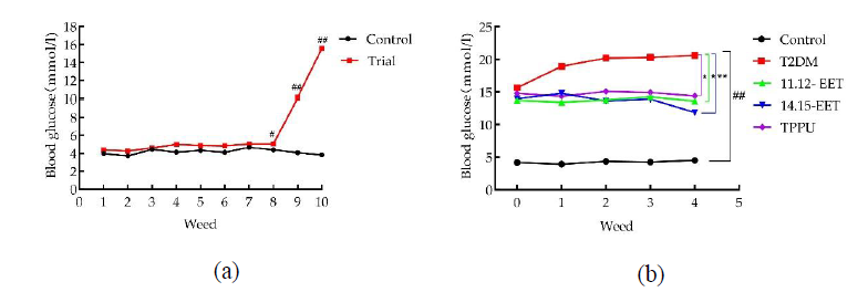
As illustrated in Fig. 1 a, after eight weeks of a high-fat diet, the FBS levels in the test group were significantly higher than those in the control group (P < 0.05). Following two intraperitoneal injections of STZ in the test group, FBS levels were not only extremely significantly elevated compared to the control group (P<0.01), but also reached values of ≥11.1 mmol/L. This indicates that the establishment of the T2DM rat model was successful.
As shown in Fig. 1 b, at the time of intervention, FBS levels are significantly increased in all experimental groups compared to the control group (p < 0.01). The FBS in the T2DM group exhibited a continuous upward trend, while the intervention groups demonstrated a significant reduction in FBS levels when compared to the T2DM group after treatment (P < 0.05), with the 14,15-EET group showing the most pronounced effect.
According to Fig. 2a and b, during the intervention period, body weight in the T2DM group decreased, whereas the three intervention groups were able to mitigate this weight loss in T2DM rats.
Fig. 2c and d further demonstrate that both the epididymal fat and hepatic indices were significantly higher in the T2DM group than in the control group. However, the intervention groups exhibited a reduction in both the epididymal fat and liver coefficients, indicating a beneficial effect of the interventions on these parameters.
The impact of EETs and TPPU on glucose metabolism in T2DM rats was evaluated using the OGTT. As shown in Fig. 3b, treatment with 11,12-EET, 14,15-EET and TPPU significantly promoted the recovery of blood glucose levels. Additionally, analysis of the area under the OGTT curve (AUC) revealed that the AUC values for the treatment groups were significantly lower than those for the T2DM group following drug intervention (P<0.05).
As depicted in Fig. 4a, the insulin content in the intervention groups of T2DM rats was significantly elevated (P < 0.01). IR levels were assessed using the Homeostasis Model Assessment of Insulin Resistance (HOMA-IR) index. Fig. 4b illustrates that IR levels in the experimental groups were significantly elevated (P < 0.01) compared to the control group, but a statistically significant reduction in IR was observed in the intervention groups (P < 0.01).
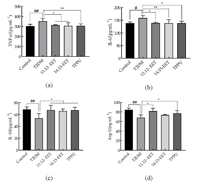
Fig.5a and b demonstrate that, compared to the T2DM group, the intervention groups significantly reduced the concentrations of TNF-α and IL-6 (P < 0.05). Similarly, in Fig.5c and b show that the 11,12-EET, 14,15-EET, and TPPU groups significantly elevated the concentrations of IL-10 and Arg-1 compared to the T2DM group (P < 0.05).
HE staining revealed that the livers of rats in the T2DM group exhibited steatosis, characterized by pale cytoplasm of hepatocytes, rounded vacuoles of varying sizes, narrowed hepatic sinusoids due to lipid droplet extrusion, and disorganization of the hepatic cord structure. In contrast, the liver histological structure in the intervention groups showed considerable improvement, including a reduction in lipid droplet infiltration (Fig. 7).
The morphology of adipocytes was assessed using HE staining. As illustrated in Fig.7a, adipocytes in the T2DM group exhibited uneven sizes and a larger average diameter compared to those in the control group. Following the intervention, the size disparity of adipocytes was partially ameliorated, and the average diameter of adipocytes was significantly reduced.
A prominent feature of chronic inflammation in adipose tissue is the aggregation of macrophages around dysfunctional or necrotic adipocytes, leading to the formation of crown-like structures (CLS). Immunohistochemical analysis of the macrophage marker F4/80 revealed substantial macrophage infiltration in the epididymal adipose tissue of the T2DM group. However, the intervention group displayed a significant reduction in macrophage aggregation in the adipose tissue(Fig.7b).
The qPCR assay demonstrated that the concentration of TNF-α mRNA in both adipose and liver tissues of rats in the T2DM group and the intervention group was significantly elevated (P < 0.01) compared to the control group (Fig. 8a and 9a). Concurrently, the expression of IL-10 mRNA showed a significant decrease (P < 0.01) in both groups. Importantly, in the intervention group, TNF-α mRNA levels were notably reduced (P < 0.01). Additionally, the concentration of IL-10 mRNA was significantly increased in the treatment group compared to the T2DM group (P < 0.05), as depicted in Figs. 8b and 9b.
Results from the qPCR assay illustrated in Figs. 10(a、c) and 11(a、c) showed that treatment with 14,15-EET and TPPU significantly reduced the relative expression of NF-κB mRNA compared to the T2DM group (P < 0.01). Furthermore, the 11,12-EET group exhibited a notable decrease in NF-κB mRNA expression (P < 0.05).
Western blot analysis, as depicted in Figs. 10b and 11b, revealed that interventions groups significantly lowered the p/NF-κB/NF-κB protein levels compared to the T2DM group (P < 0.01).

Changes in FBS of rats in each group, (a) Changes in FBS of rats in each group during high fat; (b) Changes in FBS of rats in each group during treatment. #vs. control group and *vs. T2DM group, #* (P<0.05), ##** (P<0.01).

Changes in body weight and organ coefficient of rats in each group, (a) changes in body weight of rats in each group during high-fat period; (b) changes in body weight of rats in each group during intervention; (c)epididymal fat coefficient; (d) liver coefficient. # vs. control group # (P<0.05) ##(P<0.01).

The effects of EETs and TPPU on OGTT in T2DM rats, (a)Changes in blood glucose at each time point of OGTT test in rats of each group, (a)AUC of OGTT. ##vs. control group and*vs. T2DM group, ##(P<0.01), * (P<0.05).

Insulin content and insulin resistance index of rats in each group, (a)Changes in insulin content of rats in each group, (b)HOMA-IR. ##vs. control group and*vs. T2DM group, ##**(P<0.01), * (P<0.05).

Changes in serum inflammatory factor levels in each group of rats, (a-d) were the changes in serum levels of TNF-α, IL-6, IL-10 and Arg-1 in each group. #vs. control group and *vs. T2DM group, #P<0.05), ## (P<0.01), * (P<0.05), **(P<0.01).


(a)HE staining of rat epididymal adipose tissue (20 x) F4/80 and (b)immunohistochemical staining of macrophages in rat epididymal adipose tissue (20 x).

Comparison of relative expression levels of inflammatory factor mRNA in adipose tissue of rats in each groups,(a)changes in relative expression of TNF-α mRNA in rats in each group, (b)changes in relative expression of IL-10 mRNA in rats in each group.#vs. control group and *vs. T2DM group, ### (P<0.01), * (P<0.05), **(P<0.01)

Comparison of relative expression levels of inflammatory factor mRNA in liver tissue of rats in each groups, (a)changes in relative expression of TNF-α mRNA in rats in each group, (b)changes in relative expression of IL-10 mRNA in rats in each group. #vs. control group and *vs. T2DM group, ### (P<0.01), * (P<0.05), **(P<0.01)

Expression of NF-κB mRNA and protein in adipose tissue of rats in each groups,(a) changes in the relative expression of NF-(B mRNA in rats of each group,(b) wb bar graph,(c) changes in the expression level of NF-(B protein in rats of each group. #vs. control group and *vs. T2DM group, ### (P<0.01), * (P<0.05), **(P<0.01)

Expression of NF-κB mRNA and protein in liver tissue of rats in each groups,(a) changes in the relative expression of NF-κB mRNA in rats of each group,(b) wb bar graph,(c) changes in the expression level of NF-κB protein in rats of each group. #vs. control group and *vs. T2DM group, ### (P<0.01), * (P<0.05), **(P<0.01)
DISCUSSION
Streptozotocin (STZ) is a chemical commonly employed in experimental models to induce diabetes in rodents, including mice (Akinlade et al., 2021). In our study, the T2DM rat model was established via intraperitoneal injection of STZ, resulting in FBS ≥11.1mmol/L after two weeks, indicating successful model establishment.
The CYP-EETs pathway plays a crucial role in obesity-induced adipose inflammation and the development of IR (Xu et al., 2010). Notably, studies employing T2DM mouse models have shown that both sEH knockouts and sEH inhibitors promote insulin secretion and ameliorated hyperglycaemia (Luo et al., 2010). Our results indicate that treatment with EETs and TPPU in T2DM rats significantly improved mental status and alleviated severe weight loss. These treatments effectively reduced blood glucose levels and facilitated the recovery of glycemic control, as evidenced by OGTT. Additionally, serum insulin levels and IR status exhibited significant improvement, demonstrating the ability of EETs and TPPU to regulate glucose metabolism and enhance insulin sensitivity in T2DM rats.
The association between IR, T2DM, and obesity-particularly the accumulation of abdominal and intrahepatic fat-is a critical factor in the emergence of metabolic diseases (Kahn et al., 2006; Rattarasarn, 2018). In individuals or animal models exhibiting IR or T2DM, we observe elevated levels of pro-inflammatory cytokines, such as TNF-α and IL-6, in adipose tissue, liver, and serum. Recent studies have highlighted the anti-inflammatory properties of EETs across various disease models (Bashir et al., 2022). For instance, in lipopolysaccharide-induced RAW264.7 cells, treatment with exogenous EETs or TPPU reduced in inflammatory injury by inhibiting the expression of IL-1β and TNF-α (Zhou et al., 2017).Our data corroborate these findings, showing that EETs and TPPU significantly decrease serum levels of TNF-α and IL-6 while promoting levels of the anti-inflammatory cytokine IL-10 and the macrophage-activated protein Arg-1, thus mitigating the systemic inflammatory response in T2DM rats.
Furthermore, visceral fat accumulation is recognized as a significant risk factor in the development of T2DM, IR, and cardiovascular disease (Burhans et al., 2018). Studies indicate that EET levels in adipose tissue are considerably reduced in high-fat diet-induced obesity models, and exogenous EET administration can inhibit adipogenesis (Zha et al., 2014). Our experiments demonstrated that EETs and TPPU led to reductions in visceral fat weight and improvements in adipocyte hypertrophy in T2DM-induced rats, suggesting a regulatory role for EETs in adipocyte function and maturation.
The activation and infiltration of pro-inflammatory immune cells in adipose tissue-including macrophages, neutrophils, and T cells-alongside elevated levels of pro-inflammatory molecules, contribute to a complex inflammatory network that promotes IR (Apostolopoulos et al., 2016). Macrophage infiltration and the secretion of various inflammatory cytokines in white adipose tissue activate signaling pathways such as JNK and NF-κB, resulting in both local and systemic IR (Hotamisligil et al., 1994; Olefsky and Glass, 2010). In this context, EETs not only improved adipocyte hypertrophy but also inhibited the aggregation of macrophages and the overall inflammation in adipose tissue(Dai Meiyan, 2016).Our experiments demonstrated that EETs and TPPU markedly reduced macrophage aggregation, as indicated by the decreased expression of the macrophage marker F4/80. Additionally, measurements of pro-inflammatory cytokines in adipose tissue revealed a significant reduction in the relative mRNA expression of TNF-α in response to EETs and TPPU treatment.
The observed decline in relative TNF-α mRNA expression in the EETs and TPPU groups may be linked to a reduction in macrophage aggregation in adipose tissue, as macrophages are the primary source of TNF-α production in obese patients (Cai et al., 2005). The NF-κB signaling pathway is a crucial pathway associated with inflammation and immune regulation, playing a significant role in the production of pro-inflammatory cytokines and the recruitment of leukocytes. This pathway is also involved in the development of type 2 diabetes mellitus (T2DM) and insulin resistance (IR) and is highly expressed in T2DM; its inhibition has been shown to improve hyperglycemia and insulin sensitivity in T2DM rats (Zhang et al., 2010; Iskender et al., 2017; Bako et al., 2019).
NF-κB pathway-related proteins are activated by TNF-α to bind inflammation-related genes, initiating the transcription of inflammatory cytokines such as IL-6, IL-1β, and iNOS, ultimately triggering an inflammatory response (Park et al., 2022). A study by (Node et al., 1999; Dai et al., 2015; Li et al., 2015; Luo et al., 2023) demonstrated that the activation of PPARγ receptors by EETs-derived from CYP2J2 overexpression or introduced exogenously-increases glucose uptake and ameliorates IR. This effect occurs through the inhibition of NF-κB and IKK activity, along with the modulation of macrophage polarization, which can prevent the aggregation and activity of inflammatory cells, thereby exerting anti-inflammatory effects. Our experiments showed that both EETs and TPPU inhibit NF-κB mRNA and protein expression levels in rat adipose tissue, suggesting that EETs and TPPU may regulate adipose tissue inflammation in T2DM rats by inhibiting NF-κB pathway expression.
Numerous studies have indicated that pro-inflammatory cytokines and chemokines in the liver, such as TNF-α, IL-6, MCP-1 and IL-1β, play a pivotal role in the progression of metabolic disease (Cai et al., 2005). These cytokines not only disrupt insulin signaling in target tissues, leading to IR, but also activate multiple pro-inflammatory pathways (NF-κB and MAPK), exacerbating the inflammatory response (Santos et al., 2019; Caussy et al., 2021). Experimental results have demonstrated that EETs and TPPU can attenuate steatosis and damage to liver structure, as evidenced by HE staining. Furthermore, these treatments significantly reduce the concentration of TNF-α mRNA while concurrently increasing IL-10 mRNA levels in liver tissues.
Wang et al. (2014)found that administering TPPU alleviates hepatic steatosis and inflammatory injury in mice with steatohepatitis while simultaneously suppressing the expression of pro-inflammatory cytokines. Li et al. (2015) showed that the overexpression of CYP2J2 resulted in reduced gene expression levels of pro-inflammatory cytokines (TNF-α, IL-6, and IL-1β) in the liver. Our experiments supported these findings, revealing that EETs and TPPU significantly decrease the relative expression levels of NF-κB mRNA and protein. Furthermore, CYP2J2 overexpression and exogenous EETs attenuated hepatic inflammation by inhibiting the NF-κB signaling pathway, leading to a decrease in the production of pro-inflammatory cytokines and macrophage infiltration in the liver (Li et al., 2015).
In summary, these experiments confirm that EETs may alleviate chronic inflammation in T2DM rats by inhibiting the activation of the NF-κB signaling pathway, thereby improving insulin resistance. However, further mechanistic studies and clinical trials are necessary to validate these findings.
REFERENCES
- AKINLADE, O.M.; OWOYELE, B.V.; SOLADOYE, A.O. Streptozotocin-induced type 1 and 2 diabetes in rodents: A model for studying diabetic cardiac autonomic neuropathy. Afr. Health Sci., v.21, p.719-727, 2021.
- APOSTOLOPOULOS, V.; COURTEN, M.P.J.; STOJANOVSKA, L.et al. The complex immunological and inflammatory network of adipose tissue in obesity. Mol. Nutr. Food Res., v.60, p.43-57, 2016.
- BAKO, H.Y.; IBRAHIM, M.A.; ISAH, M.S.et al. Inhibition of JAK-STAT and NF-κB signalling systems could be a novel therapeutic target against insulin resistance and type 2 diabetes. Life Sci., v.239, p.117045, 2019.
- BANU, S.; SUR, D. Role of macrophage in type 2 diabetes mellitus: macrophage polarizationa new paradigm for treatment of type 2 diabetes mellitus. Endocr. Metab. Immune Disord. Drug Targets, v.23, p.2-11, 2023.
- BASHIR, H.; MAJID, S.; KHAN, M.S.et al. Inter-relationship of pro- and anti- inflammatory biomarkers with the development of type 2 diabetes mellitus. Heliyon, v.8, p.e11329, 2022.
- BOUTARI, C.; DEMARSILIS, A.; MANTZOROS, C.S. Obesity and diabetes. Diabetes Res. Clin. Pract., v.202, p.110773, 2023.
- BURHANS, M.S.; HAGMAN, D.K.; KUZMA, J.N.et al. Contribution of adipose tissue inflammation to the development of type 2 diabetes mellitus. Compr. Physiol., v.9, p.1-58, 2018.
- CAI, D.; YUAN, M.; FRANTZ, D.F.et al. Local and systemic insulin resistance resulting from hepatic activation of IKK-beta and NF-kappaB. Nat. Med., v.11, p.183-190, 2005.
- CAUSSY, C.; AUBIN, A.; LOOMBA, R. The relationship between type 2 diabetes, NAFLD, and cardiovascular risk. Curr. Diabetes Rep., v.21, p.15, 2021.
- DAI, M.; WU, L.; HE, Z.et al. Epoxyeicosatrienoic acids regulate macrophage polarization and prevent LPS-induced cardiac dysfunction. J. Cell. Physiol., v.230, p.2108-2119, 2015.
- DAI, Y.M. CYP epioxidase 2J2 and its metabolite EETs regulate macrophage polarization to ameliorate high-fat diet-induced insulin resistance. 2016. 117p. Dissertation (PhD) - Huazhong University of Science and Technology.
- GASMI, A.; NOOR, S.; MENZEL, A.et al. Obesity and insulin resistance: associations with chronic inflammation, genetic and epigenetic factors. Curr. Med. Chem., v.28, p.800-826, 2021.
- HAASE, J.; WEYER, U.; IMMIG, K.et al. Local proliferation of macrophages in adipose tissue during obesity-induced inflammation. Diabetologia, v.57, p.562-571, 2014.
- HAJER, G.R.; HAEFTEN, T.W.V.; VISSEREN, F.L.J. Adipose tissue dysfunction in obesity, diabetes, and vascular diseases. Eur. Heart J., v.29, p.2959-2971, 2008.
- HE, F.; HUANG, Y.; SONG, Z.et al. Mitophagy-mediated adipose inflammation contributes to type 2 diabetes with hepatic insulin resistance. J. Exp. Med., v.218, p.e20201416, 2021.
- HOFF, U.; BUBALO, G.; FECHNER, M.et al. A synthetic epoxyeicosatrienoic acid analogue prevents the initiation of ischemic acute kidney injury. Acta Physiol., v.227, p.e13297, 2019.
- HOTAMISLIGIL, G.S.; BUDAVARI, A.; MURRAY, D.et al. Reduced tyrosine kinase activity of the insulin receptor in obesity-diabetes. Central role of tumor necrosis factor-alpha. J. Clin. Invest., v.94, p.1543-1549, 1994.
- ISKENDER, H.; DOKUMACIOGLU, E.; SEN, T.M.et al. The effect of hesperidin and quercetin on oxidative stress, NF-κB and SIRT1 levels in a STZ-induced experimental diabetes model. Biomed. Pharmacother., v.90, p.500-508, 2017.
- KAHN, S.E.; HULL, R.L.; UTZSCHNEIDER, K.M. Mechanisms linking obesity to insulin resistance and type 2 diabetes. Nature, v.444, p.840-846, 2006.
- KODANI, S.D.; MORISSEAU, C. Role of epoxy-fatty acids and epoxide hydrolases in the pathology of neuro-inflammation. Biochimie, v.159, p.59-65, 2019.
- LAAKSO, M. Biomarkers for type 2 diabetes. Mol. Metab., v.27, p.S139-S146, 2019.
- LEE, S.H.; PARK, S.Y.; CHOI, C.S. Insulin resistance: from mechanisms to therapeutic strategies. Diabetes Metab. J., v.46, p.15-37, 2022.
- LI, R.; XU, X.; CHEN, C.et al. attenuates metabolic dysfunction in diabetic mice by reducing hepatic inflammation via the PPARγ. Am. J. Physiol. Endocrinol. Metab., v.308, p.E270-282, 2015.
- LUO, A.; WU, Z.; LI, S.et al. The soluble epoxide hydrolase inhibitor TPPU improves comorbidity of chronic pain and depression via the AHR and TSPO signaling. J. Transl. Med., v.21, p.71, 2023.
- LUO, P.; CHANG, H.H.; ZHOU, Y.et al. Inhibition or deletion of soluble epoxide hydrolase prevents hyperglycemia, promotes insulin secretion, and reduces islet apoptosis. J. Pharmacol. Exp. Ther., v.334, p.430-438, 2010.
- MARUŠIĆ, M.; PAIĆ, M.; KNOBLOCH, M.; LIBERATI PRŠO, A.M. NAFLD, insulin resistance, and diabetes mellitus type 2. Can. J. Gastroenterol. Hepatol., v.2021, p.1-9, 2021.
- MATULEWICZ, N.; KARCZEWSKA-KUPCZEWSKA, M. Insulin resistance and chronic inflammation. Postepy Hig. Med. Dosw., v.70, p.1245-1258, 2016.
- MOSSER, D.M.; EDWARDS, J.P. Exploring the full spectrum of macrophage activation. Nat. Rev. Immunol., v.8, p.958-969, 2008.
- NODE, K.; HUO, Y.; RUAN, X.et al. Anti-inflammatory properties of cytochrome P450 epoxygenase-derived eicosanoids. Science, v.285, p.1276-1279, 1999.
- OLEFSKY, J.M.; GLASS, C.K. Macrophages, inflammation, and insulin resistance. Ann. Rev. Physiol., v.72, p.219-246, 2010.
- PARK, J.E.; KANG, E.; HAN, J.S. HM-chromanone attenuates TNF-α-mediated inflammation and insulin resistance by controlling JNK activation and NF-κB pathway in 3T3-L1 adipocytes. Eur. J. Pharmacol., v.921, p.174884, 2022.
- RATTARASARN, C. Dysregulated lipid storage and its relationship with insulin resistance and cardiovascular risk factors in non-obese asian patients with type 2 diabetes. Adipocyte, v.7, p.71-80, 2018.
- REN, Q.; MA, M.; ISHIMA, T.et al. Gene deficiency and pharmacological inhibition of soluble epoxide hydrolase confers resilience to repeated social defeat stress. Proc. Nat. Acad. Sci., v.113, p.E1944-1952, 2016.
- SANTOS, R.D.; VALENTI, L.; ROMEO, S. Does nonalcoholic fatty liver disease cause cardiovascular disease? Current knowledge and gaps. Atherosclerosis, v.282, p.110-120, 2019.
- TANASE, D.M.; GOSAV, E.M.; COSTEA, C.F.et al. The intricate relationship between type 2 diabetes mellitus (T2DM), insulin resistance (IR), and nonalcoholic fatty liver disease (NAFLD). J. Diabetes Res., v.2020, p.3920196, 2020.
- TIAN, J.; ZHAO, Y.; WANG, L.et al. Role of TLR4/MyD88/NF-κB signaling in heart and liver-related complications in a rat model of type 2 diabetes mellitus. J. Int. Med. Res., v.49, p.300060521997590, 2021.
- WANG, B.; WU, L.; CHEN, J.et al Metabolism pathways of arachidonic acids: Mechanisms and potential therapeutic targets. Signal Transd. Targeted Ther., v.6, p.94, 2021.
- WANG, X.J. Role of epoxy eicostrienoic acid in nonalcoholic steatohepatitis. 2014. 77f. Dissertation (PhD) - Huazhong University of Science and Technology.
- XU, X.; LI, R.; CHEN, G.et al. The role of cytochrome P450 epoxygenases, soluble epoxide hydrolase, and epoxyeicosatrienoic acids in metabolic diseases. Adv. Nutr., v.7, p.1122-1128, 2016.
- XU, X.; ZHAO, C.X.; WANG, L.et al. Increased CYP2J3 expression reduces insulin resistance in fructose-treated rats and db/db mice. Diabetes, v.59, p.997-1005, 2010.
- ZATTERALE, F.; LONGO, M.; NADERI, J.et al. Chronic adipose tissue inflammation linking obesity to insulin resistance and type 2 diabetes. Front. Physiol., v.10, p.1607, 2020.
- ZHA, W.; EDIN, M.L.; VENDROV, K.C.et al. Functional characterization of cytochrome P450-derived epoxyeicosatrienoic acids in adipogenesis and obesity. J. Lipid Res., v.55, p.2124-2136, 2014.
- ZHANG, J.; WU, W.; LI, D.et al. Overactivation of NF-κB impairs insulin sensitivity and mediates palmitate-induced insulin resistance in C2C12 skeletal muscle cells. Endocrine, v.37, p.157-166, 2010.
- ZHAO, P.; WONG, K.I.; SUN, X.et al. TBK1 at the crossroads of inflammation and energy homeostasis in adipose tissue. Cell, v.172, p.731-743, e12, 2018.
- ZHOU, Y.; LIU, T.; DUAN, J.X.et al. Soluble epoxide hydrolase inhibitor attenuates lipopolysaccharide-induced acute lung injury and improves survival in mice. Shock, v.47, p.638-645, 2017.