Effects of red light photobiomodulation (630nm) on L929 fibroblasts: a preliminary in vitro study
ABSTRACT
Photobiomodulation (PBM) is a noninvasive and promising therapeutic approach that has attracted growing attention for its potential applications in tissue regeneration and wound healing. In this in vitro study, we investigated the biological effects of red LED light (wavelength 630nm) on L929 fibroblasts, focusing on cell viability, nitric oxide (NO) production, and collagen synthesis. Cells were exposed to varying power intensities (50, 75, and 100mW) and irradiation durations (5 and 10 seconds). PBM did not compromise cell viability under any of the tested conditions. On the contrary, it significantly stimulated both NO production and collagen synthesis, particularly at higher power settings and longer exposure times. These findings demonstrate that red light can enhance fibroblast metabolic activity, which is essential for effective tissue repair. Therefore, PBM appears to be a valuable and safe therapeutic tool for regenerative medicine. Further in vivo investigations are needed to optimize PBM parameters and confirm their clinical relevance.
Keywords:
red light; LED therapy; fibroblasts; nitric oxide; collagen synthesis
RESUMO
A fotobiomodulação (PBM) é uma abordagem terapêutica não invasiva e promissora que tem recebido crescente atenção devido às suas potenciais aplicações na regeneração tecidual e na cicatrização de feridas. Neste estudo in vitro, foram investigados os efeitos biológicos da luz LED vermelha (comprimento de onda de 630 nm) em células fibroblásticas L929, com foco na viabilidade celular, na produção de óxido nítrico (NO) e na síntese de colágeno. As células foram expostas a diferentes intensidades de potência (50, 75 e 100 mW) e tempos de irradiação (5 e 10 segundos). A PBM não comprometeu a viabilidade celular em nenhuma das condições testadas. Pelo contrário, estimulou significativamente tanto a produção de NO quanto a síntese de colágeno, especialmente em configurações de maior potência e maior tempo de exposição. Esses achados demonstram que a luz vermelha é capaz de aumentar a atividade metabólica dos fibroblastos, a qual é essencial para um reparo tecidual eficaz. Dessa forma, a PBM se apresenta como uma ferramenta terapêutica valiosa e segura para a medicina regenerativa. No entanto, são necessárias investigações in vivo adicionais para otimizar os parâmetros da PBM e confirmar sua relevância clínica.
Palavras-chave:
luz vermelha; terapia com LED; fibroblastos; óxido nítrico; síntese de colágeno
INTRODUCTION
Photobiomodulation (PBM) is a noninvasive therapy that uses specific wavelengths of light to activate beneficial biological processes in damaged or stressed tissues. It has been extensively studied for its ability to promote wound healing and tissue regeneration, mainly by modulating mitochondrial activity, reactive oxygen species (ROS), ATP production, and the release of growth factors. These effects collectively enhance cell viability, proliferation, and inflammation regulation (Kocherova et al., 2021; Rossi et al., 2021).
Among the various wavelengths used in PBM, blue light has shown potential in reducing inflammation and promoting the growth of keratinocytes and fibroblasts. However, red and near-infrared light are generally more effective at stimulating collagen production and supporting the regeneration of deeper tissue layers (Magni et al, 2022; Chellini et al., 2020; Avci et al., 2013). Specifically, red light at 630 nm has been associated with increased fibroblast survival and regulation of apoptosis, indicating both protective and regenerative effects [Kocherova et al., 2021].
Comparative studies suggest that red light may be more effective than longer wavelengths, such as 810 nm, at promoting collagen synthesis, particularly in challenging clinical contexts such as diabetic wound healing (Zhao et al., 2020). In clinical practice, PBM has proven effective for treating radiation-induced skin injuries. Red LED light has been reported to strengthen tissue resilience, accelerate healing, and reduce cellular damage, thereby improving skin integrity after radiotherapy [Mosca et al., 2020]. Additionally, PBM has demonstrated benefits in managing neuropathic pain and improving scar tissue quality, highlighting its versatile therapeutic potential in regenerative medicine [Santiago et al., 2022].
Despite substantial evidence supporting PBM’s benefits, the cellular mechanisms underlying red light’s effects-particularly on nitric oxide (NO) production and collagen synthesis-remain incompletely understood. These biomarkers are key to understanding how oxidative and structural pathways contribute to tissue repair.
Therefore, this study aims to investigate the effects of red LED light (630 nm) on fibroblast viability, NO production, and collagen synthesis using specific power settings and exposure durations. This approach may help optimize PBM parameters for future clinical protocols in wound healing and regenerative therapies
ETHICAL ASPECTS
This research was not submitted to the Ethics Committee on Animal Use.
MATERIALS AND METHODS
A mouse fibroblast cell line (L929) was obtained from the Rio de Janeiro Cell Bank. Cells were cultured in RPMI-1640 medium (Sigma-Aldrich, R6504), supplemented with 10% fetal bovine serum (Cripion, FB0010S®), L-glutamine (Gibco®, 100×), HEPES buffer (Sigma-Aldrich, H0763®), 2-mercaptoethanol (Sigma-Aldrich, M6250®), streptomycin, and penicillin. This formulation was selected based on protocols previously optimized for oxidative stress models in L929 cells, in which HEPES improves pH buffering during irradiation and 2-mercaptoethanol contributes to redox homeostasis. Cells were cryopreserved in 5% dimethyl sulfoxide (Mallinckrodt®) and stored in liquid nitrogen. Thawing was carried out in a water bath with gentle agitation. In a laminar flow hood, the cell suspension was transferred to sterile Falcon tubes and gradually diluted with complete RPMI medium. After centrifugation at 1500 rpm for 10 minutes at 10 °C, the supernatant was discarded, and the pellet was resuspended in 1 mL of medium.
Cells were seeded into six-well plates (3 mL/well) and incubated at 37 °C in a 5% CO₂ humidified atmosphere until reaching 80-90% confluence. Subsequently, they were transferred to 96-well plates at a density of 1 × 10³ cells/mL per well and incubated for 24 hours to ensure sedimentation and adhesion. LED irradiation was applied using two fixed-time protocols: 5 seconds (groups G5-50, G5-75, G5-100) and 10 seconds (G10-50, G10-75, G10-100), at power levels of 50, 75, and 100 mW, respectively. Control groups were not irradiated. All groups were analyzed in triplicate. The chosen irradiation parameters were based on prior evidence indicating that red light at 630 nm, with short exposure times and power levels between 50 and 100 mW, activates mitochondrial cytochrome c oxidase, thereby promoting ATP and collagen production without exceeding the therapeutic window (Hamblin, 2016; Silveira, Streck, Pinho, 2007).
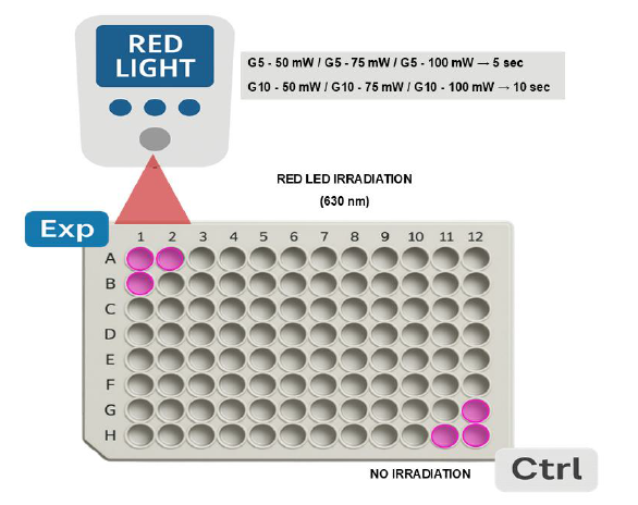
Prior to irradiation, the culture medium was replaced with 1 mL of phosphate-buffered saline (PBS) to minimize light absorption by medium components. Irradiation was conducted under controlled lighting and temperature conditions using a LINCE device (MMOptics, São Carlos, Brazil) with a red LED probe (λ = 630 nm, beam area = 9.6 cm²), operating in continuous mode. The plates remained sealed, and the light beam was positioned perpendicular to the well surface to ensure uniform energy delivery. After irradiation, PBS was replaced with complete RPMI medium, and cells were incubated for an additional 24 hours. To avoid accidental exposure, control groups were placed in the lower-right quadrant of the plate, away from the irradiation source, while experimental groups were placed in the upper-left quadrant (Fig. 1).
Experimental design for photobiomodulation of the murine fibroblast cell line L929 cultured in a 96-well microplate. Cells were exposed to red LED light (630 nm; LINCE/MMOptics, Sao Carlos, Brazil) for 5 s (G5-50, G5-75, G5-100) or 10 s (G10-50, G10-75, G10-100) at power outputs of 50, 75, and 100 mW. Non-irradiated control wells (CG/Ctrl) were maintained under the same conditions without light exposure. Pink-colored wells represent experimental and control replicates used for analysis.
Cell viability was assessed using the MTT assay (3-(4,5-dimethylthiazol-2-yl)-2,5-diphenyltetrazolium bromide; Sigma-Aldrich, St. Louis, MO, USA). The MTT solution (5mg/mL in PBS) was sterilized using a 0.22µm Millipore filter. After visual inspection under an inverted microscope, 20 µL of the MTT solution was added to each well of the 96-well plate. Plates were incubated for six hours at 37 °C. Subsequently, 60 µL of 10% sodium dodecyl sulfate (SDS) was added to solubilize the formazan crystals. Optical density was measured at 550 nm using a microplate reader at 24, 48, and 72 hours of incubation.
Nitric oxide (NO) production was quantified by measuring nitrite levels in the culture supernatant using the Griess reagent (Sigma-Aldrich). Fifty microliters of supernatant were transferred to a new 96-well plate and mixed with 50 µL of Griess reagent (0.05% N-1-naphthyl ethylenediamine dihydrochloride and 0.5% sulfanilamide in 2.5% phosphoric acid). After a 10-minute incubation at room temperature in the dark, absorbance was measured at 540 nm using a Synergy H1 microplate reader (BioTek). Nitrite concentrations were calculated based on a standard sodium nitrite curve (1.5625 to 100 µM). All measurements were performed in triplicate and expressed as a percentage relative to the control group (Pansani et al., 2015)
Collagen synthesis was evaluated using the same cell culture and irradiation protocol. After irradiation, PBS was replaced with serum-free medium, and plates were incubated for seven days without medium replacement. The experiment was conducted in triplicate for all groups. After incubation, culture media were collected and stored at −20 °C. For quantification, 500 µL of each sample was mixed with 500 µL of Direct Red 80 solution (0.1% in saturated picric acid) and incubated at 25 °C for one hour under constant agitation (400 rpm). Samples were centrifuged at 12,000 rpm for 10 minutes, and the supernatant was discarded. Next, 300 µL of 0.01 M HCl was added, followed by a second centrifugation under the same conditions. The supernatant was again removed, and the pellet was resuspended in 30 µL of 0.5 M NaOH to solubilize the bound dye. Absorbance was measured at 550nm, and results were expressed as a percentage of the mean absorbance of the control group (Barros et al., 2017).
Data normality was assessed using the Kolmogorov-Smirnov test (p < 0.05). For non-normally distributed data, the Kruskal-Wallis test was used for multiple-group comparisons, followed by the Mann-Whitney U test for pairwise comparisons, both at the 5% significance level. For normally distributed data, analysis of variance (ANOVA) was performed, followed by Tukey’s post hoc test to identify specific differences between groups.
RESULTS
A total of L929 fibroblasts were exposed to both fixed-time protocols (5 and 10 seconds) and power levels of 50, 75, and 100 mW maintained viability comparable to the control group at all evaluated time points (24, 48, and 72 hours). No statistically significant differences were observed, indicating that photobiomodulation (PBM) at these settings did not compromise cell viability.
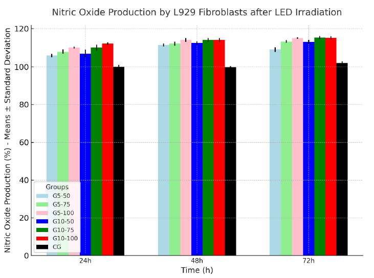
Nitric oxide (NO) production was significantly influenced by both irradiation time and power intensity. All irradiated groups showed elevated NO levels compared to the control at all time points.
At 50 mW, NO levels increased by 6% (106 ± 0.91) at 24 hours, 11% (111.29 ± 0.72) at 48 hours, and 9% (109 ± 1.07) at 72 hours, with no significant intra-group variation.
At 75 mW, levels rose by 8% (108 ± 1.00), 12% (112 ± 1.10), and 13% (113.23 ± 0.70) at the respective time points, with a significant increase between 48 and 72 hours.
At 100 mW, production increased by 10% (110 ± 0.51), 14% (114 ± 1.00), and 15% (115 ± 0.45), also showing significance between the latter time points.
Ten-second protocol:
At 50 mW, NO increased by 7% (107 ± 1.92) at 24 hours, 12% (112.40 ± 0.94) at 48 hours, and 13% (113 ± 1.07) at 72 hours, with a significant rise from 48 to 72 hours.
At 75 mW, levels rose by 10% (110 ± 1.50), 14% (114 ± 1.00), and 15% (115.06 ± 0.85), with statistically significant differences over time.
At 100 mW, values reached 12% (112 ± 0.71), 14% (114 ± 0.99), and 15% (115.06 ± 0.85), again with significant variation between 48 and 72 hours.
Complete mean values and standard deviations for nitrite production across groups and time points are presented in Table 1 and Fig. 2.
Collagen synthesis was markedly influenced by both irradiation time and power intensity. All irradiated groups exhibited significantly higher collagen levels compared to the control at all evaluated time points (24, 48, and 72 hours).

Collagen production, expressed as percentage relative to the non-irradiated control group (CG), in the murine fibroblast cell line L929 after irradiation with 630 nm red LED light. Cells were exposed for 5 s (G5) or 10 s (G10) at power outputs of 50, 75, or 100 mW. Data are presented as mean +- standard deviation. All irradiated groups showed increased collagen synthesis compared with the control group, with the highest values observed in G10-100.
Five-second protocol:
At 50 mW, collagen levels increased by 60% (160 ± 1.91) at 24 hours, 62% (161.98 ± 1.80) at 48 hours, and 65% (165 ± 1.00) at 72 hours, with no significant variation over time.
At 75 mW, values rose by 69% (169 ± 1.40), 72% (172 ± 1.18), and 71% (171 ± 1.90), also without significant intra-group differences.
At 100 mW, production reached 79% (179 ± 2.51) at 24 hours, 78% (178 ± 1.00) at 48 hours, and 79% (179.06 ± 1.45) at 72 hours, remaining stable across time points.
Ten-second protocol:
At 50 mW, collagen increased by 75% (175 ± 1.00) at 24 hours, 73% (173.08 ± 1.30) at 48 hours, and 79% (179 ± 0.98) at 72 hours, showing a significant elevation at the last time point.
At 75 mW, levels rose by 89% (189 ± 1.60), 88% (188 ± 1.08), and 93% (193 ± 1.50), with significant differences observed between 48 and 72 hours.
At 100 mW, collagen synthesis peaked at 97% (197 ± 1.50) at 24 hours, 95% (195.07 ± 1.20) at 48 hours, and 100% (200.76 ± 0.25) at 72 hours, with no significant intra-group variation.
Mean values and standard deviations for all groups are presented in Table 2 and Fig. 3.
DISCUSSION
The clinical potential of photobiomodulation (PBM) for tissue regeneration is supported by extensive research highlighting its wide-ranging applications. Previous studies have demonstrated that red light affects cellular metabolism, enhances tissue repair, and has anti-inflammatory properties (Mussttaf et al., 2023; Melo et al., 2022; Wang et al. 2022). Our in vitro study contributes to this growing body of evidence by systematically examining how different irradiation parameters-specifically, power levels (50, 75, and 100mW) and exposure times (5 and 10 seconds)-affect key fibroblast responses. Notably, our results show that PBM with red light (630 nm) significantly increased nitric oxide (NO) and collagen production in L929 fibroblasts without harming cell viability at any tested time point. These findings support the concept of a therapeutic window in laboratory settings, where PBM may exert biostimulatory effects without inducing cellular stress or toxicity. The lack of cytotoxicity, even at the highest dose tested (100mW for 10 seconds), indicates that these parameters remain within a safe and effective range in vitro.
Nitric oxide production, expressed as nitrite percentage relative to the non-irradiated control group (CG), in the murine fibroblast cell line L929 after irradiation with 630 nm red LED light. Cells were exposed for 5 s (G5) or 10 s (G10) at power outputs of 50, 75, or 100 mW. Data are presented as mean +- standard deviation. All irradiated groups showed higher nitrite levels than the control group, with the greatest responses observed in the 10 s and higher-power protocols.
The increase in NO production aligns with prior reports suggesting that PBM enhances vasodilation, tissue oxygenation, and cellular signaling pathways involved in healing (Zhang et al., 2023; Chung et al., 2012). Importantly, NO production followed a dose-dependent pattern, with higher power levels and longer irradiation times producing greater responses. These findings are consistent with the results of Chung et al. (2012) emphasizing the importance of precise dosimetry for achieving optimal cellular outcomes. Similarly, the significant upregulation of collagen synthesis highlights the potential of 630 nm light to promote extracellular matrix remodeling. This is especially relevant in wound healing and tissue engineering, where collagen organization and quantity directly influence functional recovery. Studies by Zhang et al. (2023) support these findings, reporting improved collagen deposition and fiber orientation with red-light PBM. Furthermore, PBM’s anti-inflammatory capacity-well documented by Zhevago and Samoilova (2006) enhances its potential clinical relevance, particularly in managing chronic wounds or conditions characterized by excessive inflammation. However, such clinical applications still need to be confirmed in vivo.
We acknowledge the limitations of our methods. While the MTT assay and the Griess method provide useful insights, more advanced techniques such as flow cytometry, NO-specific electrodes, and immunodetection of collagen subtypes could deepen understanding of the underlying mechanisms. Future research should incorporate these tools to clarify specific signaling pathways in PBM-mediated regeneration. Lastly, emerging strategies that combine PBM with stem cell therapies hold promise for regenerative medicine. Recent findings by Oyebode and Houreld (2022). Highlight the synergistic effects of these approaches, which could enhance future therapeutic outcomes, pending further validation in preclinical and clinical studies.
CONCLUSION
This study demonstrates that red light photobiomodulation (630 nm), applied at 50 to 100 mW for 5 to 10 seconds, safely enhances nitric oxide production and collagen synthesis in L929 fibroblasts without compromising cell viability. These in vitro findings reinforce the existence of a dose-dependent therapeutic window and provide a valuable foundation for future translational protocols in regenerative medicine. Further in vivo and clinical investigations are needed to validate these effects under physiological conditions and optimize PBM-based therapeutic strategies
REFERENCES
- AVCI, P.; GUPTA, A.; SADASIVAM, M. et al Low-level laser (light) therapy (LLLT) in skin: stimulating, healing, restoring. Semin. Cutan. Med. Surg., v.32, p.41-52, 2013.
- BARROS, T.P.; TATINI F, DE SIENA G. et al Quantitative assessment of collagen and elastic fibers in healing skin wounds treated with low-level laser therapy. Lasers Med. Sci., v.32, p.1033-1041, 2017.
- CHELLINI, F.; TANI, A.; ZECCHI-ORLANDINI, S. et al In vitro evidence of different fibroblast morpho-functional responses to red, near-infrared and violet-blue photobiomodulation: clues for addressing wound healing. Appl. Sci., v.10, p.5218, 2020.
- CHUNG, H.; DAI, T.; SHARMA, S. K. et al The nuts and bolts of low-level laser (light) therapy. Ann. Biomed. Eng., v.40, p.516-533, 2012.
- HAMBLIN, M.R. Photobiomodulation or low-level laser therapy. J. Biophotonics, v.9, p.1122-1124, 2016.
- KOCHEROVA, I.; BRYJA, A.; BŁOCHOWIAK, K. et al Photobiomodulation with red and near-infrared light improves viability and modulates expression of mesenchymal and apoptotic-related markers in human gingival fibroblasts. Materials, v.14, p.3427, 2021.
- MAGNI, G.; TATINI, F.; SIENA, G. et al Blue-LED-light photobiomodulation of inflammatory responses and new tissue formation in mouse-skin wounds. Life, v.12, p.1564, 2022.
- MELO, W.W.P.; ARAGÃO, W.A.B.; BAIA-DA-SILVA, D.C. et al Effects of photobiomodulation on oral mucositis: visualization and analysis of knowledge. Life, v.12, p.1940, 2022.
- MOSCA, R.C.; SANTOS, S.N.; NOGUEIRA, G.E.C. et al The efficacy of photobiomodulation therapy in improving tissue resilience and healing of radiation skin damage. Photonics, v.9, p.10, 2021.
- MUSSTTAF, R.A.; AL-SAHLANEE, M.H.R.; DAWOOD, N.S. et al Photobiomodulation effect of 850 nm near-infrared diode laser on the release of M1-related cytokines in monocytic THP-1 cells. J. Bioautom., v.27, p.5-14, 2023.
- PANSANI, T.S.A.; OLIVEIRA, R.C.; SANTOS, M.F. et al Platelet-rich plasma in bone repair: posttranslational histological and immunohistochemical analysis. J. Appl. Oral Sci., v.23, p.280-289, 2015. ~
- OYEBODE, O.A.; HOURELD, N.N. Photobiomodulation at 830nm stimulates migration, survival and proliferation of diabetic wounded fibroblast cells via TGF-β/Smad signaling pathway. Diabetes Metab. Syndr. Obes, v.15, p.2885-2900, 2022.
- ROSSI, F.; MAGNI, G.; TATINI, F. et al Photobiomodulation of human fibroblasts and keratinocytes with blue light: implications in wound healing. Biomedicines, v.9, p.41, 2021.
- SANTIAGO, R.; GOMES, S.; OZSARFATI, J.; ZITNEY, M. Photobiomodulation for neuropathic pain modulation and scar tissue improvement. Scars Burns Healing, v.8, p.20, 2022.
- SILVEIRA, P.C.; STRECK, E.L.; PINHO, R.A. Evaluation of mitochondrial respiratory chain activity in wound healing by low-level laser therapy. J. Photochem. Photobiol. B, v.86, p.279-282, 2007.
- WANG, Y.; HUANG, Y.; WANG, Y. et al The safety of red light (633 ± 6 nm) on human dermal fibroblasts: no genotoxic effects observed. Photodiagnosis Photodyn. Therap., v.38, p.102804, 2022.
- ZHANG, W.; ZHANG, J.; WANG, W. et al Photobiomodulation at 660 nm promotes collagen synthesis via downregulation of HIF-1α expression without photodamage in human scleral fibroblasts under hypoxic conditions. Photobiomodul. Photomed. Laser Surg., v.41, p.235-244, 2023.
- ZHEVAGO, N.A.; SAMOILOVA, K.A. Pro- and anti-inflammatory cytokine content in human peripheral blood after its transcutaneous (in vivo) and direct (in vitro) irradiation with polychromatic visible and infrared light. Photomed. Laser Surg, v.24, p.129-139, 2006.
- ZHAO, H.; JI, T.; SUN, T.; LIU, H. et al Comparative study on photobiomodulation between 630 nm and 810 nm LED in diabetic wound healing both in vitro and in vivo J. Innov. Opt. Health Sci, v.15, p.2250010, 2022.
The research data are available within the article itself.