Natural infection by Rangelia vitalii pestana, 1910, in dog from the state of Minas Gerais, Brazil - a case report
ABSTRACT
Rangeliosis is a severe, emerging hemoparasitic disease affecting dogs in South America, caused by the protozoan Rangelia vitalii and transmitted by Amblyomma aureolatum, an ixodid tick found in the Atlantic Forest biome. Although cases are commonly reported in southern Brazil and in southeastern states such as São Paulo and Rio de Janeiro, reports from the state of Minas Gerais remain scarce. This study describes a natural infection by R. vitalii in a dog from the metropolitan region of Belo Horizonte, Minas Gerais, Brazil. Peripheral blood smears revealed intra- and extra-erythrocytic piroplasms. Molecular testing detected DNA from the order Piroplasmida (18S rRNA and hsp70 genes). Genetic sequencing of the amplified piroplasmid fragments revealed 99.83% similarity with R. vitalii. This is the first study to report the ante-mortem molecular diagnosis of R. vitalii in a dog, along with clinical treatment and recovery, in the state of Minas Gerais, Brazil.
Keywords:
hemopathogens; hemoparasites; epidemiology; canids
RESUMO
A rangeliose é uma doença hemoparasitária grave e emergente, que afeta cães na América do Sul, causada pelo protozoário Rangelia vitalii e transmitida pelo carrapato ixodídeo Amblyomma aureolatum, encontrado no bioma da Mata Atlântica. Casos são comumente relatados na região Sul do Brasil e em estados do Sudeste, como São Paulo e Rio de Janeiro, enquanto em Minas Gerais os relatos ainda são escassos. Este estudo descreve uma infecção natural por R. vitalii em um cão da região metropolitana de Belo Horizonte, Minas Gerais, Brasil. Esfregaços de sangue periférico revelaram piroplasmas intra e extraeritrocitários. Testes moleculares detectaram DNA da ordem Piroplasmida (genes 18S rRNA e hsp70). O sequenciamento genético dos fragmentos amplificados revelou 99,83% de similaridade com R. vitalii. Este é o primeiro estudo a relatar o diagnóstico molecular ante mortem de R. vitalii em um cão, juntamente com o tratamento clínico e a recuperação, no estado de Minas Gerais, Brasil.
Palavras-chave:
hemopatógenos; hemoparasitas; epidemiologia; canídeos
INTRODUCTION
Rangeliosis is a hemoparasitic disease caused by the protozoan Rangelia vitalii, a piroplasm capable of parasitizing erythrocytes, neutrophils, monocytes, and endothelial cells of both wild and domestic canids (Fighera et al., 2010; Carniel et al., 2025). The ixodid tick Amblyomma aureolatum, the vector of R. vitalii, is found in South America-in countries such as Brazil, Argentina, Uruguay, Paraguay, French Guiana, and Suriname-with its occurrence predominantly recorded in higher-altitude regions of the Atlantic Forest biome (Soares, 2018; Borrás et al., 2020). For this reason, rangeliosis has only been reported on this continent, mainly in the cooler regions of southern Brazil (Oliveira et al., 2023).
In southeastern Brazil, cases have also been reported, primarily in the states of São Paulo (Silva et al., 2019b) and Rio de Janeiro (Palmer et al., 2024). Although the prevalence and geographic distribution of the disease are well established, sporadic cases have been documented in other regions of the country (Silveira et al., 2016). This suggests that, like other vector-borne pathogens, R. vitalii may be considered a re-emerging disease, expanding into non-endemic areas and remaining underdiagnosed (Soares et al., 2018).
In this context, the objective of this study is to describe the molecular diagnosis of rangeliosis in a naturally infected female dog from the state of Minas Gerais, Brazil, a non-endemic region.
ETHICAL ASPECTS
This study was not submitted to the Animal Use Ethics Committee (CEUA), as it consists of a clinical case report without experimental procedures. The animal described herein underwent only the diagnostic and therapeutic interventions clinically indicated for its condition, with no additional procedures or modifications to clinical management performed for the purposes of this study.
CASUISTRY
A six-year-old, 11.5-kg female French Bulldog presented with a history of inappetence, vomiting, diarrhea, and hematuria. The owner reported that, seven days prior to the consultation, the dog had accessed a forested area in the municipality of Nova Lima, Minas Gerais, Brazil. There was no history of ectoparasite infestation or travel. On physical examination, the dog was lethargic, with an adequate body condition score, mildly icteric mucous membranes, splenomegaly, hypothermia, and dehydration.
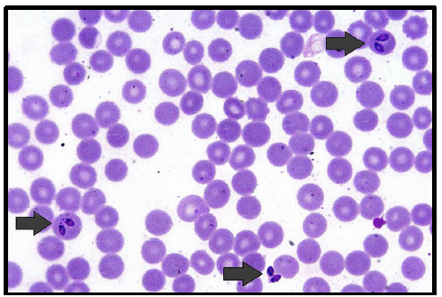
Hematological analysis revealed normocytic normochromic anemia and thrombocytopenia. Peripheral blood smears showed intraerythrocytic structures suggestive of Babesia spp., with an estimated parasitemia rate of 0.48% (Fig. 1). Serum biochemistry revealed hypoglobulinemia and increased urea and AST levels.
Blood smears from a female dog naturally infected with Rangelia vitalii. Intra- and extraerythrocytic piroplasm merozoites morphologically suggestive of Babesia spp. (red arrows). Hematoxylin and eosin stain, 100× objective.
Blood samples were submitted to the Protozoology Laboratory for molecular diagnosis of hemopathogens. DNA was extracted from 300 µL of whole blood using the Wizard® DNA extraction kit (Promega, USA). To verify the quality of the extraction, DNA integrity, and the presence of possible reaction inhibitors, the samples were tested for amplification of the gene encoding glyceraldehyde-3-phosphate dehydrogenase (gapdh) (Birkenheuer et al., 2003). Assays were performed for the detection of agents belonging to the order Piroplasmida/Hepatozoon spp., Ehrlichia spp., Anaplasma spp., Leishmania spp., and hemotropic Mycoplasma.
All primers, nucleotide sequences, target genes, fragment sizes, and cycling conditions used in this study are detailed in the works cited in Table 1. Ultrapure sterile water (Life Technologies®, USA) was used as a negative control, and DNA from samples of naturally infected hosts, previously confirmed by PCR and genetic sequencing (Castillo et al., 2024), was used as a positive control.
The sample tested positive for the gapdh gene, and subsequent molecular assays detected the presence of DNA from the order Piroplasmida (18S rRNA and hsp70) and hemotropic Mycoplasma. To confirm the identity of the piroplasm, the amplified product (hsp70 gene) was purified using the QIAquick PCR Purification Kit (Qiagen Biotechnology, Brazil) and sequenced (Sanger et al., 1977) on an ABI 3130 Genetic Analyzer (Applied Biosystems, USA). Sequencing was performed at the Oswaldo Cruz Foundation (Fiocruz) using the BigDye® Direct Cycle Sequencing Kit v3.1 (Applied Biosystems) with the same primers employed in the PCR and the POP-7™ polymer.
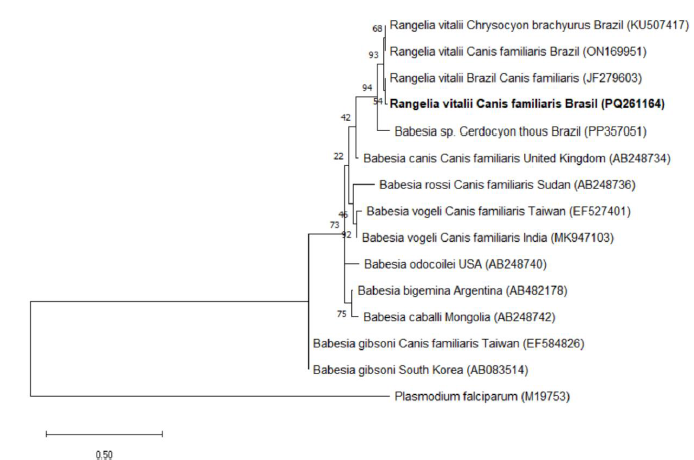
The obtained sequence (GenBank submission number: PQ261164) exhibited 99.83% identity and an E-value of 0 with R. vitalii detected in domestic dogs (Canis familiaris; JF279603) and wild canids (Cerdocyon thous: PP357051; Chrysocyon brachyurus: KU507417) sampled in Brazil.
A phylogenetic tree was constructed using MEGA (Molecular Evolutionary Genetics Analysis) version 11. Sequence alignment was performed with the ClustalW algorithm, incorporating reference sequences retrieved from GenBank. The evolutionary model was selected based on the lowest Bayesian Information Criterion (BIC) and corrected Akaike Information Criterion (AICc) values. The Maximum Likelihood (ML) method, with 1,000 bootstrap replicates, was employed (Fig. 2).

Phylogenetic tree based on a 614-bp alignment of the hsp70 gene of Piroplasmida, encompassing 21 nucleotide sequences, constructed using the Maximum Likelihood (ML) method and the K2+G+I evolutionary model. Bootstrap values from 1,000 replicates are indicated at the nodes; values below 50 are not shown. The scale bar represents evolutionary distance. The sequence detected in the present study is highlighted in bold, with the GenBank accession number in parentheses. Plasmodium falciparum was used as the outgroup.
Supportive therapy was instituted in combination with the administration of imidocarb dipropionate (6mg/kg, subcutaneously), with a repeat dose after 15 days. Complete resolution of clinical signs and hematological abnormalities was observed 49 days after hospital admission.
DISCUSSION
In Brazil, R. vitalii infection in dogs has been reported in the states of Rio Grande do Sul (Lorenzo et al., 2021), Paraná (Silva et al., 2019a), Santa Catarina (Carniel et al., 2025), São Paulo (Batista et al., 2023), and Rio de Janeiro (Palmer et al., 2024). Reports from the state of Minas Gerais are scarce. Moreira et al. (2013) described a post-mortem diagnosis of rangeliosis in a dog from the municipality of Caeté; however, the animal’s history was limited. In another report, Silveira et al. (2016) presented post-mortem findings of the disease in a free-ranging maned wolf (Chrysocyon brachyurus) rescued from a peri-urban area in the municipality of Rio Acima. These data, together with the case reported here, highlight the potential underdiagnosis of this hemopathogen in Minas Gerais, where the presence of the vector A. aureolatum is recognized (Rodrigues et al., 2002). Notably, the municipality of Nova Lima, where the patient resided, is only 17 km from Rio Acima and 45 km from Caeté, all located within the metropolitan region of Belo Horizonte. This area represents a transition zone between the Cerrado and Atlantic Forest biomes, with an average altitude of 722 m and peaks reaching 1,583 m (Werneck et al., 2010). Importantly, in the present case, there was no history of travel to endemic areas, suggesting a possible autochthonous infection.
Rangeliosis is frequently reported in mixed-breed dogs from peri-urban or rural environments. In the present case, the patient, a French Bulldog, lived in an apartment but had a history of access to areas with native vegetation, which may represent a risk factor for the disease (Silva et al., 2019a; Palmer et al., 2024). Soares et al. (2018) demonstrated that exposure to forested areas and potential contact with wild animals can contribute to R. vitalii infection in dogs. In this context, it is important to emphasize that unplanned urban expansion may facilitate the emergence of wildlife-associated diseases, such as rangeliosis, by increasing contact between humans, domestic animals, wildlife, and vectors.
Although the owner denied tick infestation, certain nonspecific clinical signs presented by the patient reinforced the suspicion of infection by ixodid-transmitted hemopathogens, allowing the establishment of a diagnostic plan (Carniel et al., 2025). Coinfection with the hemoparasites R. vitalii and Mycoplasma spp., as observed in the present case, may exacerbate the clinical condition, intensifying anemia, thrombocytopenia, and systemic signs.
Compared to other hemopathogens affecting dogs, R. vitalii is recognized for causing severe disease (Soares et al., 2014). The severity of the clinical presentation and the opportunity to discuss the case with parasitologists in an academic setting were crucial for including rangeliosis as a differential diagnosis. This highlights the need to disseminate information about the disease even in areas considered non-endemic, facilitating appropriate treatment of affected animals and supporting animal health surveillance actions.
Blood smears are valuable tools for confirming the presence of piroplasm. In the case of R. vitalii, intracellular forms can be observed five days post-exposure, with a progressive increase in parasitemia until the eleventh day (Paim et al., 2012). In the present study, the hemoprotozoan was microscopically identified as intra- and extraerythrocytic piriform parasitic forms. However, according to Carniel et al. (2025), Rangelia vitalii is observed in blood smears of only approximately 61,1% of naturally infected dogs.
Blood smear evaluations may be insufficient for the diagnosis of rangeliosis. This is because R. vitalii is genetically related to hemoprotozoans of the genus Babesia, with which it shares similar morphology under light microscopy (Soares et al., 2011). Distinction between R. vitalii and Babesia species with merozoites >2.5 μm is only possible microscopically when the protozoan is identified in non-erythrocytic cells, such as leukocytes or endothelial cells, which was not observed in the present case.
In the present study, the diagnosis of rangeliosis was only possible through molecular analysis followed by nucleotide sequencing. Phylogenetic analysis placed the obtained sequence within a well-supported clade (bootstrap = 94) alongside other R. vitalii strains previously identified in C. familiaris, C. brachyurus, and C. thous from Brazil. This highly supported grouping suggests a close evolutionary relationship among the isolates, regardless of the domestic or wild origin of the hosts. Such a phylogenetic pattern reinforces the hypothesis of potential ecological interactions between domestic dogs and wild canids, facilitating vector sharing and, consequently, bidirectional pathogen flow between distinct populations.
CONCLUSION
Rangeliosis should be considered in the differential diagnosis of dogs presenting with anemia, jaundice, and fever in the state of Minas Gerais, Brazil, where the disease is not yet recognized as endemic despite favorable conditions for the tick vector. This report describes the first confirmed antemortem, autochthonous diagnosis of rangeliosis in a dog from this state, supporting the hypothesis of either geographic expansion of the parasite or underdiagnosis of the infection in the region.
REFERENCES
- BATISTA, T.F.; CORTEZ, A.; LABRUNA, M.B. et al Dogs naturally infected by Rangelia vitalii, Babesia canis vogeli, and Ehrlichia canis in São Paulo, Brazil. Braz. J. Vet. Res. Anim. Sci., v.60, p.1-5, 2023.
- BIRKENHEUER, A.D.; LEVY, M.G.; BREITSCHWERDT, E.B. Development and evaluation of a seminested PCR for detection and differentiation of Babesia gibsoni (Asian genotype) and Babesia canis vogeli J. Clin. Microbiol. , v.41, p.4172-4177, 2003.
- BORRÁS, P.; SALVADOR, F.; RINALDI, V. et al Use of molecular tools for the diagnosis of rangeliosis by Rangelia vitalii in Argentina: a case report. Vet. Parasitol. Reg. Stud. Rep., v.21, p.e100426, 2020.
- CARNIEL, F.; PANDOLFO, G.W.; FERIAN, P.E. et al Rangelia vitalii in naturally infected dogs in southern Brazil: clinical classification of the disease into acute and subacute phases. Vet. Parasitol. Reg. Stud. Rep., v.64, p.e101333, 2025.
- CASTILLO, A.P.; COLÁCIO, N.; RODRIGUES, P.H.C. et al Parasitic protozoa and other vector-borne pathogens in captive mammals from Brazil. J. Zool. Bot. Gardens, v.5, p.754-773, 2024.
- FIGHERA, R.A.; SOUZA, T.M.; KOMMERS, G.G. et al Patogênese e achados clínicos, hematológicos e anatomopatológicos da infecção por Rangelia vitalii em 35 cães (1985-2009). Pesqui. Vet. Bras. , v.30, p.974-987, 2010..
- KAWAHARA, M.; TAJIMA, T.; TORII, H. et al. Ehrlichia chaffeensis infection of sika deer, Japan. Emerg. Infect. Dis, v.15, p.1991-1993, 2009.
- LORENZO, C.; BIANCHI, M.V.; EHLERS, L.P. et al Rangelia vitalii molecular and histological quantification in tissues comparing crab-eating foxes (Cerdocyon thous) and domestic dogs. Ticks Tick Borne Dis, v.12, p.e101731, 2021.
- MASSUNG, R.F.; SLATER, K.; OWENS, J.H. et al. Nested PCR assay for detection of granulocytic ehrlichiae. J. Clin. Microbiol, v.36, p.1090-1095, 1998.
- MOREIRA, L.; GUIMARÃES, L.B.; SILVA, J.F. et al Infecção por Rangelia vitalii em um cão em Minas Gerais. Arch. Vet. Sci, v.18, p.3, 2013.
- OLIVEIRA, Á.F.X.; CALCHI, A.C.; STOCCO, A.V. et al Expanding the universe of piroplasmids: morphological detection and phylogenetic positioning of putative novel piroplasmids in black-eared opossums (Didelphis aurita) from southeastern Brazil, with description of “South American Marsupialia Group” of Piroplasmida. Parasitol. Res., v.122, p.1519-1530, 2023.
- PAIM, C.B.; PAIM, F.C.; SILVA, A.S. et al Thrombocytopenia and platelet activity in dogs experimentally infected with Rangelia vitalii. Vet. Parasitol, v.185, p.131-137, 2012.
- PALMER, J.P.S.; GAZÊTA, G.S.; ANDRÉ, M.R. et al Piroplasmid infections among domestic dogs in the mountain city of Rio de Janeiro, Brazil. Acta Parasitol, v.69, p.1172-1191, 2024.
- RODRIGUES, D.S.; CARVALHO, H.A.; FERNANDES, A.A. et al Biology of Amblyomma aureolatum (Pallas, 1772) (Acari: Ixodidae) on some laboratory hosts in Brazil. Memórias Inst. Oswaldo Cruz, v.97, p.853-856, 2002.
- SANGER, F.; NICKLEN, S.; COULSON, A.R. DNA sequencing with chain-terminating inhibitors. Proc. Natl Acad. Sci. USA, v.74, p.5463-5467, 1977.
- SILVA, B.R.D.; FERREIRA, M.F.K.; MAFFEZZOLLI, G. et al Detection molecular of Rangelia vitalii in dogs from Paraná State, southern Brazil. Rev. Bras. Parasitol. Vet, v.28, p.310-313, 2019a.
- SILVA, B.R.F.; LABRUNA, M.B.; MARCILI, A. et al Rangelia vitalii infection in a dog from São Paulo city, Brazil: case report. Braz. J. Vet. Res. Anim. Sci, v.56, p.e150791, 2019b.
- SILVEIRA, J.A.; RABELO, E.M.; RIBEIRO, M.F. Detection of Theileria and Babesia in brown brocket deer (Mazama gouazoubira) and marsh deer (Blastocerus dichotomus) in the State of Minas Gerais, Brazil. Vet. Parasitol, v.177, p.61-66, 2011.
- SILVEIRA, J.A.G.; D’ELIA, M.L.; AVELAR, I.O. et al Rangelia vitalii in a free-ranging maned wolf (Chrysocyon brachyurus) and co-infections. Int. J. Parasitol. Parasite. Wildl, v.5, p.280-285, 2016.
- SOARES, J.F.; COSTA, F.B.; GIROTTO-SOARES, A. et al Evaluation of the vector competence of six ixodid tick species for Rangelia vitalii (Apicomplexa, Piroplasmorida), the agent of canine rangeliosis. Ticks Tick Borne Dis, v.9, p. 1221-1234, 2018.
- SOARES, J.F.; DALL’AGNOL, B.; COSTA, F.B. et al Natural infection of the wild canid, Cerdocyon thous, with the piroplasmid Rangelia vitalii in Brazil. Vet. Parasitol., v.202, p.156-163, 2014.
- SOARES, J.F.; GIROTTO, A.; BRANDÃO, P.E. et al Detection and molecular characterization of a canine piroplasm from Brazil. Vet. Parasitol, v.180, p.203-208, 2011.
- WERNECK, M.S.; REZENDE, S.G.; BRINA, A.E.; FRANCESCHINELLI, E.V. Composição florística do componente arbóreo e afinidade fitogeográfica de uma floresta semidecídua em Nova Lima, MG. Rev. Bras. Bot, v.33, p.547-561, 2010.
- ZAHLER, M.; RINDER, H.; SCHEIN, E.; GOTHE, R. Detection of a new pathogenic Babesia microti-like species in dogs. Vet. Parasitol, v.89, p.241-248, 2000.
-
FUNDING
This study was financed by scholarships from Coordenação de Aperfeiçoamento de Pessoal de Nível Superior (CAPES), financial support for the field, lab work and Research productivity grant from Conselho Nacional de Desenvolvimento Científico e Tecnológico (CNPq) (315466/2021-9; CNPq/SECTICS/ CAPES/FAPs no 46/2024 and 308527/2025-9), Fundação de Amparo à Pesquisa do Estado de Minas Gerais (FAPEMIG) (APQ-02531-24).
-
DATA AVAILABILITY STATEMENT
The research data are available upon request.
The research data are available upon request.