
Abstracts
Purpose:
We reported on the clinical approaches of ophthalmology and otorhinolaryngology departments in the treatment of the orbital complications of sinusitis. We also included an in-depth literature review.
Methods:
We retrospectively reviewed the medical files of 51 patients from January 2008 to January 2014. The records were evaluated for age, gender, type of orbital complications, symptoms, predisposing factors, imaging studies, medical and surgical management, culture results, and follow-up information. SPSS version 15.0 software (Statistical Analysis, The Statistical Package for Social Sciences Inc, Chicago, IL) was used for the statistical analysis.
Results:
Fifty-one patients met the criteria, with available medical records, for the study (29 male, 22 female). Thirty-two (62.7%) were diagnosed with preseptal cellulitis and 19 (37.3%) with postseptal cellulitis. After a detailed evaluation, 15 were diagnosed with a subperiosteal abscess (SPA), and 4 were diagnosed with orbital cellulitis. The age and gender was similar for the two groups. Five patients with medial SPA were treated with endoscopic sinus surgery, one patient with inferior SPA was treated with external surgery, and six patients with other localizations were treated with a combination of endoscopic sinus surgery and external surgery. All patients presented with periorbital erythema and edema. The length of hospitalization and duration of symptoms were similar in both groups. Visual acuity was between 1/10 to 10/10 (mean 7/10) and statistically significant for preseptal and postseptal cellulitis groups (p<0.001). All patients received intravenous antibiotics upon the first day of admission.
Conclusion:
Orbital complications of acute sinusitis required intensive follow-up and a multidisciplinary approach. A contrast-enhanced paranasal sinus computerized tomography (CT) scan can detect the extent of the infection. An initial trial of intravenosus (IV) antibiotics may be appropriate when close monitoring is possible. Surgery may be indicated when there has been no improvement within 48 hours of intravenous treatment, loss of visual acuity (under 8/10), and a non-medial abscess.
Sinusitis/complications; Orbital diseases/etiology; Abscess
Objetivo:
Relatamos abordagens clínicas dos departamentos de oftalmologia e otorrinolaringologia para tratar complicações orbitais da sinusite. Uma revisão em profundidade literatura é discutida.
Métodos:
Foram analisados retrospectivamente os prontuários de 51 pacientes no período de janeiro de 2008 a janeiro de 2014. Os registros foram avaliados quanto à idade, sexo, tipo de complicação orbital, sintomas, fatores predisponentes, estudos de imagem, tratamento médico e cirúrgico, resultados da cultura microbiológica e seguimento. Foi utilizado o programa SPSS versão 15.0 (Statistical Analysis, The Statistical Package for Social Sciences Inc, Chicago, IL) para a análise estatística.
Resultados:
Cinquenta e um pacientes preencheram os critérios, com os registros médicos disponíveis, para o estudo (29 do sexo masculino, 22 do sexo feminino). Trinta e dois (62,7%) foram diagnosticados com celulite presseptal e 19 (37,3%), com celulite posseptal. Depois de uma avaliação detalhada, 15 foram diagnosticados como abscesso subperiosteal (SPA), 4 eram celulite orbitária. A idade e sexo foi similar para ambos os grupos. Cinco pacientes com abscesso subperiosteal medial foram tratados com cirurgia endoscópica, um paciente com abscessso subperiosteal inferior foi tratado com cirurgia externa, 6 pacientes com outras localizações foram tratados com a combinação de cirurgia endoscópica e cirurgia externo. Todos os pacientes apresentaram eritema e edema periorbital. Tempo de internação hospitalar e a duração dos sintomas foi similar em ambos os grupos. A acuidade visual foi entre 1/10 a 10/10 (média de 7/10) e estatisticamente significante para os grupos celulite presseptal e posseptal (p<0,001). Todos os pacientes receberam antibióticos por via intravenosa, no primeiro dia de admissão.
Conclusão:
A complicação orbital da sinusite aguda exige um acompanhamento intensivo e multidisciplinar. A tomografia computadorizada dos seios paranasais com contraste pode detectar a extensão da infecção. Uma tentativa inicial com o uso de antibióticos intravenosos é adequada quando um acompanhamento rigoroso é possível. A cirurgia é indicada quando não houver melhora dentro de 48 horas de tratamento clínico, diminuição da acuidade visual (em 8/10) e abscesso não-medial.
Sinusite/complicações; Doenças orbitárias/etiologia; Abscesso
INTRODUCTION
Sinusitis is a common disease and is typically managed without complications. In some cases, infection can spread to the orbit. If left untreated, several complications of orbital cellulitis can manifest including blindness, meningitis, and even death(11 Chandler JR, Langenbrunner DJ, Stevens ER. The pathogenesis of orbital complications in acute sinusitis. Laryngoscope. 1970;80(9):1414-28. Comment in: Ann Otol Rhinol Laryngol. 1977;86(Pt 1):616-22; Laryngoscope. 1997;j107(4):441-6.). Kanra et al. reported that orbital cellulitis was caused by sinusitis in 43% of cases(22 Kanra G, Secmeer G, Gonc EN, Ceyhan M, Ecevit Z. Periorbital cellulitis: a comparison of different treatment regimens, Acta Paediatr Jpn. 1996;38(4)339-42.). Other etiologies were dacryocystitis, orbital foreign body, periocular trauma, history of surgery, odontogenic infection, endophthalmitis, orbital tumors, and local skin inflammation(22 Kanra G, Secmeer G, Gonc EN, Ceyhan M, Ecevit Z. Periorbital cellulitis: a comparison of different treatment regimens, Acta Paediatr Jpn. 1996;38(4)339-42.,33 Vairaktaris E, Moschos MM, Vassiliou S, Baltatzis S, Kalimeras E, Avgoustidis D, et al. Orbital cellulitis, orbital subperiosteal and intraorbital abscess: report of three cases and review of the literature. J Craniomaxillofac Surg. 2009;37(3):132-6.). Before the antibiotic era, 20% of patients with periorbital cellulitis had permanent loss of vision, and 17% died(11 Chandler JR, Langenbrunner DJ, Stevens ER. The pathogenesis of orbital complications in acute sinusitis. Laryngoscope. 1970;80(9):1414-28. Comment in: Ann Otol Rhinol Laryngol. 1977;86(Pt 1):616-22; Laryngoscope. 1997;j107(4):441-6.). Nowadays, the vision loss has improved (3 to 11%) and mortality rates have declined (1 to 2.5%)(44 Younis RT, Lazar RH, Bustillo A, Anand VK. Orbital infection as a complication of sinusitis: are diagnostic and treatment trends changing? Ear Nose Throat J. 2002;81(11): 771-5.). Because of the urgent situation in orbital infections, a multidisciplinary approach is needed.
The orbital septum originates from the periosteum and, arising from the anterior extension of the periosteum from the orbital margins into the eyelids, separates the superficial portion of eye (preseptal region) from the deeper orbital structures (postseptal region)(55 Soon VT. Pediatric subperiosteal orbital abscess secondary to acute sinusitis: a 5-year review. Am J Otolaryngol. 2011;32(1):62-8.).
Chandler et al. described orbital complications as five stages(11 Chandler JR, Langenbrunner DJ, Stevens ER. The pathogenesis of orbital
complications in acute sinusitis. Laryngoscope. 1970;80(9):1414-28. Comment in: Ann
Otol Rhinol Laryngol. 1977;86(Pt 1):616-22; Laryngoscope.
1997;j107(4):441-6.): preseptal cellulitis (stage I),
orbital cellulitis (stage II), subperiosteal abscess (stage III), orbital abscess (stage
IV), and cavernous sinus thrombosis (stage V). However, identifying the stages in
children with swollen and/or painful eyes can be more ambiguous. To simplify the
classification criteria, complications of the orbital septum including preseptal
cellulitis and postseptal infections have been proposed(66 Sobol SE, Marchand J, Tewfik TL, Manoukian JJ, Schloss MD. Orbital
complications of sinusitis in children. J Otolaryngol.
2002;31(3):131-6.). When sinusitis is the primary cause, infection
usually spreads from the ethmoid sinuses; however, infection can also spread through the
floor of the frontal sinus or the roof of the maxillary antrum(77 Swift AC, Charlton G. Sinusitis and the acute orbit in children. J
Laryngol Otol. 1990;104(3):213-6.
8 Wald ER, Pang D, Milmore GJ, Schramm VL Jr. Sinusitis and its
complications in paediatric patients. Paediat. Clin North Am.
1981;28(4):777-96.-99 Uzcategui N, Warman R, Smith A, Howard CW. Clinical practice guideline
for the anagement of orbital cellulitis. J Paediatr Ophhalmol Strabismus.
1998;35(2):73-9; quizz 110-1.). The orbital complications may be a result of extension of the
infection through bony defects, osteitic bone destruction, or through thrombophlebitis
of the communicating veins(1010 Suhaili DN, Goh BS, Gendeh BS. A ten year retrospective review of
orbital complications secondary to acute sinusitis in children. Med J Malaysia.
2010;65(1):49-52.).
In this study, we have discussed the clinical approaches for ophthalmology and otorhinolaryngology, and their use in treating and managing orbital complications of sinusitis. We also completed a literature review.
METHODS
We retrospectively reviewed the medical files of 51 patients who had been followed at Dicle University Medical School Department of Otorhinolaryngology and Opthalmology with a diagnosis of orbital inflammation secondary to acute sinusitis from January 2008 to January 2014. The study protocol was approved by the ethics committee. The records were evaluated for age, gender, type of orbital complication, symptoms, predisposing factors, imaging studies, medical and surgical management, culture results, and follow-up information. The diagnosis of acute sinusitis with orbital complications was based on clinical symptoms and signs as well as laboratory tests and imaging. Sinusitis was defined by the presence of sinus opacification or air-fluid levels using computerized tomography (CT) imaging. Ophthalmologic and otorhinolaryngologic examinations and CT scans were obtained on all patients. The patients were grouped according to the basic classifications of preseptal and postseptal cellulitis. Patients with evidence of a subperiosteal mass were treated with surgery (endoscopic sinus and/or external surgery) or medical therapy. In addition, patients with systemic diseases and/or edema or erythema arising from an etiology other than sinusitis were excluded from the study.
SPSS version 15.0 software (Statistical Analysis, The Statistical Package for Social Sciences Inc, Chicago, IL) was used for the statistical analysis. Descriptive statistics were used for the data, chi-square test was used for the comparison of categorical variables, and the Mann-Whitney U test was used to compare the numeric values. The results were considered significant if the p value was less than 0.05.
RESULTS
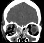
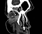
Fifty-one patients met the criteria for the study and had available medical records (29 male, 22 female). Thirty patients were treated on the otorhinolaryngology service and twenty-one patients were treated on the opthalmology service. The age range was 1 to 63 years (mean, 19.92 ± 17.98). Approximately 70% patients (n=36) were younger than 18 years of age. Thirty-two (62.7%) were diagnosed with preseptal cellulitis and 19 (37.3%) were diagnosed with postseptal cellulitis. After detailed evaluations, 15 were diagnosed with a subperiosteal abscess (SPA) and 4 with orbital cellulitis. No one had an intraorbital abscess or a cavernous sinus thrombosis. The age and gender was similar for the two groups. SPA was localized as medial (n=8) (Figure 1), superiomedial (n=4) (Figure 2), inferior (n=1) (Figure 3), and inferiomedial (n=1) (Figure 4). In the postseptal cellulitis group, bilateral pansinusitis was found in 5, unilateral pansinusitis in 6, unilateral maxillary, ethmoid, and frontal sinusitis in 5, unilateral maxillary and ethmoid sinusitis in 2, and unilateral maxillary sinusitis in 1. In the preseptal cellulitis group, bilateral pansinusitis was present in 9, unilateral pansinusitis in 10, unilateral maxillary, ethmoid, and frontal sinusitis in 4, unilateral maxillary, ethmoid sinusitis in 5, and unilateral ethmoid sinusitis in 4 patients. Five patients with medial SPA were treated with endoscopic sinus surgery, one patient with inferior SPA was treated with external surgery, and six patients with other localizations were treated with combination of endoscopic sinus surgery and external surgery. The seasonal distribution of this pathology is shown in figure 5.


The right side was involved in 27 patients and left side was involved in 24. All patients presented with periorbital erythema and edema. In postseptal cellulitis group, 13 (68.4%) patients had additional proptosis, 17 (89.4%) had limited eye movements, 15 (78.9%), and 15 (78.9%) had visual loss (under 8/10). The average length of hospitalization was 8.94 ± 3.93 (range 4 to 20 d) for all patients. The mean duration of symptoms before admission was 5.69 ± 3.19 days (range, 2 to 15 d). The length of hospitalization and duration of symptoms were similar in both groups. Visual acuity was between 1/10 to 10/10 (mean 7/10) and statistically significant for both the preseptal and postseptal celulitis groups (p<0.001). In the preseptal group, only one patient had a visual acuity less than 8/10. This patient was 63 years old and was suffering from visual loss associated with macular degeneration. All patients received intravenous antibiotics beginning on the first day of admission. The most common antibiotics used were ceftriaxone (84.3%), ornidazol (68.6%), clindamycin (13.7%), vancomycin (5.8%), meropenem (3.9%), and gentamicin (3.9%).
Multiple antibiotics were used in 72.5% of patients. Purulent material was analyzed from 9 patients and blood cultures were analyzed from 12 patients. Cultures were inoculated and bacterial growth was not observed. Patients were discharged on oral antibiotic treatment to be completed in the 2 weeks following the office visit. Other findings are presented in table 1.
DİSCUSSİON
The cause of 74% of orbital infections is sinusitis(1111 Schramm VL Jr, Curtin HD, Kennerdell JS. Evaluation of orbital cellulites and results of treatment. Laryngoscope. 1982;92(7):732-8.). Infections mostly originate from ethmoid sinuses; however, other sinuses may also be involved(1212 Noordzij JP, Harrison SE, Mason JC, Hashisaki GT, Reibel JF, Gross CW. Pitfalls in the endoscopic drainage of subperiosteal orbital abscesses secondary to sinusitis. Am J Rhinol. 2002;16(2):97-101.). In the presence of ethmoiditis, the infection spreads to the orbital region through the bony dehiscences, neurovascular foramina, or by septic thrombophlebitis of ophthalmic venous system(1212 Noordzij JP, Harrison SE, Mason JC, Hashisaki GT, Reibel JF, Gross CW. Pitfalls in the endoscopic drainage of subperiosteal orbital abscesses secondary to sinusitis. Am J Rhinol. 2002;16(2):97-101.). In our series, most of the infected sinuses were the ethmoid sinus (98%).
If left untreated, complications of orbital infections can cause blindness, meningitis, brain abscess, and death(11 Chandler JR, Langenbrunner DJ, Stevens ER. The pathogenesis of orbital complications in acute sinusitis. Laryngoscope. 1970;80(9):1414-28. Comment in: Ann Otol Rhinol Laryngol. 1977;86(Pt 1):616-22; Laryngoscope. 1997;j107(4):441-6.). Visual loss may be secondary to the elevation of the intraorbital pressure resulting in retinal ischemia or optic neuritis due to the spread of infection(33 Vairaktaris E, Moschos MM, Vassiliou S, Baltatzis S, Kalimeras E, Avgoustidis D, et al. Orbital cellulitis, orbital subperiosteal and intraorbital abscess: report of three cases and review of the literature. J Craniomaxillofac Surg. 2009;37(3):132-6.).
Medial SPA is the most common postseptal orbital complication of sinusitis(1313 Manning SC. Endoscopic management of medial subperiosteal orbital abscess. Arch Otolaryngol Head Neck Surg. 1993;119(7):789-91.). In our study, medial SPA was observed in 53.4% of the cases.
Patients with preseptal or orbital cellulitis present with similar symptoms, like
periorbital swelling and fever. In preseptal cellulitis, the patients have intact
extraocular movements and proptosis yet, visual loss is not seen. Conversely, orbital
cellulitis is pronounced by decreased extraocular movements, proptosis, and loss of
vision(1414 Donahue SP, Schwartz G. Preseptal and orbital cellulitis in childhood. A
changing microbiologic spectrum. Ophthalmology. 1998;105(10):1902-6; discussion
1905-6.
15 Noel LP, Clarke WN, MacDonald N. Clinical management of orbital
cellulitis in children. Can J Ophthalmol. 1990;25(1):11-6.-1616 Lessner A, Stern GA. Preseptal and orbital cellulitis. Infect Dis Clin
North Am. 1992; 6(4):933-52.). The distinction between preseptal
cellulitis and orbital involvement is important and can not be shown with clinical
examination alone. Rahbar et al. presented three cases of a SPA (15.7%) with periorbital
cellulitis, without any proptosis, restriction of gaze, or vision
changes(1717 Rahbar R, Robson CD, Petersen RA, DiCanzio J, Rosbe KW, McGill TJ, et
al. Management of orbital subperiosteal abscess in children. Arch Otolaryngol Head
Neck Surg. 2001;127(3):281-6.). All of the
patients in our study had eyelid edema and erythema. Other ophthalmologic manifestations
included protosis (33.4%), visual loss less than 8/10 (31.3%), and limited ocular
motility (29.4%).
The length of hospital stay in preseptal infections and postseptal infections was similar (8.28 and 10.05 d). A similar result was reported by Souliere et al. and Liu et al.(1818 Souliere CR Jr, Antoine GA, Martin MP, Blumberg AI, Isaacson G. Selective non-surgical management of subperiosteal abscess of the orbit: computerized tomography and clinical course as indication for surgical drainage. Int J Pediatr Otorhinolaryngol. 1990;19(2):109-19. Commnet in: Int J Pediatr Otorhinolaringol. 1993;25(1-3):277-8; Int J Pediatr Otorhinolaringol. 1991;21(3):277-9.-1919 Liu CH, Lin CD, Cheng YK, Lin HC, Tsai MH. Orbital complications of acute sinusitis in children. Mid Taiwan J Med. 2005;10(3):144-9.).
Streptococcus species are more frequently seen in preseptal and orbital cellulitis(2020 Schwartz GR, Wright SW. Changing bacteriology of periorbital cellulitis. Ann Emerg Med. 1996;28(6):617-20.). In an analysis of 30 patients with orbital SPA, the predominant pathogens were S. pneumoniae, S pyogenes, and H. influenza(2121 Dudin A, Othman A. Acute periorbital swelling: evaluation of management protocol. Paediatr Emerg Care 1996;12(1):16-20.). Anaerobic organisms are generally associated with chronic sinusitis(2222 Frederick J, Braude AI. Anaerobic infections of the paranasal sinusues. N Engl J Med. 1974;290(3):135-7.). In our studies, purulent material from 9 patients, and blood cultures from 12 patients were analyzed but no bacterial growth was seen. The reason may be due to the use of antibiotics before the sample cultures were taken.
According to Howe et al., a CT scan was necessary if an evaluation of the eye was not possible because of gross edema. In addition, patients with proptosis, ophthalmoplegia, impaired visual acuity, or central symptoms may require a CT scan(2323 Howe L, Jones NS. Guidelines for the management of periorbital cellulitis/abscess. Clin Otolaryngol Allied Sci. 2004;29(6):725-8.). But in some cases of an SPA, there will be no proptosis, restriction of gaze, or vision changes(1717 Rahbar R, Robson CD, Petersen RA, DiCanzio J, Rosbe KW, McGill TJ, et al. Management of orbital subperiosteal abscess in children. Arch Otolaryngol Head Neck Surg. 2001;127(3):281-6.). Therefore, close follow-up will be needed in all patients with orbital infections. We routinely obtained CT scans of all patients.
The combination of ampicillin-sulbactam or ceftriaxone-metranidazole is the advised antibiotic therapy for preseptal and orbital cellulitis(2424 American Academy of Pediatrics. Subcommittee on Management of Sinusitis and Committee on Quality Improvement. Clinical practice guideline: management of sinusitis. Pediatrics. 2001;108(3):798-808. Erratum in: Pediatrics. 2002;109(5):40; Pediatrics. 2001;108(5):A24. Comment in: Pediatrics. 2003;111(4 Pt 1):922; Pediatrics. 2002;109(3):557-8; Pediatrics. 2002;110(1 Pt 1):192-3; author reply 192-3; Pediatrics. 2002;110(1 Pt 1):195; author reply 195.). Other suggested initial intravenous antibiotics for orbital cellulitis are ampicillin(2525 Smith TF, O'Day D, Wright PF. Clinical implications of preseptal (periorbital) cellulitis in childhood. Pediatrics. 1978;62(6):1006-9.), cefuroxime(1616 Lessner A, Stern GA. Preseptal and orbital cellulitis. Infect Dis Clin North Am. 1992; 6(4):933-52.), ampicillin and nafcillin(2626 Goldberg F, Berne AS, Oski FA. Differentiation of orbital cellulitis from preseptal cellulitis by computed tomography. Pediatrics. 1978;62(6):1000-5.), ampicillin and methicillin(2525 Smith TF, O'Day D, Wright PF. Clinical implications of preseptal (periorbital) cellulitis in childhood. Pediatrics. 1978;62(6):1006-9.), ampicillin and chloramphenicol(1111 Schramm VL Jr, Curtin HD, Kennerdell JS. Evaluation of orbital cellulites and results of treatment. Laryngoscope. 1982;92(7):732-8.), vancomycin and chloramphenicol(1515 Noel LP, Clarke WN, MacDonald N. Clinical management of orbital cellulitis in children. Can J Ophthalmol. 1990;25(1):11-6.), cloxacillin and chloramphenicol(1515 Noel LP, Clarke WN, MacDonald N. Clinical management of orbital cellulitis in children. Can J Ophthalmol. 1990;25(1):11-6.), or imipenem and vancomycin(2727 Duarte Reis M, Freitas IP, Sousa Coutinho V, Guerra Rodrigo F. Facial V periorbital cellulitis with orbital involvement. J Eur Acad Dermatol Venereol. 2002;16(2):156-8.). Multiple antibiotics were used in 72.5% of our patients and the most common combination was ceftriaxone and metronidazole.
According to Oxford et al., the criteria for medical management of medial SPA were the absence of vision loss, a normal pupillary reflex, no retinal pathologies, no restricted directions of gaze, intraocular pressure <20 mmHg, proptosis of 5 mm or less, and width of 4 mm or less on CT(2828 Oxford LE, McClay J. Medical and surgical management of subperiosteal orbital abscess secondary to acute sinusitis in children. Int J Pediatr Otorhinolaryngol. 2006; 70(11):1853-61.). In our study, 3 of 15 (20%) subperiosteal abscesses (SPA) were treated with antibiotics, while others were treated with surgery. The indication for surgery in our patients was visual loss (under 4/10), and unresponsive towards antibioterapy for 48 hours.
Historically, the classic external approach for SPA is the external ethmoidectomy via a Lynch incision(2929 Fakhri S, Pereira K. Endoscopic management of orbital abscesses. Otolaryngol Clin North Am. 2006;39(5):1037-47.). In 1993, Manning reported drainage via an intranasal, endoscopic approach(1313 Manning SC. Endoscopic management of medial subperiosteal orbital abscess. Arch Otolaryngol Head Neck Surg. 1993;119(7):789-91.). Since then, many studies have confirmed the endoscopic approach as the most used method for the management of SPA(2929 Fakhri S, Pereira K. Endoscopic management of orbital abscesses. Otolaryngol Clin North Am. 2006;39(5):1037-47.). In medial or medial-inferior SPA, a transnasal endoscopic approach is possible, but in superior orbital abscess an external incision is required(33 Vairaktaris E, Moschos MM, Vassiliou S, Baltatzis S, Kalimeras E, Avgoustidis D, et al. Orbital cellulitis, orbital subperiosteal and intraorbital abscess: report of three cases and review of the literature. J Craniomaxillofac Surg. 2009;37(3):132-6.). In our study, five patients with medial SPAs were treated with endoscopic sinus surgery, one patient with an inferior SPA was treated with external surgery, and six patients with other localizations were treated with a combination of endoscopic sinus surgery and external surgery.
In the study of Devrim et al., they found an relationship between seasonal distrubition and the orbital complication of sinusitis but we did not find any relationship(3030 Devrim I, Kanra G, Kara A, Cengiz AB, Orhan M, Ceyhan M, et al. Preseptal and orbital cellulitis: 15-year experience with sulbactam ampicillin treatment. Turk J Pediatr. 2008; 50(3):214-8.).
CONCLUSION
The orbital complication of the acute sinusitis requires an intensive follow-up and multidisciplinary approach by a pediatrician, otolaryngologist, and ophthalmologist to avoid undesirable complications. The clinical findings in patients with preseptal cellulitis or orbital cellulitis/abscess do not always correlate with each other. Therefore a contrast-enhanced paranasal sinus CT scan can be useful to detect the extent of the infection through the orbit. An initial trial of IV antibiotics can be appropriate when close monitoring is possible. Loss of visual acuity (under 8/10) and non-medial abscess appear to be indicators for surgical treatment. Surgery may also indicated when there is no improvement within 48 hours of IV treatment. In medial or mediale inferior SPA, a transnasal approach is possible, but in superior and inferior orbital abscesses, an external approach is required.
-
Funding: No specific financial support was available for this study.
-
Study conducted at Dicle University.
ACKNOWLEDGEMENT
We are grateful to Dicle University DUBAP for their sponsorship for the English editing of this manuscript.
REFERENCES
-
1Chandler JR, Langenbrunner DJ, Stevens ER. The pathogenesis of orbital complications in acute sinusitis. Laryngoscope. 1970;80(9):1414-28. Comment in: Ann Otol Rhinol Laryngol. 1977;86(Pt 1):616-22; Laryngoscope. 1997;j107(4):441-6.
-
2Kanra G, Secmeer G, Gonc EN, Ceyhan M, Ecevit Z. Periorbital cellulitis: a comparison of different treatment regimens, Acta Paediatr Jpn. 1996;38(4)339-42.
-
3Vairaktaris E, Moschos MM, Vassiliou S, Baltatzis S, Kalimeras E, Avgoustidis D, et al. Orbital cellulitis, orbital subperiosteal and intraorbital abscess: report of three cases and review of the literature. J Craniomaxillofac Surg. 2009;37(3):132-6.
-
4Younis RT, Lazar RH, Bustillo A, Anand VK. Orbital infection as a complication of sinusitis: are diagnostic and treatment trends changing? Ear Nose Throat J. 2002;81(11): 771-5.
-
5Soon VT. Pediatric subperiosteal orbital abscess secondary to acute sinusitis: a 5-year review. Am J Otolaryngol. 2011;32(1):62-8.
-
6Sobol SE, Marchand J, Tewfik TL, Manoukian JJ, Schloss MD. Orbital complications of sinusitis in children. J Otolaryngol. 2002;31(3):131-6.
-
7Swift AC, Charlton G. Sinusitis and the acute orbit in children. J Laryngol Otol. 1990;104(3):213-6.
-
8Wald ER, Pang D, Milmore GJ, Schramm VL Jr. Sinusitis and its complications in paediatric patients. Paediat. Clin North Am. 1981;28(4):777-96.
-
9Uzcategui N, Warman R, Smith A, Howard CW. Clinical practice guideline for the anagement of orbital cellulitis. J Paediatr Ophhalmol Strabismus. 1998;35(2):73-9; quizz 110-1.
-
10Suhaili DN, Goh BS, Gendeh BS. A ten year retrospective review of orbital complications secondary to acute sinusitis in children. Med J Malaysia. 2010;65(1):49-52.
-
11Schramm VL Jr, Curtin HD, Kennerdell JS. Evaluation of orbital cellulites and results of treatment. Laryngoscope. 1982;92(7):732-8.
-
12Noordzij JP, Harrison SE, Mason JC, Hashisaki GT, Reibel JF, Gross CW. Pitfalls in the endoscopic drainage of subperiosteal orbital abscesses secondary to sinusitis. Am J Rhinol. 2002;16(2):97-101.
-
13Manning SC. Endoscopic management of medial subperiosteal orbital abscess. Arch Otolaryngol Head Neck Surg. 1993;119(7):789-91.
-
14Donahue SP, Schwartz G. Preseptal and orbital cellulitis in childhood. A changing microbiologic spectrum. Ophthalmology. 1998;105(10):1902-6; discussion 1905-6.
-
15Noel LP, Clarke WN, MacDonald N. Clinical management of orbital cellulitis in children. Can J Ophthalmol. 1990;25(1):11-6.
-
16Lessner A, Stern GA. Preseptal and orbital cellulitis. Infect Dis Clin North Am. 1992; 6(4):933-52.
-
17Rahbar R, Robson CD, Petersen RA, DiCanzio J, Rosbe KW, McGill TJ, et al. Management of orbital subperiosteal abscess in children. Arch Otolaryngol Head Neck Surg. 2001;127(3):281-6.
-
18Souliere CR Jr, Antoine GA, Martin MP, Blumberg AI, Isaacson G. Selective non-surgical management of subperiosteal abscess of the orbit: computerized tomography and clinical course as indication for surgical drainage. Int J Pediatr Otorhinolaryngol. 1990;19(2):109-19. Commnet in: Int J Pediatr Otorhinolaringol. 1993;25(1-3):277-8; Int J Pediatr Otorhinolaringol. 1991;21(3):277-9.
-
19Liu CH, Lin CD, Cheng YK, Lin HC, Tsai MH. Orbital complications of acute sinusitis in children. Mid Taiwan J Med. 2005;10(3):144-9.
-
20Schwartz GR, Wright SW. Changing bacteriology of periorbital cellulitis. Ann Emerg Med. 1996;28(6):617-20.
-
21Dudin A, Othman A. Acute periorbital swelling: evaluation of management protocol. Paediatr Emerg Care 1996;12(1):16-20.
-
22Frederick J, Braude AI. Anaerobic infections of the paranasal sinusues. N Engl J Med. 1974;290(3):135-7.
-
23Howe L, Jones NS. Guidelines for the management of periorbital cellulitis/abscess. Clin Otolaryngol Allied Sci. 2004;29(6):725-8.
-
24American Academy of Pediatrics. Subcommittee on Management of Sinusitis and Committee on Quality Improvement. Clinical practice guideline: management of sinusitis. Pediatrics. 2001;108(3):798-808. Erratum in: Pediatrics. 2002;109(5):40; Pediatrics. 2001;108(5):A24. Comment in: Pediatrics. 2003;111(4 Pt 1):922; Pediatrics. 2002;109(3):557-8; Pediatrics. 2002;110(1 Pt 1):192-3; author reply 192-3; Pediatrics. 2002;110(1 Pt 1):195; author reply 195.
-
25Smith TF, O'Day D, Wright PF. Clinical implications of preseptal (periorbital) cellulitis in childhood. Pediatrics. 1978;62(6):1006-9.
-
26Goldberg F, Berne AS, Oski FA. Differentiation of orbital cellulitis from preseptal cellulitis by computed tomography. Pediatrics. 1978;62(6):1000-5.
-
27Duarte Reis M, Freitas IP, Sousa Coutinho V, Guerra Rodrigo F. Facial V periorbital cellulitis with orbital involvement. J Eur Acad Dermatol Venereol. 2002;16(2):156-8.
-
28Oxford LE, McClay J. Medical and surgical management of subperiosteal orbital abscess secondary to acute sinusitis in children. Int J Pediatr Otorhinolaryngol. 2006; 70(11):1853-61.
-
29Fakhri S, Pereira K. Endoscopic management of orbital abscesses. Otolaryngol Clin North Am. 2006;39(5):1037-47.
-
30Devrim I, Kanra G, Kara A, Cengiz AB, Orhan M, Ceyhan M, et al. Preseptal and orbital cellulitis: 15-year experience with sulbactam ampicillin treatment. Turk J Pediatr. 2008; 50(3):214-8.
Publication Dates
-
Publication in this collection
Oct 2014
History
-
Received
27 June 2014 -
Accepted
20 Aug 2014


