Abstracts
PURPOSE: To evaluate an experimental model for anorectal anomalies and their principal associated malformations induced by ethylene thiourea (ETU). METHODS: Rat fetuses were utilized, divided into two groups: experimental group - fetuses from rats that received ETU on the 11th day of gestation at the dose of 125 mg/kg, diluted in distilled water to 1% concentration (12.5 ml/kg); and control group - fetuses from rats that received distilled water alone, at a volume of 12.5 ml/kg. On the 21st day of gestation, the animals were sacrificed by hypoxia in a carbon dioxide chamber, followed by laparotomy to remove the fetuses. These were initially examined externally to determine the sex and whether anorectal anomalies and malformations of the vertebral column and tail were present. Then, with the aid of microscopy, the fetuses underwent exploratory laparotomy to characterize the type of anorectal anomaly and investigate urological malformations. RESULTS: None of the fetuses in the control group presented anorectal anomaly, vertebral column malformation or urological structural alterations. In the experimental group, 71% presented anorectal anomaly, 80% presented vertebral column alterations and 35% presented urological alterations. CONCLUSION: The model described was shown to be easy to implement and presented results that allow its use in studying anorectal anomalies and associated malformations.
Anus imperforate; Ethylenethiourea; Rats
OBJETIVO: Avaliar o modelo experimental de AAR, induzido pela Etilenotiouréia (ETU), quanto à ocorrência de anomalia anorretal e das principais malformações associadas. MÉTODOS: Foram utilizados fetos de ratos distribuídos em 2 grupos: Grupo experimental - Fetos provenientes de ratas que receberam ETU no décimo primeiro dia de gestação na dose de 125 mg/Kg, diluída em água destilada na concentração de 1% (12,5 ml/Kg) e Grupo controle - Fetos de ratas que receberam somente água destilada num volume de 12,5 ml/Kg. No 21° dia de gestação, os animais foram submetidos à eutanásia por hipóxia em câmara de gás carbônico e laparotomia para retirada dos fetos. Os fetos foram, inicialmente, examinados externamente para determinação do sexo, presença de AAR, de malformações de coluna vertebral e da cauda. A seguir, com o auxílio de microscopia, os fetos foram submetidos a laparotomia exploradora para caracterização do tipo de AAR e investigação de malformações urológicas. RESULTADOS: Nenhum dos fetos do grupo controle apresentou AAR, malformações de coluna vertebral e alterações urológicas estruturais. No grupo experimental, 71% apresentaram anomalia anorretal, 80% apresentaram alterações de coluna vertebral e 35% apresentaram alterações urológicas. CONCLUSÃO: O modelo descrito se mostrou de fácil execução e apresentou resultados que permite o seu emprego no estudo das anomalias anorretais e das malformações associadas.
Anus imperfurado; Etilenotiouréia; Ratos
ORIGINAL ARTICLE
Evaluation of an experimental model for anorectal anomalies induced by ethylenethiourea1 1 Research performed at Surgery Department of Federal University of São Paulo (UNIFESP). São Paulo, Brazil.
Avaliação de um modelo experimental de anomalia anorretal induzida pela etilenotiouréia
Maurício MacedoI; José Luiz MartinsII; Karine Furtado MeyerI
IMD, Fellow PhD degree. Post-graduation Program in Experimental Surgery of UNIFESP. São Paulo, Brazil
IIAssociate Professor, Division of Pediatric Surgery, Surgery Department of UNIFESP. São Paulo, Brazil
Correspondence Correspondence: Maurício Macedo Rua Comandante Garcia D´Ávila, 37 05654-040 São Paulo SP Brazil mmmacedo@uol.com.br
ABSTRACT
PURPOSE: To evaluate an experimental model for anorectal anomalies and their principal associated malformations induced by ethylene thiourea (ETU).
METHODS: Rat fetuses were utilized, divided into two groups: experimental group fetuses from rats that received ETU on the 11th day of gestation at the dose of 125 mg/kg, diluted in distilled water to 1% concentration (12.5 ml/kg); and control group fetuses from rats that received distilled water alone, at a volume of 12.5 ml/kg. On the 21st day of gestation, the animals were sacrificed by hypoxia in a carbon dioxide chamber, followed by laparotomy to remove the fetuses. These were initially examined externally to determine the sex and whether anorectal anomalies and malformations of the vertebral column and tail were present. Then, with the aid of microscopy, the fetuses underwent exploratory laparotomy to characterize the type of anorectal anomaly and investigate urological malformations.
RESULTS: None of the fetuses in the control group presented anorectal anomaly, vertebral column malformation or urological structural alterations. In the experimental group, 71% presented anorectal anomaly, 80% presented vertebral column alterations and 35% presented urological alterations.
CONCLUSION: The model described was shown to be easy to implement and presented results that allow its use in studying anorectal anomalies and associated malformations.
Key words: Anus imperforate. Ethylenethiourea. Rats.
RESUMO
OBJETIVO: Avaliar o modelo experimental de AAR, induzido pela Etilenotiouréia (ETU), quanto à ocorrência de anomalia anorretal e das principais malformações associadas.
MÉTODOS: Foram utilizados fetos de ratos distribuídos em 2 grupos: Grupo experimental - Fetos provenientes de ratas que receberam ETU no décimo primeiro dia de gestação na dose de 125 mg/Kg, diluída em água destilada na concentração de 1% (12,5 ml/Kg) e Grupo controle - Fetos de ratas que receberam somente água destilada num volume de 12,5 ml/Kg. No 21° dia de gestação, os animais foram submetidos à eutanásia por hipóxia em câmara de gás carbônico e laparotomia para retirada dos fetos. Os fetos foram, inicialmente, examinados externamente para determinação do sexo, presença de AAR, de malformações de coluna vertebral e da cauda. A seguir, com o auxílio de microscopia, os fetos foram submetidos a laparotomia exploradora para caracterização do tipo de AAR e investigação de malformações urológicas.
RESULTADOS: Nenhum dos fetos do grupo controle apresentou AAR, malformações de coluna vertebral e alterações urológicas estruturais. No grupo experimental, 71% apresentaram anomalia anorretal, 80% apresentaram alterações de coluna vertebral e 35% apresentaram alterações urológicas.
CONCLUSÃO: O modelo descrito se mostrou de fácil execução e apresentou resultados que permite o seu emprego no estudo das anomalias anorretais e das malformações associadas.
Descritores: Anus imperfurado. Etilenotiouréia. Ratos.
Introduction
Anorectal anomalies include a spectrum of malformations that affect the terminal intestine and other systems such as the urinary, skeletal and cardiovascular systems. The term "imperforate anus", which has become recognized over time, merely reflects the most marked appearance of this entity. Approximately one in every 5,000 live newborns presents anorectal anomaly 1-3. The embryology of the normal terminal intestine, and also the embryology of anorectal anomalies, is controversial and is mainly based on unproven theories. Given the difficulty of obtaining human embryos with anorectal anomaly, studies on their embryogenesis require the use of experimental animal models 4. In addition to demonstrating morphological aspects of the gastrointestinal tract, animal models also allow study of the intrinsic enteric innervation, the striated musculature and urological and skeletal malformations. The experimental models for anorectal anomalies include those due to mutations, those that occur spontaneously and those induced by drugs. The objective of the present study was to evaluate the experimental model for anorectal anomalies and associated malformations induced by ethylene thiourea (ETU).
Methods
Four pregnant rats of Wistar lineage OUT-B EPM-1 (Rattus norvegicus albinus, Rodentia Mammalia), coming from the central vivarium of the Federal University of São Paulo, Escola Paulista de Medicina, were utilized. During the experiment, the animals received commercial feed and water ad libitum, under constant environmental conditions of temperature and humidity, with 12-hour day and night cycles using artificial light, controlled automatically. After one night of mating, the animals that presented a vaginal smear with the presence of spermatozoids were considered to be potentially fertilized. This was considered to be day zero (D0) of gestation. From this time onwards, the rats were kept in individual cages. On the 11th day of gestation (D11), three pregnant rats received ethylene thiourea (2-imidazolidinethione C3H6N2S 98%, from batch no. 24251-089 from Sigma-Aldrich, Brazil), diluted in distilled water at 1% concentration, at a dose of 125 mg/kg (12.5 ml/kg), by intragastric gavage. The fourth animal received distilled water alone, without the addition of ethylene thiourea, at a volume of 12.5 ml/kg. From this time onwards, the animals were defined as two groups:
Control group (C): the animal that received distilled water alone, without the addition of ethylene thiourea (ETU); and
Experimental group (E): the animals (subgroups E1, E2 and E3) that received ETU.
On the 21st day of gestation, the animals were sacrificed by means of hypoxia in a carbon dioxide chamber, and underwent laparotomy to remove the fetuses. The fetuses were initially examined externally to determine their sex and whether there were anorectal anomalies and/or malformations of the vertebral column and tail. Following this, with the aid of microscopy, the fetuses underwent exploratory laparotomy to characterize the type of anorectal anomaly and investigate any urological malformations.
Ethics
This experiment received approval from the Research Ethics Committee of the Federal University of São Paulo (UNIFESP-EPM), with protocol no. 0406/05. The experiment also received funding from FAPESP, as case no. 05/56062-8.
Results
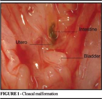
The numbers of fetuses obtained per rat are presented in Table 1. None of the fetuses in the control group presented anorectal anomalies. In the experimental group, nine fetuses from E1 (90%), ten from E2 (91%) and three from E3 (30%) presented anorectal anomalies. The model utilized provoked anorectal anomalies in 71% of all the fetuses studied. The types of anorectal anomaly found were: 14 cloacal malformations (Figure 1), four perineal fistulas and four urethral fistulas (Figures 2-3) (Table 2).

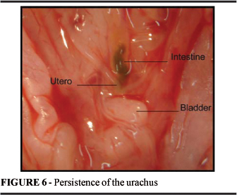
None of the fetuses in the control group presented malformations of the vertebral column. However, these were found in 80% of the fetuses in the experimental group (all the fetuses from E1 and E2, and four from E3). The abnormalities found were tail agenesis (Figure 4) or short tail (Table 3). None of the fetuses in the control group presented urological structural alterations. Among the fetuses in the experimental group, 35% presented alterations. Ureteral hydronephrosis (Figure 5) was found in five fetuses, unilateral kidney agenesis in four fetuses, hypoplasia of the left kidney in one fetus, and persistence of the urachus (Figure 6) in one fetus (Table 4). Among the fetuses from subgroup E1, there was an association between anorectal anomaly and malformation of the vertebral column in six fetuses (60%) and an association of anorectal anomaly with both malformation of the vertebral column and urological malformation in three fetuses (30%). Malformation of the column alone was present in one fetus (10%) (Table 5). Among the fetuses in subgroup E2, there was an association between anorectal anomaly and malformation of the vertebral column in four fetuses (36%) and an association of anorectal anomaly with both malformation of the vertebral column and urological malformation in six fetuses (60%). One fetus presented malformation of the column with urological abnormality but without anorectal anomaly (9%) (Table 6). Among the fetuses in subgroup E3, there was an association between anorectal anomaly and malformation of the vertebral column in three fetuses (30%). There was no association of anorectal anomaly with both malformation of the vertebral column and urological malformation. There was malformation of the column alone in one fetus (10%) and urological malformation alone in one fetus (10%) (Table 7).
Discussion
The embryology of anorectal malformations is a controversial subject. It is difficult to study this in human beings because of the scarcity of fetuses with anorectal anomalies. Hence, there is a need for an animal model 4. The experimental models for anorectal anomalies include those due to mutations, those that occur spontaneously and those induced by drugs. The most classical and oldest model is the one described by Danforth, which is known as "Danforth's short-tail mouse" 5. This is a natural mutation that presents a series of malformations, and among these is imperforate anus. More recently, a mouse with a mutation involving the "sonic hedgehog" was discovered. This is a signaling molecule that performs a variety of roles in the development of vertebrates, and which exhibits imperforate anus in its phenotype 6. These animals are difficult to obtain and extremely expensive. The spontaneous occurrence of anorectal malformations is not uncommon among domestic animals, especially among pigs, with incidence of around 0.2%. They present similarities with the malformations in humans, but with a low rate of associated malformations 7. By crossing affected animals, Hori et al8 obtained an incidence of 62% of the offspring presenting anorectal anomalies. The advantage of this model is in the size of the animal, which allows more in-depth anatomical and histological study. However, it is a model that requires a long development time for obtaining affected offspring, in addition to the longer gestation and greater length of time these animals need to be kept in a vivarium. The utilization of inducing drugs in small rodents such as mice and rats is the most common technique. The gestation period is short (21 days) and these animals are easily kept in vivariums. Moreover, inducement by drugs is easy to perform, since the only procedure utilized is the administration of the drug by means of gavage. The drugs most utilized are vitamin A derivatives 9 and ETU. Vitamin A derivatives only present an effect in mice, whereas ETU only presents an effect in rats. The utilization of rats enables study of larger fetuses, which is an advantage. Khera, in 1973 10, and Ruddick & Khera, in 1975 11, published results from experiments in which ETU administration (the degradation product from the fungicide ethylene-bis-thiocarbonate) to pregnant rats and rabbits was capable of inducing anomalies in various organs of the fetuses generated. The potential for inducing anorectal anomalies was studied by Hirai & Kuwabara, in 1990 12. The data obtained indicated that the ideal dose for inducing anorectal anomalies was 125 mg/kg. The anomalies most often found were rectourethral fistula in males and rectocloacal fistula in females. Other studies, such as those conducted by Qui et al 13-14 and Yuan et al 15, utilized this drug at the same dose and achieved the same result, thereby demonstrating the reproducibility of the method. The mechanism for the action of ETU is not well established. Qui et al 13 suggested that ETU might cause disturbances in the mechanism for the action of the "sonic hedgehog" signaling molecule. ETU is an easily obtained drug that is cheaper than vitamin A derivatives, thus facilitating the utilization of rats, and these animals therefore formed the subject of our study. In addition to the incidence of anorectal anomalies, we also investigated the presence of urological and vertebral column malformations. These are the malformations that are most frequently associated with anorectal anomalies in humans, with incidence ranging from 20 to 54% and 13 to 50%, respectively 1-3. We obtained an overall incidence of 71% for anorectal anomalies. Associations with other malformations were also present: vertebral column malformations in 80% of the fetuses and urological malformations in 35% of the fetuses. The incidence of anorectal anomalies was comparable with what has been found in other studies, ranging from 55 to 85% 12-15. This rate of incidence is less than what is obtained through utilizing vitamin A derivatives, from which 95% of the offspring present the malformation 16. The types of anomalies encountered were similar to those found in the human species, but with different incidence. In our model, cloacal alterations were the most frequent type, whereas in humans this is one of the rarest types 1-3. The 80% incidence of abnormalities of the vertebral column was somewhat lower than found by other authors, who have reported tail agenesis or rudimentary tails in 100% of the fetuses, of which 48 to 70% exhibited myeloschisis. We did not find this in our animals 12,14,15. Urological alterations, which in the human species occur in 13 to 50% of the patients, occurred in 35% of our fetuses. The principal abnormalities found were kidney agenesis and ureteral hydronephrosis, similar to findings among humans. There is no corresponding data in the literature for comparison. The presence of these malformations takes on special interest, since they are the cause of great morbidity and mortality in humans.
Conclusion
The model described was shown to be easy to implement and presented results that allow its utilization for studying anorectal anomalies and their associated malformations.
Received: November 21, 2006
Review: December 14, 2006
Accepted: January 10, 2007
Conflict of interest: none
Financial source: FAPESP
- 1. Stephens FD, Smith ED. Ano-rectal malformations in children. Chicago IL: Year Book Medical; 1971.
- 2. Peña A. Anomalias anorretais. In: Maksoud JG. Cirurgia pediátrica. Rio de Janeiro: Livraria e Editora Revinter Ltda; 1998. p.809-35.
- 3. Macedo M, Martins JL, Freitas-Filho LG. Sacral Ratio and fecal continence in children with anorectal malformations. BJU Int. 2004;94:893-4.
- 4. Qui BQ, Beasley SW, Frizelle FA. Clarification of the process that lead to anorectal malformations in the ETU-induced rat model of imperforate anus. J Pediatr Surg. 2002;37(9):1305-12.
- 5. Danforth CH. Developmental anomalies in a special strin of mice. Am J Anat. 1930;45:275-87.
- 6. Mo R, Kim JH, Zhang J, Chiang C, Hui C, Kim PCW. Anorectal Malformations caused by defects in sonic hedgehog signaling. Am J Pathol. 2001;159:765-74.
- 7. Lambrecht W, Lierse W. The internal sphincter in anorectal malformation: Morphologic investigations in neonatal pigs. J Pediatr Surg. 1987;22(12):1160-8.
- 8. Hori T, Giuffra E, Anderson L, Ohkawa H. Mapping loci causing susceptibility to anal atresia in pigs, using a resource pedigree. J Pediatr Surg. 2001;36:1370-4.
- 9. Kubota Y, Shimotake T, Iwai N. Congenital anomalies in mice induced by etretinate. Eur J Pediatr Surg. 2000;10:248-51.
- 10. Khera KS. Ethylenethiourea: Teratogenicity study in rats and rabbits. Teratology. 1973;7:243-52.
- 11. Ruddick JA, Khera KS. Pattern of anomalies following single oral doses of ethylenethiourea to pregnant rats. Teratology. 1975;12:277-82.
- 12. Hirai Y, Kuwabara N. Transplacentally induced anorectal malformations in rats. J Pediatr Surg. 1990; 25(7):812-6.
- 13. Qui BQ, Beasley SW, Frizelle FA. Clarification of the process that lead to anorectal malformations in the ETU-induced rat model of imperforate anus. J Pediatr Surg. 2002;37(9):1305-12.
- 14. Qui BQ, Beasley SW, Frizelle FA. Evidence that the notochord may be pivotal in the development of sacral and anorectal malformations. J Pediatr Surg. 2003;38(9):1310-6.
- 15. Yuan ZW, Lui VCH, Tam PKH. Deficient motor innervation of the sphincter mechanism in fetal rats with anorectal malformation: a quantitative study by fluorogold retrograde tracing. J Pediatr Surg. 2003;38(9):1383-8.
- 16. Bitoh Y, Shimotake T, Kubota Y, Kimura O, Iwai N. Impaired distribution of retinoic acid receptors in the hindgut-tailgut region of murine embryos with anorectal malformations. J Pediatr Surg 2001; 36:377-80.
Publication Dates
-
Publication in this collection
14 Mar 2007 -
Date of issue
Apr 2007
History
-
Reviewed
14 Dec 2006 -
Received
21 Nov 2006 -
Accepted
10 Jan 2007