Abstracts
PURPOSE: To identify the types of malformations resulting from the administration of retinoic acid (RA) to Swiss mice on different days of pregnancy. METHODS: Twenty-four pregnant Swiss mice were divided into 4 groups of 6 animals each. The experimental groups received a single intraperitoneal injection of RA (70 mg/kg) on gestational days 7, 8 and 9 (D7, D8 and D9), while control animals (C) received only saline solution. RESULTS: Were obtained: exencephaly (C:0; D7:16.1%; D8:25.4%; D9:0), myelomeningocele (C:0; D7:25.8%, D8:30.9%, D9:0), spina bifida occulta (C:0, D7:29%, D8:41.8%, D90), gastroschisis (C:0, D7:6.4% D8:5.4%, D9:0), omphalocele (C:0, D7:6.4%, D8:14.5%, D9:0), lower limb alterations (C:0, D7:74.1%, D8:80%, D9:0), imperforated anus (C:0, D7:100%, D8:100%, D9:100%), and tail agenesis/alteration (C: D7:100%, D8:100%, D9:100%). CONCLUSION: The experimental model using Swiss mice proved to be efficient in the induction of the different types of defects, with the eighth gestational day being the one that most favored the induction of neural tube defect, omphalocele, gastroschisis, lower limb defects, imperforated anus and tail agenesis/alteration. On this basis, this is a useful model for future investigation of neural development and of the formation of the appendicular skeleton.
Neural Tube Defects; Tretinoin; Mice
OBJETIVO: Identificar os tipos de malformação resultantes da administração do ácido retinóico (AR) a camundongos Swiss em diferentes dias gestacionais. MÉTODOS: Foram utilizados 24 camundongos fêmeas, linhagem Swiss, prenhes, divididos em 4 grupos com 6 animais cada. Os grupos experimentais receberam uma única injeção intraperitoneal de AR (70mg/Kg) nos dias gestacionais 7, 8 e 9 (D7, D8 e D9), enquanto que os animais do grupo controle (C) receberam apenas solução salina. RESULTADOS: Foram encontrados: exencefalia (C:0; D7:16.1%; D8:25.4%; D9:0); mielomeningocele (C:0; D7:25.8%; D8:30.9%; D9:0); Espina Bífida Oculta (C:0; D7:29%; D8:41.8%; D90); gastrosquise (C:0; D7:6.4% D8:5.4%; D9:0); onfalocele (C:0; D7:6.4%; D8:14.5%; D9:0); alterações do membro inferior (C:0; D7:74.1%; D8:80%; D9:0); imperfuração anal (C:0; D7:100%; D8:100%; D9:100%) e agenesia/alteração de cauda (C: D7:100%; D8:100%; D9:100%). CONCLUSÕES: O modelo experimental utilizando camundongo Swiss mostrou-se eficiente na indução dos diferentes tipos de defeitos, sendo o oitavo dia gestacional o mais propicio na indução de DFTN, onfalocele, gastrosquise, defeitos de membro inferior, imperfuração anal e agenesia/alteração de cauda, tornando este um modelo útil para futuras investigações do desenvolvimento neural e no processo de formação do esqueleto apendicular.
Defeitos do Tubo Neural; Tretinoína; Camundongos
ORIGINAL ARTICLE
EFFECTS OF DRUGS
Teratogenic effect of retinoic acid in swiss mice1 1 . Research performed at Department of Pathology, Faculty of Medicine, University of São Paulo, Ribeirão Preto (FMRP-USP), São Paulo, Brazil.
Efeito teratogênico do ácido retinóico em camundongo swiss
Paulo Roberto Veiga QuemeloI; Charles Marques LourençoII; Luiz Cesar PeresIII
IFellow PhD degree, Experimental Pathology, FMRP-USP, São Paulo, Brazil
IIMD. Resident in Genetics, FMRP-USP, São Paulo, Brazil
IIIPhD, Associate Professor, Department of Pathology, FMRP-USP, São Paulo, Brazil
Correspondence Correspondence: Luiz Cesar Peres Department of Pathology Faculty of Medicine Ribeirão Preto - USP 14049-900 Ribeirão Preto SP Brazil Phone: (55 16)3602-3072 Fax: (55 16)3633-1068 pquemelo@usp.br | pquemelo@hotmail.com
ABSTRACT
PURPOSE: To identify the types of malformations resulting from the administration of retinoic acid (RA) to Swiss mice on different days of pregnancy.
METHODS: Twenty-four pregnant Swiss mice were divided into 4 groups of 6 animals each. The experimental groups received a single intraperitoneal injection of RA (70 mg/kg) on gestational days 7, 8 and 9 (D7, D8 and D9), while control animals (C) received only saline solution.
RESULTS: Were obtained: exencephaly (C:0; D7:16.1%; D8:25.4%; D9:0), myelomeningocele (C:0; D7:25.8%, D8:30.9%, D9:0), spina bifida occulta (C:0, D7:29%, D8:41.8%, D90), gastroschisis (C:0, D7:6.4% D8:5.4%, D9:0), omphalocele (C:0, D7:6.4%, D8:14.5%, D9:0), lower limb alterations (C:0, D7:74.1%, D8:80%, D9:0), imperforated anus (C:0, D7:100%, D8:100%, D9:100%), and tail agenesis/alteration (C: D7:100%, D8:100%, D9:100%).
CONCLUSION: The experimental model using Swiss mice proved to be efficient in the induction of the different types of defects, with the eighth gestational day being the one that most favored the induction of neural tube defect, omphalocele, gastroschisis, lower limb defects, imperforated anus and tail agenesis/alteration. On this basis, this is a useful model for future investigation of neural development and of the formation of the appendicular skeleton.
Key words: Neural Tube Defects. Tretinoin. Mice.
RESUMO
OBJETIVO: Identificar os tipos de malformação resultantes da administração do ácido retinóico (AR) a camundongos Swiss em diferentes dias gestacionais.
MÉTODOS: Foram utilizados 24 camundongos fêmeas, linhagem Swiss, prenhes, divididos em 4 grupos com 6 animais cada. Os grupos experimentais receberam uma única injeção intraperitoneal de AR (70mg/Kg) nos dias gestacionais 7, 8 e 9 (D7, D8 e D9), enquanto que os animais do grupo controle (C) receberam apenas solução salina.
RESULTADOS: Foram encontrados: exencefalia (C:0; D7:16.1%; D8:25.4%; D9:0); mielomeningocele (C:0; D7:25.8%; D8:30.9%; D9:0); Espina Bífida Oculta (C:0; D7:29%; D8:41.8%; D90); gastrosquise (C:0; D7:6.4% D8:5.4%; D9:0); onfalocele (C:0; D7:6.4%; D8:14.5%; D9:0); alterações do membro inferior (C:0; D7:74.1%; D8:80%; D9:0); imperfuração anal (C:0; D7:100%; D8:100%; D9:100%) e agenesia/alteração de cauda (C: D7:100%; D8:100%; D9:100%).
CONCLUSÕES: O modelo experimental utilizando camundongo Swiss mostrou-se eficiente na indução dos diferentes tipos de defeitos, sendo o oitavo dia gestacional o mais propicio na indução de DFTN, onfalocele, gastrosquise, defeitos de membro inferior, imperfuração anal e agenesia/alteração de cauda, tornando este um modelo útil para futuras investigações do desenvolvimento neural e no processo de formação do esqueleto apendicular.
Descritores: Defeitos do Tubo Neural. Tretinoína. Camundongos.
Introduction
Vitamin A and its metabolites, collectively called retinoids, are essential for adequate embryo development. Retinoids are important signaling molecules for the regulation of cell differentiation, proliferation and morphogenesis. Inadequate levels of these compounds (excess or deficiency) may result in a set of defects denoted retinoic acid embryopathy which may provoke defects in the development of the neural crista1, 2, 3, in addition to limb malformation4 and other skeletal manifestations5. Retinoic acid (RA) is a biologically active vitamin A derivative6 which plays an important role in the development of the central nervous system2, 7, 8 and in the maintenance of various tissues in adult vertebrates, and is essential in various embryologic processes by interacting with RRA and RXR nuclear receptors. It may also imply changes in the expression of the Hox genes which are fundamental in the process of cell migration, differentiation and regionalization during embryogenesis9, 10, 11. Animal models have been used to investigate several questions related to the teratogenic effects of RA such as dose and temporal and spatial correlation3, 12. Since there is variation in the effects, sensitivity and critical period in different species and even in different races of the same species, it is useful to define the effects of excess RA on Swiss mice in order to plan studies directed at specific teratogenic aspects.
Methods
The experiment was approved by the Ethics Committee for Animal Experimentation (CETEA) of the Faculty of Medicine of Ribeirão Preto (FMRP) (Nº 018/2005). Twenty-four pregnant Swiss mice from the colony of the Central Animal House of the Ribeirão Preto Campus, University of São Paulo, were used. The animals, young adults weighing on average 40 g, were allowed to habituate to the Animal Facilities of the Department of Pathology, FMRP for a few days, with free access to appropriate chow and water. The environment in which the animals were housed throughout the experiment is sound protected and is kept under rigorous control with 12 hour light/dark cycles, with low luminosity, a constant temperature of 22ºC and an automatic exhaust with several changes of air throughout the day. The females were mated and tested for the presence of a vaginal plug, which represented day zero of pregnancy. After mating, the pregnant females were housed in individual plastic boxes. The dose of all-trans RA was first standardized on different gestational days, with doses from 30 to 120 mg/kg being used3, 12, 13. We observed that doses of 80 mg/kg or higher provoked many reabsorptions, while doses of less than 60 mg/kg caused few defects. Thus, we determined that the ideal dose for Swiss mice was 70 mg/kg. After RA standardization, the pregnant dams were divided at random into 4 groups of 6 animals each: control (C), 7th gestational day (D7), 8th gestational day (D8), and 9th gestational day (D9). The experimental groups received a single intraperitoneal injection of RA (70 mg/kg) diluted with sunflower oil administered on gestational day 7, 8 or 9, according to the group. Control animals received only saline solution by the same route and in an equal volume on the 8th gestational day. The animals were killed in a CO2 chamber on the 17th day of gestation and submitted to cesarian section by surgical incision along the abdominal midline, with exposure of the peritoneal cavity. Both uterine horns were removed, place on a clean Petri dish, and opened lengthwise to expose the gestational sacs for the removal of live fetuses. Fetuses who suffered late fetal death and reabsorptions, when present, were also removed. The material obtained was examined externally with a magnifying glass, photographed and fixed in 10% buffered formalin for 24 hours. Next, some of the fetuses were fixed in 70% ethanol, defatted with acetone, again passed through 95% ethanol, and transferred to 2% potassium hydroxide until the bones became visible. The fetuses were then stained with 0.01% Alizarin Red (H2O) in 1% potassium hydroxide until the bones were stained red. Next, the fetuses were placed in a pure glycerin solution, analyzed under a stereoscopic microscope and photographed. The number of implants, reabsorptions and late fetal deaths was recorded throughout the experiment. Data were analyzed statistically by ANOVA and by the Kruskal-Wallis and Tukey tests, with the level of significance set at p≤0.05. All analyses were carried out using the GraphPad Prism 4.00 software.
Results
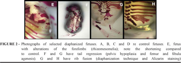
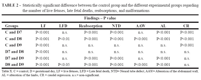
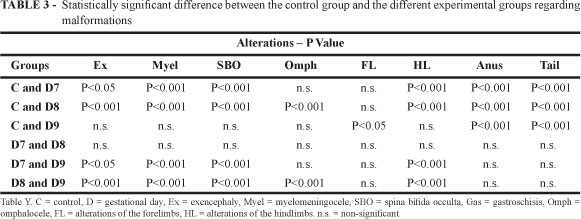
Regarding live fetuses, all groups differed significantly from one another (p<0.001). The number of reabsorptions was higher in the D7 and D8 groups compared to the D9 and C groups (p<0.001), although there was no significant difference between groups D7 and D8 or groups D9 and C. Group D8 also showed an increase in the number of late fetal deaths compared to groups C and D9 (p<0.05), as shown in Tables 2 and 3. Congenital anomalies were more common in groups D7 and D8 than in group D9 (Table 1). NTD were detected in 17 fetuses (54.8%) in group D7 and in 42 fetuses (76.3%) in group D8, but were not detected in any fetus in group D9. Among the neural tube defect (NTD) there was a preponderance of fetuses with spina bifida occulta (Figure 1-I), followed by myelomeningocele (Figure 1-J) and exencephaly (Figures 1-E, 1-F, 1-G and 1-H). There was a statistically significant difference between the number of fetuses with NTD in groups D7 and D8 compared to groups C and D9, but not between groups D7 and D8 (Table 2). Congenital anomalies of the abdominal wall omphalocele, (Figure 1-N) and gastroschisis (Figure 1-O) were observed only in groups D7 and D8, with the latter differing significantly from groups C and D9 (p<0.001). Hindlimb defects (Figures 1-I and 1-M) occurred in groups D7 and D8 (p<0.001), whereas forelimb defects were observed in group D9 (p<0.05). Limb defects included digital defects such as syndactyly (Figure 1-P), polysyndactyly (Figure 1-Q), brachydactyly, and even phocomelia, as shown in Figure 1-L. Tail regression (imperforated anus, tail agenesis/reduction (Figures 1-I and 1-J)) was observed in all fetuses of all experimental groups (p<0.001). External examination of diaphanized fetuses with a magnifying lens revealed bone changes, especially of the forelimbs (rhizomesomelia) (Figures 2-E and 6-G) in group D9 and hindlimbs (Figures 2-F and 2-G) in groups D7 and D8. Fusions of inferior contiguous ribs (Figures 2-G and 2-H) were also observed in groups D7 and D8.
Discussion
Embryopathy due to RA is being intensely investigated in view of the teratogenic potential of retinols and of the crucial role played by their receptors in embryo development. Our results with Swiss mice focus on the effect of this teratogen on structures derived from the neural crista and from the neural tube on different gestational days. However, we also observed other types of anomalies such as omphalocele, gastroschisis, limb and rib alterations and tail regression. The NTD detected in our study indicate that groups D7 and D8 were the most affected ones, especially the latter, perhaps because this is the critical period in the migratory process of cells of the cranial neural crystal in Swiss mice1. An adequate pattern of migration of these cells is essential for craniofacial morphogenesis and is highly conserved among vertebrates. Their migration is intimately related to early segmentation of the neural tube, so that they migrate discretely in small flows14. The present results indicate that this migration occurs until the 8th gestational day since no fetus with NTD was observed on the 9th gestational day, in agreement with data reported by Kuno, et al. Among NTD, spina bifida occulta prevailed in the fetuses studied here, a fact also observed in other studies15, followed by myelomeningocele and exencephaly, in agreement with the multisite closure theory16. Exencephaly is the most serious defect and is commonly incompatible with life. We observed that the administration of RA to Swiss mice provoked exencephaly in 25% of cases, in agreement with the literature. Experimental studies involving RA administration provoked exencephaly in SWC/Bc and 22% ICR/Bc mice13. However, other genetically modified lines (SELH/Bc) and ICR mice can induce greater amounts of fetuses with exencephaly, i.e., 53% and 81%, respectively12, 13. Although very interesting results have been obtained with knock-out experimental models, these animals are expensive, require special care and are difficult to manipulate, whereas Swiss mice provide an inexpensive experimental model of easy manipulation and efficient for the induction of different types of defects. The present results also indicate that a larger number of fetuses with limb alterations (p<0.001) occurred in groups D7 and D8, once again emphasizing that these days are critical periods in embryo formation and indicating the role of retinoids in the process of formation of the appendicular skeleton11, 17. RA interacts with the nuclear receptors RRA and RXR, which recognize the sequence within the target genes, called response to RA elements (RRAE), affecting the proximal-distal regulation and formation of the limbs and changing the expression of various genes (Fgf8, Shh, Ptc1, Gli3, dHand). These elements activate the expression of Hox genes, which participate in the anteroposterior orientation of the axis, the neural crista, the paraxial mesoderm, and the limbs. Thus, excess RA can provoke changes in the expression of Hox genes, causing posteriorization of anterior structures. Since the anterior regions usually are more sensitive to RA, they do not develop normally under these conditions, with the occurrence of limb defects9-11. Additionally, the expression of CYP26B1 is also implicated in the limb anomalies detected in animal models since it occurs in the distal part of the limb bud and its action is influenced by circulating RA levels11. The fusion of lower thoracic ribs, observed only in groups D7 and D8, is a skeletal anomaly belonging to the disruptive spectrum of the teratogenic effects of RA, which also includes hemivertebrae and pelvic anomalies both in humans and in animals. This is a recognized anomaly but its physiopathologic mechanism has not been fully elucidated, even though the effects of this teratogen on Hox genes have also been implicated in this process in some experimental studies18. The pathogenetic mechanism of the tail regression observed in all the experimental groups is unknown, although a vascular etiology has been proposed. The animals treated with RA presented tail anomalies in addition to spina bifida occulta, with extreme presentations such as sirenomelia being observed in some cases19. Omphalocele and gastroschisis, observed in some animals of groups D7 and D8, are not common anomalies in RA-induced embryopathy, although they have been reported in the literature19. These findings may be explained by the critical period of development of the abdominal wall, which starts at about the eighth gestational day and can extend up to the 12th day, when the primary abdominal wall of the mouse is formed20. However, RA seems to have a teratogenic effect only during the beginning of the formation of the abdominal wall since no alteration was detected in group D9. The spectrum of malformations associated with RA is wide and, at present, with the use of experimental knock-out models for certain genes, the mechanisms underlying this process are beginning to be understood. The present study conducted on Swiss mice demonstrated that the effects of this teratogen are temporally and spatially correlated in the embryos, but also revealed a marked phenotypic overlap of these cases, suggesting that, in fact, the molecular interactions of RA are more extensive than previously thought and represent a field of excellence for experimental teratology. The experimental model with Swiss mice proved to be efficient for the induction not only of NTD but also of changes in the appendicular skeleton and tail regression. We observed that in group D9 the anomalies were milder, possibly because the critical period of formation of various tissues had already passed, although signs of tail regression did occur. In the present study, even though both D7 and D8 were appropriate for the induction of developmental defects, D8 was the one showing the largest number of defects.
Conclusion
In the experimental model using Swiss mice the RA dose of 70 mg/kg administered by the intraperitoneal route on the eighth gestational day proved to be more efficient in the induction of NTD, omphalocele, gastroschisis, hindlimb defects, imperforated anus and tail agenesis/alteration, representing a useful model for future investigations of these defects.
Acknowledgment
To Deise Lucia Chesca Simões for her technical assistance.
Received: April 12, 2007
Review: June 14, 2007
Accepted: July 17, 2007
Conflict of interest: none
Financial source: FAPESP (Proc. nº05/51158-7) and CNPq
- 1. Maden M. Retinoids and spinal cord development. J Neurobiol. 2006;66:726-38.
- 2. Ross SA, Mccaffery PJ, Dräger VC, DE Luca LM. Retinoids in embryonal development. Physiol. Rev 2000;80:1021-54.
- 3. Mulder GB, Manley N, Grant S, Schimidt K, Zeng W, Eckhoff C, Maggio-Price L. Effects of excess vitamin A on development of cranial neural crist-derived structures: a neonatal and ebryologic study. Teratology. 2000;62:214-26.
- 4. Ali-khan S, Hales B. Retinoid receptor antagonists alter the pattern of apoptosis in organogenesis stage mouse limbs.Toxicol Sci. 2006;90:208-20.
- 5. Zile MH. Vitamin A and embryonic development: an overview. J Nutr. 1998;128:455-8.
- 6. Means A.L, Gudas LJ. The roles of retinoids in vertebrate development. Ann Rev Biochem. 1995;64:201-33.
- 7. White JC, Highland M, Kaiser M, Clagett-Dame M. Vitamin A deficiency results in the dose-dependent acquisition of anterior character and shortening of the caudal hindbrain of the rat embryo. Dev Biol. 2000;220:263-84.
- 8. Wendling O, Dennefeld C, Chambon P, Mark M. Retinoid signaling is essential for patterning the endoderm of the third and fourth pharyngeal arches. Development. 2000;127:1553-62.
- 9. Marshall H, Morrinson A, Michčle S, Pöpperi H, Krumlauf R. Retinoids and Hox genes. FASEB J. 1996;9:969-78.
- 10. Sporn MB, Roberts AB, Goodman DS. The retinoids: biology, chemistry and medicine. New York: Raven Press; 1994.
- 11. Yashiro K, Zhao MU, Yamashita K, Nishijima M, Nishino J, Saijoh Y, Sakai Y, Hamada H. Regulation of retinoic acid distribution is required for proximodistal patterning and outgrowth of the developing mouse limb. Dev Cell. 2004;6:411-22.
- 12. Kuno N, Kadomatsu K, Muramatsu T. Determination of the optimal time and dosage of all-trans acid retinoic for induction of murine excencephaly. Teratology. 1999;60:63-7.
- 13. Tom C, Juriloff DM, Harris MJ. Studies of the effect of retinoic acid on anterior neural tube closure in mice genetically liable to exencephaly. Teratology. 1991;43:24-40.
- 14. Kulesa P, Ellis DL, Trainor PA. Comparative analysis of neural crest cell death, migration, and function during vertebrate embryogenesis. Dev Dyn. 2004;229:14-29.
- 15. Diez-Pardo JAA, Marino JM, Baoquan Q, Delgado-Baeza E, Fernaneez A, Morales MC, Tovar JA. Neural tube defects: an experimental model in the fetal rat. Eur J Pediatr Surg. 1995;5:198-02.
- 16. Padmanabhan R. Etiology, pathogenesis and prevention of neural tube defects. Congenit Anom. 2006;46:55-67.
- 17. Niswander L. Pattern formation: old models out on limb. Nat Rev Get. 2003;4:133-43.
- 18. Kawanishi CY, Hartig P, Bobseine KL, Schmid J, Cardon M, Massenburg G, Chemoff N. Axial skeletal and Hox expression domain alterations induced by retinoic acid, valproic acid, and bromoxynil during murine development. J Biochem Mol Toxicol. 2003;17:346-56.
- 19. Padmanabhan R. Retinoic acid-induced caudal regression syndrome in the mouse fetus. Reprod Toxicol. 1998;12:139-51.
- 20. Brewer S, Williams T. Finally, a sense of closure? Animal models of human ventral body wall defects. Bioessays. 2004;26:1307-21.
Publication Dates
-
Publication in this collection
22 Jan 2008 -
Date of issue
Dec 2007
History
-
Accepted
17 July 2007 -
Reviewed
14 June 2007 -
Received
12 Apr 2007


