Abstracts
PURPOSE: To analyze mucosal proliferation and its characteristics, through specific models of duodenogastric reflux, in the stomach of Wistar rats. METHODS: Seventy-five healthy and adult male rats were divided into three groups: group I - control (n = 25 animals), submitted to gastrotomy of the posterior wall of the glandular stomach; group II - DGR (n = 25 animals), submitted to duodenogastric reflux through latero-lateral gastrojejunal anastomosis in the posterior wall of the glandular stomach and group III - DGR-P (n = 25 animals), submitted to duodenogastric reflux through the pylorus following the same procedure of group II, sectioning and closing the afferent loop. The animals were observed during 36 weeks and subsequently the mucosal lesions were analyzed, with macroscopic and microscopic examination of the prepyloric, the gastrojejunostomy and the squamous area of the stomach. RESULTS: Group I did not present any kind of lesion. Macroscopic lesions of the prepyloric area in groups II and III were 0% and 20%, respectively. Macroscopic lesions of the gastrojejunal stoma in groups II and III were 36% and 88%, respectively, and 12% and 28%, respectively, in the squamous area. Microscopically, adenomatous hyperplasia (AH), squamous hyperplasia (SH) and adenocarcinoma (AC) were diagnosed. The occurrence of AH at the prepyloric area in groups II and III was 0% and 40%, respectively, and in the gastrojejunal stoma, 40% and 72%, respectively. The occurrence of SH in the squamous area in groups II and III was 12% and 20%, respectively, without statistical differences between the groups. AC was found only in three animals of groups III (12%). CONCLUSIONS: The duodenogastric reflux in this experimental model caused high frequency of proliferative lesions of the gastrojejunal stoma and in the prepyloric area, while adenocarcinoma was a rare occurrence.
Duodenogastric Reflux; Hyperplasia; Adenocarcinoma; Rats
OBJETIVO: Avaliar as lesões proliferativas que se desenvolvem na mucosa gástrica de ratos Wistar após modelo específico de refluxo duodeno-gástrico. MÉTODOS: Foram utilizados 75 ratos adultos machos divididos em três grupos experimentais: o grupo I (controle) submetido a gastrotomia na parede posterior do estômago glandular (25 animais); o grupo II (RDG), foi submetido a gastrojejunoanastomose látero-lateral na parede posterior do estômago glandular (25 animais) e o grupo III (RDG-P) submetido a gastrojejunoanastomose látero-lateral na parede posterior do estômago glandular, com secção e fechamento da alça (25 animais). Os animais foram observados durante 36 semanas, após o que foram realizados estudos macroscópicos e microscópicos da anastomose gastrojejunal, da região pré-pilórica e região escamosa do estômago. RESULTADOS: Os animais do Grupo I não apresentaram nenhum tipo de lesão. No grupo II observou-se 40% de lesões do tipo hiperplasia adenomatosa na anastomose e 12% de hiperplasia escamosa. No grupo III obteve-se 40% de hiperplasia adenomatosa na mucosa pré-pilórica, 72 % de hiperplasia adenomatosa na mucosa da anastomose, 20% de hiperplasia escamosa e 12 % de adenocarcinoma. CONCLUSÕES: O refluxo duodeno-gástrico induz a alta freqüência de lesões proliferativas na mucosa adjacente à anastomose gastrojejunal ou na mucosa pré-pilórica e o adenocarcinoma é um evento raro neste modelo experimental.
Refluxo Duodenogástrico; Hiperplasia; Adenocarcinoma; Ratos
ORIGINAL ARTICLE
Gastric proliferative lesions induced by duodenogastric reflux in rats11 Master's Thesis presented at the Post Graduation Program in Surgery of the School of Medical Science (FCM) of the State University of Campinas (UNICAMP). São Paulo, Brazil.
Lesões proliferativas gástricas induzidas pelo refluxo duodenogástrico em ratos
Rosângela Lucinda Rocha MonteiroI; Nelson Adami AndreolloII; Maria Aparecida Marchesan RodriguesIII; Marina Raquel AraujoIV
IAssistant Professor at the School of Medicine of Pouso Alegre, University of Vale do Sapucaí (UNIVAS). Brazil
IIAssociate Professor of the Department of Surgery, UNICAMP. Brazil
IIIFaculty Member of the Department of Pathologic Anatomy at the School of Medicine of Botucatu (UNESP). Brazil
IVBiologist of the Laboratory of Enzymology and Experimental Carcinogenesis (UNICAMP). Brazil
Correspondência Correspondência: Rosângela Rocha Monteiro Rua Amadeu Bertolacini, 20 37.564-000 Borda da Mata MG Brazil mrclin@uol.com.br
ABSTRACT
PURPOSE: To analyze mucosal proliferation and its characteristics, through specific models of duodenogastric reflux, in the stomach of Wistar rats.
METHODS: Seventy-five healthy and adult male rats were divided into three groups: group I control (n = 25 animals), submitted to gastrotomy of the posterior wall of the glandular stomach; group II DGR (n = 25 animals), submitted to duodenogastric reflux through latero-lateral gastrojejunal anastomosis in the posterior wall of the glandular stomach and group III DGR-P (n = 25 animals), submitted to duodenogastric reflux through the pylorus following the same procedure of group II, sectioning and closing the afferent loop. The animals were observed during 36 weeks and subsequently the mucosal lesions were analyzed, with macroscopic and microscopic examination of the prepyloric, the gastrojejunostomy and the squamous area of the stomach.
RESULTS: Group I did not present any kind of lesion. Macroscopic lesions of the prepyloric area in groups II and III were 0% and 20%, respectively. Macroscopic lesions of the gastrojejunal stoma in groups II and III were 36% and 88%, respectively, and 12% and 28%, respectively, in the squamous area. Microscopically, adenomatous hyperplasia (AH), squamous hyperplasia (SH) and adenocarcinoma (AC) were diagnosed. The occurrence of AH at the prepyloric area in groups II and III was 0% and 40%, respectively, and in the gastrojejunal stoma, 40% and 72%, respectively. The occurrence of SH in the squamous area in groups II and III was 12% and 20%, respectively, without statistical differences between the groups. AC was found only in three animals of groups III (12%).
CONCLUSIONS: The duodenogastric reflux in this experimental model caused high frequency of proliferative lesions of the gastrojejunal stoma and in the prepyloric area, while adenocarcinoma was a rare occurrence.
Key words: Duodenogastric Reflux. Hyperplasia. Adenocarcinoma. Rats.
RESUMO
OBJETIVO: Avaliar as lesões proliferativas que se desenvolvem na mucosa gástrica de ratos Wistar após modelo específico de refluxo duodeno-gástrico.
MÉTODOS: Foram utilizados 75 ratos adultos machos divididos em três grupos experimentais: o grupo I (controle) submetido a gastrotomia na parede posterior do estômago glandular (25 animais); o grupo II (RDG), foi submetido a gastrojejunoanastomose látero-lateral na parede posterior do estômago glandular (25 animais) e o grupo III (RDG-P) submetido a gastrojejunoanastomose látero-lateral na parede posterior do estômago glandular, com secção e fechamento da alça (25 animais). Os animais foram observados durante 36 semanas, após o que foram realizados estudos macroscópicos e microscópicos da anastomose gastrojejunal, da região pré-pilórica e região escamosa do estômago.
RESULTADOS: Os animais do Grupo I não apresentaram nenhum tipo de lesão. No grupo II observou-se 40% de lesões do tipo hiperplasia adenomatosa na anastomose e 12% de hiperplasia escamosa. No grupo III obteve-se 40% de hiperplasia adenomatosa na mucosa pré-pilórica, 72 % de hiperplasia adenomatosa na mucosa da anastomose, 20% de hiperplasia escamosa e 12 % de adenocarcinoma.
CONCLUSÕES: O refluxo duodeno-gástrico induz a alta freqüência de lesões proliferativas na mucosa adjacente à anastomose gastrojejunal ou na mucosa pré-pilórica e o adenocarcinoma é um evento raro neste modelo experimental.
Descritores: Refluxo Duodenogástrico. Hiperplasia. Adenocarcinoma. Ratos.
Introduction
The gastric mucosa is particularly susceptible to the onset of proliferative lesions when submitted to reflux of the duodenal content 1,2. Several investigations were carried out to clarify whether patients undergoing surgery with techniques that promote duodenogastric reflux, such as partial gastrectomies in case of benign disorders, would be at higher risk than the non-gastrectomized population for development of cancer of the remaining gastric stump3,4,5. Since the start of the 80s decade, by means of well-conducted research, it has been definitively established that patients submitted to techniques that promote duodenogastric reflux, such as Billroth II reconstruction, are more susceptible to neoplasia after a postoperative period longer than 20 years6,7,8,9 .
At present, authors endeavor to better evaluate the potential relationship between the primary duodenogastric reflux that takes place through the pylorus in fasting and postprandial conditions, in non-operated stomachs, and neoplastic development10,11,12,13. Most of stomach neoplasias appear in an environment of chronic atrophic gastritis with intestinal metaplasia 11. The intensity of glandular atrophy and intestinal metaplasia is directly related to the reflux of duodenal secretions in the gastric mucosa14.
Considering the divergent results in the literature regarding the frequency of gastric adenocarcinoma in experimental models that produce duodenogastric reflux, we proposed to carry out an experimental study with the objective of diagnosing the nature of these proliferative lesions. Our model investigates one of the variations, since the gastrojejunal anastomosis is situated in the posterior wall of the glandular stomach.
Methods
Seventy-five male Wistar rats, approximately three months old and weighing between 180 and 280 g were divided into three experimental groups. The number of participating animals was calculated based on proportional sample size (descriptive study qualitative variable). The groups were organized as follows:
Group I (control) - 25 animals submitted to median laparotomy, followed by gastrotomy of the posterior wall of the glandular stomach with manipulation of intestinal loops, closure of the stomach by means of continuous polypropylene suture and India ink markings for posterior identification (Figure 1).
Group II (DGR) - 25 animals submitted to median laparotomy with identification of the duodenum, at a 4 cm distance from the pylorus, with opening of the intestinal loop and gastric mucosa, with gastrojejunal anastomosis in the isoperistaltic direction of the loop and the posterior wall of the glandular stomach (Figure 2).
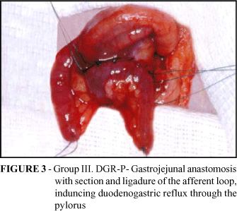
Group III (DGR-P) - 25 animals submitted to gastrojejunal anastomosis similar to group II, but with section and ligature of the afferent loop, inducing duodenogastric reflux through the pylorus (Figure 3).
All of the animals were observed and sacrificed after 36 weeks. The surgical pieces were removed under ether anesthesia and median laparotomy, following exploration of the peritoneal cavity. The stomach was opened at its greater curvature, and the duodenum and the jejunum segment at the mesenteric border. The pieces were then washed in saline, identified and photographed. The pieces were extended on polystyrene plaques with the serous surface down, secured with pins, fixed in 10% formaldehyde solution and plugged with a tampon for 24 hours and then in 70% alcohol.The squamous mucosa and the gastric mucosa were then transversally cut at the anastomosis and pylorus level, obtaining 2 fragments of about 2 mm from each segment. The fragments were identified, placed in vessels with 10% formaldehyde solution and processed by the usual methods to obtain histological cuts, which were stained by hematoxylin and eosin.
The protocol for the histological analysis was prepared, including the following criteria:
Squamous Hyperplasia - diagnosed when the stratified squamous epithelium is two or more times thicker than normal, with hyperkeratosis.
Adenomatous Hyperplasia characterized by proliferation of glandular structures, with endophytic or exophytic growth regarding the submucosa and absence of cellular atypia.
Adenocarcinoma differentiated from the previously described lesion by the mandatory presence of severe cellular and structural atypias, as well as by invasive growth.
The statistical analysis employed the Chi-square test and Fischer's exact tests, with a 5% level of significance 15.
Results
The macroscopic alterations found were identified as polypoid vegetative lesions or sessile, of varied sizes, at the gastrojejunal anastomosis level and some in the prepyloric mucosa in group III. In group I, no macroscopic lesions were identified except for discrete prominence of the mucosa at the gastrotomy location.
Table 1 demonstrates the percentages of macroscopic lesions found in the studied groups:
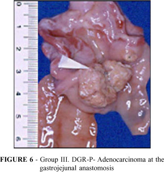
Figures 4, 5 and 6 below show the stomachs of opened animals and the aspect of the macroscopic lesions found:
It should be noted that the macroscopic lesions were more numerous both in the squamous stomach, in the anastomosis and in the prepyloric region of Group III (DGR-P), with statistical difference when compared to Group II (DGR) (p < 0.05).
The histopathological analysis was quite detailed and the analyzed gastric regions were:
- Mucosa at the anastomosis level (glandular stomach);
- Mucosa at the level of the squamous region;
- Prepyloric mucosa
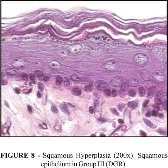
The proliferative lesions found and analyzed were adenomatous hyperplasia, squamous hyperplasia and adenocarcinoma, as can be seen in Figures 7, 8 and 9 below:
Table 2 demonstrates the percentages of histological lesions (%) in the prepyloric mucosa and in anastomosis in the studied groups.
It should be noted that in group III (DGR-P), adenomatous hyperplasia was more frequent when compared with group II (DGR), both in the prepyloric region and at the anastomosis site, with a significant statistical difference (p < 0.05).
Table 3 below shows the percentages of squamous hyperplasia found in the studied groups.
The percentages of squamous hyperplasia did not differ between groups GII (DGR) and GIII (DGR-P) (p > 0.05).
Three lesions of the mucous adenocarcinoma type were diagnosed at the gastrojejunal anastomosis site in group III, during the histological examination. These lesions are characterized by atypical columnar cells disposed in small blocks or strings, amid abundant stroma composed of mucous lakes. Table 4 demonstrates the percentage of mucous adenocarcinoma found in the gastrojejunal anastomosis mucosa.
It may be observed that adenocarcinoma occurred only in the gastrojejunal anastomosis mucosa of group III (DGR-P) with duodenogastric reflux through the pylorus.
Discussion
A review of the experimental research literature on the induction of duodenogastric reflux shows great divergence in relation to the results, probably due to the diversity of surgical techniques used, which alter the degree of reflux, as well as the histopathological criteria adopted.
The spontaneous neoplastic development rarely occurs in the glandular stomach of Wistar rats. Most of the experimental studies employ N-nitrous compounds or nitrosamines to induce stomach carcinomas in experimental animals and the rat offers the most facilities for research, both of proliferative lesions and carcinogenesis4,16.
The duodenogastric reflux may be obtained through gastric resection or not. Gastric resection models simulate the existing techniques in surgical clinic (Billroth I or II), accompanied or not by truncal vagotomy3,4,17,18.
In 1991, Kobayasi et al. 19 investigated the influence of the duodenogastric reflux in four groups of Wistar rats subjected to Billroth II gastrectomy, Billroth II gastrectomy with conversion to Roux-en-Y at 24 weeks, Billroth II gastrectomy with conversion to Roux-en-Y at 36 weeks and basic gastrectomy with Roux-en-Y reconstruction. In that study, the selected histological criteria were identical to those of the present investigation, considering the degree of cellular atypias for diagnosis of adenocarcinoma and the benign lesions classified as squamous hyperplasia or adenomatous hyperplasia. The authors have concluded, through histochemical examination, that the benign proliferative lesions presented the phenotype of gastric cells and the malignant lesions presented the phenotype of small intestine cells. They also concluded that the benign proliferative lesions do not progress to adenocarcinoma and that their incidence decreases if the duodenogastric reflux is interrupted with the conversion to Roux-en-Y.
The possible mechanisms through which the duodenogastric reflux would cause stomach cancer are controversial. One of them would be the proliferation of bacteria in the lumen of the organ, which would lead to an increase in nitrate-reducing bacteria and induce N-nitrous compound formation. The action of billiary acids and lysolecithin in the duodenal juice could also destroy the lipoprotein membrane that protects the mucosa1,16.
In 2002, Rodrigues et al. 20 investigated the effect of duodenal secretion reflux in groups of Wistar rats, with 3 experimental groups: G1 (control), G2 (submitted to gastrojejunal anastomosis and 2 weeks later to ligature of the afferent loop, deviating secretions through the pylorus) and G3 (gastrojejunal anastomosis; after the 36th week, alimentary transit was reestablished by latero-lateral anastomosis between the afferent and efferent loops. After a period of 54 weeks, the author concluded that the reflux through the pylorus favored the development of predominantly benign proliferative lesions in the gastric mucosa. The interruption of reflux caused an inhibiting effect on the growth of these lesions, confirming the benign histological characteristic of these lesions. The neoplastic development was rare in this experimental model.
Recent publications emphasized the mutagenic potential of duodenogastric reflux, both for the gastric and for the esophageal mucosa, associating it even to the occurrence of Barrett's esophagus and adenocarcinoma that appears on this epithelium2,5,13. Ovrebo et al. 21 studied the histological alterations in the gastric mucosa of rats 24, 36 and 52 weeks after gastrojejunal anastomosis, concluding that the ulcers and carcinomas appear more frequently in the body than in the antrum, with ulcers preceding the onset of carcinomas
In this research, the anastomosis was located in the posterior wall of the glandular stomach and its purpose was to compare with data in the literature whether the anastomosis location would influence the incidence of proliferative lesions in the gastric mucosa. Adenomatous hyperplasia was diagnosed predominantly at the gastrojejunal anastomosis level, presenting benign characteristics and in compliance with data in the literature. It was much more frequent in group III, in which the animals had the duodenum closed, so that the duodenal content had to pass through the stomach. Its benign characteristics suggest that if the animal were operated again AH might regress, as previously demonstrated by other authors19,20. In the squamous epithelium that lines the proximal third of the stomach the presence of squamous hyperplasia was found, with a frequency that did not significantly differ between the animals in groups II and III. These results differ from those obtained by Miwa et al. 11 that reported development of squamous cell carcinoma in a model of induction of duodenogastric reflux similar to the present study, although the follow-up period was 50 weeks and the anastomosis was located close to the greater curvature of the squamous stomach.
The adenocarcinoma diagnosed was of the mucous type and was found in 12% of the animals in group III at the gastrojejunal anastomosis level. These results were similar to those found by the authors that used the same technique for induction of duodenogastric reflux 18,19,22. Detailed studies by Rodrigues et al. 23 conclude that the adenocarcinoma induced by duodenogastric reflux is morphologically and histologically different from that occurring in the stomachs of animals following the use of nitrosamines.
In other experimental models employing Billroth II gastrectomies the incidence is high, while practically nonexistent in Roux-en-Y reconstructions, demonstrating that the determining factor is the duodenogastric reflux. The time of exposure to reflux is also a determining factor for the onset of such lesions, as found by Liu et al. 24 in their 2003 study, who did not diagnose adenocarcinomas after a 3-week follow-up.
The results of this research show that the duodenogastric reflux induced high frequency of proliferative lesions with benign histopathological characteristics in the mucosa adjacent to the gastrojejunal anastomosis or the prepyloric mucosa, and that carcinomas are rare in this experimental model.
The research in the area of experimental gastric carcinogenesis has been achieving significant advancement with the aid of molecular biology, opening new perspectives to future studies and contributing for the elucidation of the complex neoplastic transformation process.
Conclusions
The duodenogastric reflux after gastrojejunal anastomosis in the posterior wall of the stomach of Wistar rats induces a high frequency of proliferative lesions in the mucosa adjacent to the anastomosis or in the prepyloric mucosa. The histopathological characteristics of these lesions are benign and the occurrence of carcinomas not frequent in this experimental model.
Received: January 10, 2006
Review: February 18, 2006
Accepted: March 21, 2006
Conflict of interest: none
Financial source: none
How to cite this article: Monteiro RLR, Andreollo NA, Rodrigues MAM, Araujo MR. Gastric proliferative lesions induced by duodenogastric reflux in rats. Acta Cir Bras. [serial on the Internet] 2006 July-Aug;21(4). Available from URL: http://www.scielo.br/acb
- 1. Miwa K, Hasegawa H, Fugimura T, Matsumoto H, Mitaya R, Kosaka T, Miyasaki I, Hattori T. Duodenal reflux trought the pylorus induces gastric adenocarcinoma in the rat. Carcinogenesis. 1992;13: 2313-6.
- 2. Mukaisho K, Miwa K, Kumagai H, Bamba M, Sugihara H, Hattori T. Gastric carcinogenesis by duodenal reflux through gut regenerative cell lineage. Dig Dis Sci. 2003;48(11):2153-8.
- 3. Houghton PWJ, Mortensen NJM, Williamson RCN. Effect of duodenogastric reflux on gastric mucosal proliferation after gastric surgery. Br J Surg. 1987;74:288-91.
- 4. Andreollo NA, Brandalise NA, Lopes LR, Leonardi LS, Alcantara F. Are the nitrites and nitrates responsible for the carcinoma in the operated stomach ? Acta Cir Bras. 1995;10(3):103-6.
- 5. Theisen J, Peters JH, Fein M, Hughes M, Hagen JA, Demeester SR, Demeester TR, Laird PW. The mutagenic potential of duodenoesophageal reflux. Ann Surg. 2005;241(1):63-8.
- 6. Cayagill CPJ, Hill MJ, Hall CN, Kirkhan JS, Northfield, TC. Mortality from gastric cancer following gastric surgery for peptic ulcer. Lancet. 1986;1:929-31.
- 7. Lundegardh G, Adami HD, Helmick C, Zack M, Meirik O. Stomach cancer after partial gastrectomy for benign ulcer disease. N Engl J Med. 1988;319:195-200.
- 8. Tersmette KWF, Stijnen TH, Hoedemaecker J, Vanderbroucke JP, Tytgat GN. Mortality caused by stomach cancer after remote partial gastrectomy for benign conditions: 40 years of follow-up of an Amsterdam cohort of 2633 postgastrectomy patients. Gut. 1988;29:1588-9.
- 9. Toftgaard C. Gastric cancer after peptic ulcer surgery: a historic prospective cohort investigation. Ann Surg. 1989;210:159-64.
- 10. Attwood SEA, Smyrk TC, De Meester TR, Mirvish SS, Stein HJ, Hinder RA. Duodenoesophageal reflux and development of esophageal adenocarcinoma in rats. Surgery. 1992;111:503-10
- 11. Miwa K, Hasegawa M, Takano Y, Matsumoto H, Sahara H, Yagi M, Miyasaki I, Hattori T. Induction of oesophageal and forestomach carcinomas in rats by reflux of duodenal contents. Br J Cancer. 1994;70:185-9.
- 12. Miwa K, Hattori T, Miyasaki I. Duodenogastric reflux and foregut carcinogenesis. Cancer. 1995;75:1426-32.
- 13. Koek GH, Vos R, Sifrim D, Cuomo R, Janssens J, Tack J. Mechanisms underlying duodeno-gastric reflux in man. Neurogastroenterol Motil. 2005;17(2):191-9.
- 14. Sobala GM, O´Connor HJ, Dewar EP, King RFG, Axon ATR, Dixon NF. Bile reflux and intestinal metaplasia in gastric mucosa. J Clin Pathol. 1993;46:235-40.
- 15. Fonseca JL, Martins GA. Curso de estatística. 5ed. São Paulo: Editora Atlas; 1994.
- 16. Mirvish S S. The etiology of gastric cancer: intragastric nitrosamine and other theories. J Natl Cancer Inst. 1983;71:629-47.
- 17. Langhans P, Heger RA, Stegemann B. The cancer risk in the stomach subjected to nonresecting procedures. Scand J Gastroenterol. 1984;92,13841.
- 18. Saad LHC. Lesões da mucosa do coto gástrico após reconstrução a Billroth II e em Y de Roux, em ratos [Dissertação Mestrado]. Botucatu: Faculdade de Medicina, Universidade Estadual Paulista "Júlio de Mesquita Filho"; 1993.
- 19. Kobayasi S, Tatematsu M, Ogawa K, Camargo JLV, Rodrigues MAM, Ito N. Reversibility of adenomatous hyperplasia in the gastric stump after diversion of the bile reflux in rats. Carcinogenesis. 1991;12:1437-43.
- 20. Rodrigues PA, Naresse LE, Leite CVS, Rodrigues MAM, Kobayasi S. O refluxo duodeno-gástrico (RDG), através do piloro, induz lesões proliferativas gástricas em ratos ? Acta Cir Bras. 2002;17(3):160-7.
- 21. Ovrebo KK, Aase S, Grong K, Viste A, Svanes K, Sorbye H. Ulceration as a possible link between duodenogastric reflux and neoplasms in the stomach of rats. J Surg Res. 2002;107(2):167-78.
- 22. Rodrigues PA. Prevenção das lesões proliferativas da mucosa do coto gástrico de ratos gastrectomizados à Billroth II, através de derivação em Y de ROUX com ou sem vagotomia [Dissertação Mestrado]. Botucatu: Faculdade de Medicina, Universidade Estadual Paulista "Júlio de Mesquita Filho"; 1995.
- 23. Rodrigues MA, Kobayasi S, Naresse LE, de Souza Leite CV, Nakanishi H, Imai T, Tatematsu M. Biological differences between reflux stimulated proliferative stomal lesions and N-methyl-N'-nitro-N-nitrosoguanidine induced carcinomas in Wistar rats. Cancer Lett. 1999;145(1-2):85-91.
- 24. Liu JX, Liu XG, Dai Y, Tang XY, Li J, Wang HH. An experimental on gastric mucosal damage induced by duodenogastric reflux in rats. Zhonghua Nei Ke Za Zhi. 2003;42(12):837-9.
Publication Dates
-
Publication in this collection
20 Sept 2006 -
Date of issue
Aug 2006
History
-
Accepted
21 Mar 2006 -
Received
10 Jan 2006 -
Reviewed
18 Feb 2006


