Abstracts
Thyrty-six rats werw implanted with endometrial squares (2 X 2mm) to the left parietal peritoneum and the right uterine horn was tied. The rats were mated for 20 days and then sacrificed. Topic and ectopic endometrial histological pattern were compared. There was a correlation between the histological pattern of the autografted endometrium and the eutopic endometrium im most rats, pregnant or not. This suggests that both responded in a similar way to the same endocrine stimuli. The experimental model for endometriosis admiting the histological correlation between the uterine endometrium and the ectopic is factible and it is easy to be repetead.
Endometriosis; Endometrial histology; Rats
Trinta e seis ratas foram submetidas a implante cirúrgico de retalho endometrial quadrangular (2 x 2mm) no peritônio parietal esquerdo e ligadura do corno uterino direito. As ratas foram acasaladas por 20 dias e logo após sacrificadas. Foram comparados o padrão histológico do endométrio eutópico e do ectópico.Houve correspondência entre o padrão histológico do endométrio auto-transplantado e o endométrio eutópico na maioria das ratas, prenhes ou não, sugerindo que ambos responderam de forma semelhante aos mesmo estímulos endócrinos. O modelo experimental de endometriose na rata, permitindo a correlação histológica entre o endométrio uterino e o ectópico, é viável e de fácil repetição.
Endometriose; Histologia de endométrio; Ratas
3 - ARTIGO ORIGINAL
EXPERIMENTAL MODEL FOR ENDOMETRIOSIS. COMPARATIVE HISTOLOGICAL STUDY BETWEEN THE ECTOPIC AND EUTOPIC ENDOMETRIUM1 1 Trabalho realizado no Curso de Pós-Graduação em Cirurgia Abdominal da Faculdade de Medicina da UFMG - Belo Horizonte. Apoio da Fundação de Amparo à Pesquisa do Estado de Minas Gerais (FAPEMIG). 2 Professor Adjunto Doutor do Departamento de Ginecologia e Obstetrícia da Faculdade de Medicina da UFMG. 3 Professor Titular de Cirurgia do Aparelho Digestivo do Departamento de Cirurgia da Faculdade de Medicina da UFMG. 4 Professor Adjunto Doutor do Departamento de Ginecologia e Obstetrícia da Faculdade de Medicina da UFMG. 5 Professor Adjunto Doutor do Departamento de Anatomia Patológica e Medicina Legal da Faculdade de Medicina da UFMG. 6 Aluna do Curso de Pós-Graduação da Faculdade de Medicina da UFMG.
Cézar Alencar de Lima Rezende2 1 Trabalho realizado no Curso de Pós-Graduação em Cirurgia Abdominal da Faculdade de Medicina da UFMG - Belo Horizonte. Apoio da Fundação de Amparo à Pesquisa do Estado de Minas Gerais (FAPEMIG). 2 Professor Adjunto Doutor do Departamento de Ginecologia e Obstetrícia da Faculdade de Medicina da UFMG. 3 Professor Titular de Cirurgia do Aparelho Digestivo do Departamento de Cirurgia da Faculdade de Medicina da UFMG. 4 Professor Adjunto Doutor do Departamento de Ginecologia e Obstetrícia da Faculdade de Medicina da UFMG. 5 Professor Adjunto Doutor do Departamento de Anatomia Patológica e Medicina Legal da Faculdade de Medicina da UFMG. 6 Aluna do Curso de Pós-Graduação da Faculdade de Medicina da UFMG.
Alcino Lázaro da Silva3 1 Trabalho realizado no Curso de Pós-Graduação em Cirurgia Abdominal da Faculdade de Medicina da UFMG - Belo Horizonte. Apoio da Fundação de Amparo à Pesquisa do Estado de Minas Gerais (FAPEMIG). 2 Professor Adjunto Doutor do Departamento de Ginecologia e Obstetrícia da Faculdade de Medicina da UFMG. 3 Professor Titular de Cirurgia do Aparelho Digestivo do Departamento de Cirurgia da Faculdade de Medicina da UFMG. 4 Professor Adjunto Doutor do Departamento de Ginecologia e Obstetrícia da Faculdade de Medicina da UFMG. 5 Professor Adjunto Doutor do Departamento de Anatomia Patológica e Medicina Legal da Faculdade de Medicina da UFMG. 6 Aluna do Curso de Pós-Graduação da Faculdade de Medicina da UFMG.
João Lúcio dos Santos Júnior4 1 Trabalho realizado no Curso de Pós-Graduação em Cirurgia Abdominal da Faculdade de Medicina da UFMG - Belo Horizonte. Apoio da Fundação de Amparo à Pesquisa do Estado de Minas Gerais (FAPEMIG). 2 Professor Adjunto Doutor do Departamento de Ginecologia e Obstetrícia da Faculdade de Medicina da UFMG. 3 Professor Titular de Cirurgia do Aparelho Digestivo do Departamento de Cirurgia da Faculdade de Medicina da UFMG. 4 Professor Adjunto Doutor do Departamento de Ginecologia e Obstetrícia da Faculdade de Medicina da UFMG. 5 Professor Adjunto Doutor do Departamento de Anatomia Patológica e Medicina Legal da Faculdade de Medicina da UFMG. 6 Aluna do Curso de Pós-Graduação da Faculdade de Medicina da UFMG.
Helenice Gobbi5 1 Trabalho realizado no Curso de Pós-Graduação em Cirurgia Abdominal da Faculdade de Medicina da UFMG - Belo Horizonte. Apoio da Fundação de Amparo à Pesquisa do Estado de Minas Gerais (FAPEMIG). 2 Professor Adjunto Doutor do Departamento de Ginecologia e Obstetrícia da Faculdade de Medicina da UFMG. 3 Professor Titular de Cirurgia do Aparelho Digestivo do Departamento de Cirurgia da Faculdade de Medicina da UFMG. 4 Professor Adjunto Doutor do Departamento de Ginecologia e Obstetrícia da Faculdade de Medicina da UFMG. 5 Professor Adjunto Doutor do Departamento de Anatomia Patológica e Medicina Legal da Faculdade de Medicina da UFMG. 6 Aluna do Curso de Pós-Graduação da Faculdade de Medicina da UFMG.
Madalena Maria Ferreira Martins6 1 Trabalho realizado no Curso de Pós-Graduação em Cirurgia Abdominal da Faculdade de Medicina da UFMG - Belo Horizonte. Apoio da Fundação de Amparo à Pesquisa do Estado de Minas Gerais (FAPEMIG). 2 Professor Adjunto Doutor do Departamento de Ginecologia e Obstetrícia da Faculdade de Medicina da UFMG. 3 Professor Titular de Cirurgia do Aparelho Digestivo do Departamento de Cirurgia da Faculdade de Medicina da UFMG. 4 Professor Adjunto Doutor do Departamento de Ginecologia e Obstetrícia da Faculdade de Medicina da UFMG. 5 Professor Adjunto Doutor do Departamento de Anatomia Patológica e Medicina Legal da Faculdade de Medicina da UFMG. 6 Aluna do Curso de Pós-Graduação da Faculdade de Medicina da UFMG.
REZENDE, C.A.L.; SILVA, A.L.; JÚNIOR, J.L.S.; GOBBI, H.; MARTINS, M.M.F. Experimental model for endometriosis - comparative histological study between the ectopic and eutopic endometrium - Acta. Cír. Bras. 12(4):226-30, 1997.
SUMMARY: Thyrty-six rats werw implanted with endometrial squares (2 X 2mm) to the left parietal peritoneum and the right uterine horn was tied. The rats were mated for 20 days and then sacrificed. Topic and ectopic endometrial histological pattern were compared. There was a correlation between the histological pattern of the autografted endometrium and the eutopic endometrium im most rats, pregnant or not. This suggests that both responded in a similar way to the same endocrine stimuli. The experimental model for endometriosis admiting the histological correlation between the uterine endometrium and the ectopic is factible and it is easy to be repetead.
SUBJECTS HEADINGS: Endometriosis. Endometrial histology. Rats.
INTRODUCTION
The high frequency of endometriosis in infertile patients and the existing controversies concerning the pathophysiology of endometriosis and its influence on pregnancy and vice versa demonstrate the need for research in laboratory animals, aiming at the development of well-defined, easily reproducible experimental models of the disease. The histologic and functional behavior of endometriotic implants during pregnancy is still a controversial subject in the literature.9
The ability of endometriotic tissue to remain viable and to proliferate after autotransplantation to various ectopic locations has been documented in monkeys, rabbits, rats and other animals.8,12
The rat has several attributes that make is appealing for use as an experimental model for endometriosis. Like humans, it has spontaneous ovulation, whereas as the rabbit is an induced ovulator and, therefore, does not have a discernible estrous cycle. The double uterus of the rat allows for altering one side and studying the effects of these alterations, as compared with the contralateral side.
The intent of this study was to obtain an experimental model of endometriosis in rats which, in addition to being easily performed and readily reproducible, permits a morphological and functional comparison to be made between the ectopic endometrium (uterine horn) and the eutopic endometrium (implant) in pregnant and nonpregnant rats.
METHOD
Thirty-six adult albino rats ("Rattus norvegicus albinus") of verified fertility, weighing 250 g on average, supplied by the Central Vivarium of Faculdade de Medicina da UFMG, were selected for study.
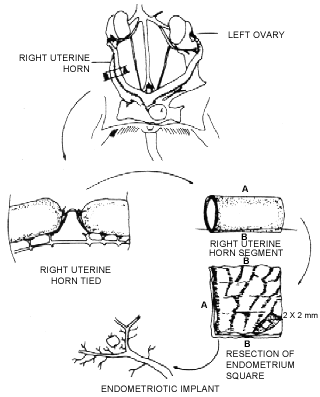
Anesthesia was induced by placing the rat in a bell jar, in contact with 2% sulfuric ether. The operative field was bounded by a fenestrated sheet, and a longitudinal, midline supra-public incision approximately 5 cm in length was performed. After the laparotomy, the abdominal cavity was examined for identification of the right uterine horn, which was exposed and isolated by a small sterilized green plastic sheet. With the aid of a surgical microscope, the mesenteric vascular arcade was identified 2 cm away from the junction with the uterine cervix, and cauterization was performed over and approximate length of 1.5 cm, using special microsurgical nippers connected to a bipolar cautery. The uterine horn was sectioned perpendicularly to its axis, at a point 2 cm away from its junction with the uterine cervix, and a 1 cm segment was resected and immediately placed in 9% saline solution (Figure 1).
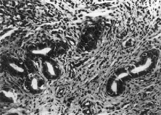
- Viable endometrial implant containing five straight tubular glands, lined with vacuumlinized columnar epithelium. The stroma contains several newly-formed capillaries (Case of group 2; HE; 260 X).
The stumps of the uterine horn were tied 5-0 nylon suture, with interposition of mesometrium between them.
The resected segment of the horn was transferred to a slide with a millimetric scale, opened longitudinally and kept humid. Resection of the endometrium was performed, with straight microsurgical scissors being used for separating the basal layer from the muscle layer. A 2 x 2 mm square was then removed from the fragment of the endometrium.
For performing the implant, the intestinal loops were separated and kept apart, to allow visualization by transparency and exposure of the vascular-nervous hypogastric bundle under the left parietal peritoneum. With the aid of a surgical microscope , the endometrial square was sutured onto the peritoneum, at a point just above the bifurcation of the hypogastric vessels. A single stitch using 8-0 nylon suture was applied at two opposite sides of the "square", with the basal portion of the endometrium contacting the peritoneum (Figure 1). Finally, following inspection of the abdominal cavity, the abdominal wall was closed with crossed continuous total stitches, using no. 1 silk suture.
After a period of 15 days under the initial feeding and hydration conditions, the rats were mated in the proportion of 4 females to one male of proven fertility.
Twenty days after mating began, the peritoneal cavity of the rats was inspected. The site of the implant, both horns, and the ovaries were then resected. The wole (left) horn was opened longitudinally for the identification and counting of embryos, in case the rat was pregnant. The animals were sacrificed by pneumothorax induced by thoracophrenectomy.
The fragments containing the endometriotic implant were sectioned into two equal parts. The uterine horns were sectioned at three levels: at the junction with the uterine cervix, across the medial portion, and across the extremity near the ovarium, with 3 fragments being obtained from each horn. The blocks were submitted to microtomy at six different levels.
The glands in the ectopic and eutopic endometrium were evaluated, with regard to: glandular pattern (straight tubular or tortuous); type of lining epithelium; cytoplasmic and nuclear features; and the presence of mitotic figures. The stromal pattern (dense or loose) and the presence of inflammatory cells were also studied.
RESULTS
Endometriotic implants that were considered viable showed glands and stroma of the endometrial type, with epithelial lining and luminal formation (Figure 2). Endometrial-like glands were observed in 19 (52.8%) animals. Of 19 rats with endometrial implants considered viable, 11 became pregnant. In 14 (73.7%) of these animals, there was histologic similarity between the glandular pattern of the implant and that of the uterine horns in the same animal. In addition to occuring in a smaller number, the endometrial glands in the implant showed less pronounced functional and gravidic changes than those in the endometrium of the whole horn in the same rat.
- Viable implant in a nonpregnant rat, showing straight and tortuous tubular glands, lined with non-vacuumlinized columnar epithelium. Presence of mitosis in the glandular epithelium (arrow) (Case of group 2; HE; 400 X).
In six (66.7%) rats that became pregnant histologic similarity was found between the gland pattern of the implant and that of the tied horn.
In general, there was correpondence between the type of lining epithelium and the endometrial glandular pattern, i. e., where the glands were tortuous, the epithelial lining was vacuolized columnar; where the glands were of the straight tubular type, the epithelial lining was simple columnar.
Stroma of the endometrial type was observed in all viable implants, its characteristics being similar to stroma in the uterine horns.
DISCUSSION
The animal selected for this study was the rat, not only because its physiological and endocrine functions are well-known, but also on account of its excellent reproductive capacity and great resistence to anesthetics, surgical interventions, diseases, and infections. The monkey would be the ideal animal for the induction of endometriosis, due to its physiological similarity and greater phylogenetic proximity to the human species.11 Yet, the difficulty to obtain these species, the high cost and the maintenance problems involved limit their use as research animals. The main disadvantage of rats in relation to monkeys is that the anatomy of the internal genitals and the endometrial changes in the monkey resemble more closely those in the human. However, unlike the monkey, the rat has a bicornuat uterus, which was an important anatomical feature in this study, as it permitted one of the horns to be tied, with the other one remaining intact. A comparative study of the endometrium in three different situations was thus possible, upon the rat becoming pregnant: 1) an ectopic endometrium; 2) a topic endometrium which, although under the influence of the hormonal milieu of pregnancy, did not receive the embryo, and; 3) another topic endometrium which, in addition to the hormonal stimuli of pregnancy, also received the embryos and was under local influences. A more adequate comparison was therefore possible between the ectopic and eutopic endometrium, both influenced by the same hormonal milieu, yet without the embryonic implantations.
The histopathologic finding of endometrial glands and stroma in the surgical site of implantation of endometrial squares in 52.8% of the animals allowed the diagnosis of experimental endometriosis, as well as the viability of the implants, to be established.
The small number of glands in the endometrial implant did not allow a quantitative comparison with the glands in the eutopic endometrium. However, a qualitative comparison was possible. RAJKUMAR et all6, in a study of experimental endometriosis in rats, also noted an evident development of endometrial stroma and vascularization, although glandular development was reduced.
In the majority (66.7%) of the animals, there was correspondence or similarity, with regard to glandular pattern (type of gland, epithelial lining), between the implant endometrium and the eutopic endometrium in the same pregnant rat. These data suggest that the ectopic endometrial implant preserves its responsiveness to the hormonal stimulation of pregnancy. The histologic correspondence noted between the implant endometrium and that of the uterine horns in the same rat suggests that both tissues react to hormonal influences in the same way. It could also indicate that vascular supply was adequate, permitting serous stimuli to be received, and that the transplanted tissue remained reasonably sensitive to hormonal stimulation.
Correlative histologic studies of the eutopic endometrium and pelvic endometriosis in women have produced conflicting results. Some authors state that, when the ectopic endometrium and the topic endometrium react to ovarian hormones in the same way, they have a similar histologic appearence.2,7 Other authors, however, have found a different histologic appearence in most of the cases. The cause for this variable hormonal dependence and the histologic and ultrastructural differences between endometrial implants is not clear.9 Among the possible causes, one might include a populations deficiency of steroid receptors, an altered epithelium-stroma ratio, local factors such as abnormal vascularization, collagen deposition and inflamatory changes, or atrophy from the pressure resulting from the inability of secretions to scape.4
Substantial differences appear to exist between stroma and glands in ectopic endometrium implants, with regard to the distribuition of receptors3. A normal stromal/epithelial relationship is probably necessary to generate a histologic response to a rapidly fluctuating hormonal enviroment. Thus, it is not surprising that there as in ample variability in the histologic appearence of ectopic endometrium.4
Experimental evidence concerning the hormone requirements of endometrial implants in endometriosis is still insufficient1. Normal endometrium and endometrial implants are know to contain strogen, progesterone and androgen receptors. A hormonal response pattern similar to that of normal endometrial cells is probably retained by endometrial implants1. Recent studies show that, in addition to the concentration of steroid receptors in endometrial implants being lower than that in the eutopic endometrium, over 20% of implants do not have measurable concentrations of strogen or progesteron receptors.13 The differences in the concentration of steroid receptors correlate to histologic and ultrastructural studies that demonstrate an incomplete morphological response of endometrial implants to cyclic hormonal alterations.7,10
Through controlled experiments, the rat model could be used for investigating the role of endometriosis in infertility, the formation of adhesions, ovarian and uterine hormone receptors, the production of prostaglandins, and autoimmunity. Among the factors that could be systematically evalueted are the morphological characteristics of the implant, the histologic behavior of ectopic implants as compared to the topic endometrium, under the influence or not of the endocrine stimulation of pregnancy, the effects of medication, and the different forms of treatment.
CONCLUSIONS
1 - The experimental rat model of endometriosis, allowing histologic correlation between the uterine endometrium and the ectopic endometrium, is feasible and can be easily repeated.
2 - The growth and development of ectopic endometrial tissue transplanted into rats provides a suitable model for studying those aspects of endometriosis that cannot be adequately investigated in the human.
3 - In most the cases, there was correspondence between the histologic pattern of the implant endometrium and that of the eutopic endometrium, which demonstrates that both respond in a similar way to the same endocrine stimuli.
4 - Responsiveness to the hormonal stimulation of pregnancy was preserved by the ectopic endometrium square, whose glandular pattern was similar to that of the topic endometrium.
REZENDE, C.A.L.; JÚNIOR, J.L.S.; GOBBI, H.; MARTINS, M.M.F. Modelo experimental de endometriose: estudo histológico comparativo entre o endométrio estópico e o eutópico - Acta. Cír. Bras. 12(4): 226-30, 1997.
RESUMO: Trinta e seis ratas foram submetidas a implante cirúrgico de retalho endometrial quadrangular (2 x 2mm) no peritônio parietal esquerdo e ligadura do corno uterino direito. As ratas foram acasaladas por 20 dias e logo após sacrificadas. Foram comparados o padrão histológico do endométrio eutópico e do ectópico.Houve correspondência entre o padrão histológico do endométrio auto-transplantado e o endométrio eutópico na maioria das ratas, prenhes ou não, sugerindo que ambos responderam de forma semelhante aos mesmo estímulos endócrinos. O modelo experimental de endometriose na rata, permitindo a correlação histológica entre o endométrio uterino e o ectópico, é viável e de fácil repetição.
DESCRITORES: Endometriose. Histologia de endométrio. Ratas.
Endereço para correspondência:
Cézar Alencar de Lima Rezende
Rua Maranhão, 1061 apto. 1002
Funcionários - 30.150-331 - Belo Horizonte - MG
Fone (031) 221.3860
Data do recebimento: 15.04.97
Data da revisão: 20.05.97
Data da aprovação: 10.06.97
- 1. BARBIERI, R. L. & KISTNER, R. W. - Hormonal terapy of endometriosis. In: RAYNAUD, J. P.; OJASOO, T.; MARTIN, L. (ed) - Medical mangement of endometriosis New York, Raven Press, 1984. cap. 3, p. 27-40.
- 2. BERGQVIST, A.; KJELL, C.; JEPPSSON, S.; KULLANDER, S.; LJUNGBERG, O. - Histochemical localization of specific estrogen and progesterone binding in human endometrium and endometriotic tissue: a preliminar report. Acta. Obstet. Gynecol. Scand. (Suppl.), 123:15-19, 1984.
- 3. GOULD, S.F.; SHANNON, J.M.; CUNHA, G.R. - Nuclear estrogen binding sites in human endometriosis. Fertil. Steril., 39:520-4, 1983.
- 4. HANNEY, A. F. - Endometriosis; pathogenesis and pathophysiology. In: WILSON, E. A. (ed). Endometriosis New York, Alan R. Liss, 1987. cap. 3, p. 23-51.
- 5. NOVAK, E. & DE LIMA, O. A. - A correlative study of adenomyosis and pelvic endometriosis, with special reference to the hormonal reaction of ectopic endometrium. Am. J. Gynecol., 56:634-44, 1948.
- 6. RAJKUMAR, K.; SCHOTT, P.; SIMPSON, C. W. - The rat as an animal model for endometriosis to examine recurrence of ectopic endometrial tissue after regression. Fertil. Steril. ,53:921-5, 1990.
- 7. RODDICK, J.W.; CONKEY, G.; JACOBS, E. J. - The hormonal response of endometrium in endometriotic implants and its relationship to symptomatology. Am. J. Obstet. Gynecol., 79: 1173-7, 1960.
- 8. SCHENKEN, R. S. & ASCH, R. H. - Surgical induction of endometriosis in the rabbit: effect on fertility and concentration of peritoneal fluid prostaglandins. Fertil. Steril., 34: 581-7, 1980.
- 9. SCHENKEN, R. S. - Pathogenesis. In: SCHENKEN, R. S. Endometriosis, Philadelphia, J. B. Lippincott, 1989. cap. 1, p. 1-48.
- 10. SCHWEPPE, K. W.; WYNN, R. M.; BELLER, F. B. - Ultrastructural comparison of endometriotic implants and eutopic endometrium. Am. J. Obstet. Gynecol., 148: 1024-39, 1984.
- 11. VERNON, M. W. & HODGEN, G.D. - Experimental endometriosis. In: WILSON, E. A. (ed.). Endometriosis, New York, Alan R. Liss, 1987. cap. 12, p. 207-23.
- 12. VERNON, M. W. & WILSON, E. A. - Studies on surgical induction of endometriosis in the rat. Fertil. Steril., 44:684-94, 1985.
- 13. VIERIKKO, P.; KAUPILLA, A.; RŐNNBERG, L.; VIHKO, R. - Steroidal regulation of endometriosis tissue: lack of induction of 17-beta-hydroxysteroid dehydrogenase activity by progesterone, medroxy-progesterone acetate, or danazol. Fertil. Steril., 43:218-24, 1985.
Publication Dates
-
Publication in this collection
31 May 2001 -
Date of issue
Dec 1997
History
-
Received
15 Apr 1997 -
Reviewed
20 May 1997 -
Accepted
10 June 1997