Abstracts
PURPOSE: To evaluate the healing process of rat traumatic liver lesion corrected with the use of 2-octyl cyanoacrylate adhesive, compared to the use of biologically absorbable chromed catgut thread suture. METHODS: Thirty mail adult rats were divided into two groups (15 per group) according to the used method for liver lesion correction as follows: adhesive group (AG), and catgut group (CG); each group being divided into three subsets of five animals (7th, 14th, and 21st day), respectively, according to post-surgery evaluation. All animals were submitted to homogeneous lesion applying synthetic bonding to AG and using chromed catgut suture to CG for lesion correction. Macroscopic and microscopic parameters of healing processes were evaluated. RESULTS: Both groups of animals showed excellent abdominal wall healing, with no evidence of infection, and no abdominal cavity peritonitis or abscess. The presence of adherence was observed in both groups with no statistically significant difference. As to macroscopic evaluation, there was statistically significant difference with respect to specific factors of clinical inflammation (ischemic inflammation and giant celular inflammatory reaction) between animals evaluated on the 10th day (ischemic necrosis and giant celular inflammatory reaction) among animals evaluated on the 14th day (A14 versus C14). CONCLUSION: Applying 2-octyl-cyanoacrylate adhesive for correcting rat liver lesion does not change healing process when compared to the use of chromed catgut stitch.
Wound Healing; Liver; Adhesives; Rats
OBJETIVO: Avaliar o processo de cicatrização de lesão induzida em fígado de rato utilizando adesivo de 2-octil cianoacrilato em comparação com a sutura com catgut cromado. MÉTODOS: Utilizou-se 30 ratos adultos, machos, distribuídos em dois grupos de 15 animais denominados de grupo adesivo (GA) e grupo catgut (GC); cada grupo foi dividido em três subgrupos, cada um com cinco animais, de acordo com a data marcada para avaliação pós-operatória (sétimo, décimo quarto e vigésimo primeiro dia). Todos os animais foram submetidos à lesão homogênea, aplicando o adesivo sintético no GA e utilizando sutura com catgut cromado no GC para correção das lesões. Foram avaliados parâmetros de macroscópicos e microscópicos do processo de cicatrização. RESULTADOS: Em ambos os grupos os animais evoluíram com boa cicatrização da parede abdominal, sem evidência clínica de infecção, sem abscessos ou peritonite na cavidade abdominal; a presença de aderências foi observada em ambos os grupos sem diferenças estatisticamente significantes; quanto à avaliação microscópica só houve diferença estatisticamente significante quanto aos parâmetros específicos de inflamação crônica (necrose isquêmica e reação gigantocelular) entre os animais avaliados no décimo quarto dia (A14 vs. C14). CONCLUSÃO: O emprego de adesivo de 2-octil cianoacrilato na correção de lesão em fígado de rato não altera o processo de cicatrização quando comparado ao uso de sutura com fio de categute cromado.
Cicatrização; Fígado; Adesivos; Ratos
6 - ORIGINAL ARTICLE
MODELS, BIOLOGICAL
Use of 2-octyl cyanoacrylate adhesive in rat liver induced lesion1 1 Research performed at Department of Medicine II, School of Medicine, Federal University of Maranhao (UFMA), Sao Luis, Brazil.
Uso adesivo de 2-octil cianoacrilato em lesão induzida de fígado de ratos
Orlando José dos SantosI; Giancarlo de Souza MarquesII; Euler Nicolau Sauaia FilhoIII; Gustavo Medeiros FrotaIII; Rayan Haquim Pinheiro SantosIV; Rennan Abud Pinheiro SantosV
IPhD, Associate Professor, Department of Medicine II, UFMA, Sao Luis-MA, Brazil. Main author. Conception, design, intellectual and scientific content of the study
IIProctology Surgeon, Presidente Dutra University Hospital, UFMA, Sao Luis-MA, Brazil. Acquisition and interpretation of data
IIIMD, Presidente Dutra University Hospital, UFMA, Sao Luis-MA, Brazil. Intellectual and scientific content of the study, acquisition and interpretation of data
IVGraduate student, Federal University of Paraiba (UFPB), Joao Pessoa-PB, Brazil. Acquisition of data
VGraduate student, UFMA, Sao Luis-MA, Brazil. Acquisition of data
Correspondence Correspondence: Orlando José dos Santos Universidade Federal do Maranhão, Departamento de Medicina II Avenida dos Portugueses, s/n 65085-580 São Luis - MA Brasil orlanddojs@hotmail.com
ABSTRACT
PURPOSE: To evaluate the healing process of rat traumatic liver lesion corrected with the use of 2-octyl cyanoacrylate adhesive, compared to the use of biologically absorbable chromed catgut thread suture.
METHODS: Thirty mail adult rats were divided into two groups (15 per group) according to the used method for liver lesion correction as follows: adhesive group (AG), and catgut group (CG); each group being divided into three subsets of five animals (7th, 14th, and 21st day), respectively, according to post-surgery evaluation. All animals were submitted to homogeneous lesion applying synthetic bonding to AG and using chromed catgut suture to CG for lesion correction. Macroscopic and microscopic parameters of healing processes were evaluated.
RESULTS: Both groups of animals showed excellent abdominal wall healing, with no evidence of infection, and no abdominal cavity peritonitis or abscess. The presence of adherence was observed in both groups with no statistically significant difference. As to macroscopic evaluation, there was statistically significant difference with respect to specific factors of clinical inflammation (ischemic inflammation and giant celular inflammatory reaction) between animals evaluated on the 10th day (ischemic necrosis and giant celular inflammatory reaction) among animals evaluated on the 14th day (A14 versus C14).
CONCLUSION: Applying 2-octyl-cyanoacrylate adhesive for correcting rat liver lesion does not change healing process when compared to the use of chromed catgut stitch.
Key words: Wound Healing. Liver. Adhesives. Rats.
RESUMO
OBJETIVO: Avaliar o processo de cicatrização de lesão induzida em fígado de rato utilizando adesivo de 2-octil cianoacrilato em comparação com a sutura com catgut cromado.
MÉTODOS: Utilizou-se 30 ratos adultos, machos, distribuídos em dois grupos de 15 animais denominados de grupo adesivo (GA) e grupo catgut (GC); cada grupo foi dividido em três subgrupos, cada um com cinco animais, de acordo com a data marcada para avaliação pós-operatória (sétimo, décimo quarto e vigésimo primeiro dia). Todos os animais foram submetidos à lesão homogênea, aplicando o adesivo sintético no GA e utilizando sutura com catgut cromado no GC para correção das lesões. Foram avaliados parâmetros de macroscópicos e microscópicos do processo de cicatrização.
RESULTADOS: Em ambos os grupos os animais evoluíram com boa cicatrização da parede abdominal, sem evidência clínica de infecção, sem abscessos ou peritonite na cavidade abdominal; a presença de aderências foi observada em ambos os grupos sem diferenças estatisticamente significantes; quanto à avaliação microscópica só houve diferença estatisticamente significante quanto aos parâmetros específicos de inflamação crônica (necrose isquêmica e reação gigantocelular) entre os animais avaliados no décimo quarto dia (A14 vs. C14).
CONCLUSÃO: O emprego de adesivo de 2-octil cianoacrilato na correção de lesão em fígado de rato não altera o processo de cicatrização quando comparado ao uso de sutura com fio de categute cromado.
Descritores: Cicatrização. Fígado. Adesivos. Ratos.
Introduction
The abdominal cavity holds most of the digesting organ system as well as the adjunct glandules, being part of the urogenital system and autonomous plexus1.The increasing urban violence coupled with driving accidents has increased abdominal trauma incidence. Being the major abdominal human cavity glandule, the liver is a main lesion target. Therefore, liver traumatism is quite frequent, accounting to a high mortality rate2.
Liver trauma merits special attention as to its diagnosis and management. It is assumed that around 45% of closed abdominal trauma and 40% of open abdominal traumas involving the liver. Regarding closed abdominal trauma 84% due traffic accidents followed by 8% trauma by falling, and 2% by physical attack3.
Though presenting under several aspects, most liver traumas provoke small liver lesions that at times do not need specific management and when they do, they are solved with minor procedures. Liver lesions can be treated in several manners depending on their severity varying from simple stitching, drainage, regradas and widening resections, or even transplant. In spite of hospital improved infrastructure, aseptic means, material, technical and anatomical know-how, mortality rate is still elevated for subjects with lever lesions, though such organ is protected by the costal gradil4,5.
Lately, several treatment alternatives have been tried for liver lesion such as collagen haemostatic agent application, cellulose gelatin, as well as the use of base synthetic bondings diffused in spleen and digestive system, notably in experimental studies6.
There is scant information in the literature referring to this bonding use in parenchimatous bleeding. Aiming at collecting more information, we set out to study the use of 2-octyl cyanocrylate in rat liver provoked lesions to evaluate the healing process of the target organ lesions, comparing the use of the bonding agent with the long traditional chromed catgut suture, to evaluate both microscopic and macroscopic aspects.
Methods
The project was evaluated and approved by the by the Ethics in Research Committee at the Federal University of Maranhao (UFMA), protocol nº 00386/2004. The study followed the guidelines for animal research as set out by the Brazilian College of Animal Experimentation (COBEA).
The sample was made up of 30 male Wistar rats (Rattus norvegicus albinus, Rodentia mammalia), supplied by the UFMA vivarium. It was between 40 and 60 days old (average age 46 days). The weight was between 119g and 286g (average weight 219.1g). They were kept in groups of five per standard cage for the species at the Research Laboratory of the Physiology Department where the light-dark cycle was 12 hours and humidity was the same as a normal environment without any artificial regulation. The temperature was a constant 20 ± 2ºC. The animals were fed standard commercial food and had free access to water throughout the experiment.
An anesthetic by an intramuscular injection of 0.2ml/100g of weight from a mixture of 1ml of ketamine (50mg) and 1ml of xylazine (20mg). The trychotomy of the ventral abdominal wall was done and anti-sepsis with polyvinylpyrrolidone-iodine.
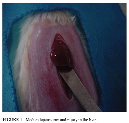
The surgical procedure consisted of a median laparotomy with four centimeters. The injury in the liver was done at 2.5cm from the peritoneal reflection, followed by simple synthesis with catgut cromate (two stitches) on the catgut groups and synthesis with cianocrilate adhesive and adhesive groups. We then moved on to the laparorraphy at two levels: the first, peritoneal-muscularaponeurotic; the second, of the skin, with continuous synthesis, utilizing 5.0 monofilament nylon (Figures 1 and 2).
After recuperating from the anesthetic, the rats were returned to their cages, where they had free access to standard commercial food and water. Immediately after surgery they were given an intramuscular dose of sodium diclofenac at a dosage of 10 mg/kg, as an analgesic and anti-inflammatory drug.
The animals were divided in two groups: adhesive group (AG) and catgut group (CG). Each group subdivided at random into three subgroups according to the date of euthanasia: catgut 7 (C7), catgut 14 (C14), catgut 21 (C21), adhesive 5 (A7), adhesive 5 (A14) and adhesive 21 (A21).The numbers correspond on the seventh, fourteenth and twenty one days after surgery. For euthanasia, a lethal dose of intraperitoneal sodium thiopental was used at 120mg/kg.
After euthanasia, another laparotomy was performed, enabling us to observe the abdominal cavity, the existence of peritonitis or abscess, adherences and the integrity of the synthesis (Figure 3).
After this evaluation, the surgical parts were opened lengthwise and extended on filter paper and set in 10% formalin and forwarded for histopathological study.
Results
Macroscopic evaluation
All animals from the experiment had their clinical course classified as good during the post-surgery period, up to the programmed of death day.
The animals presented satisfactory healing of surgical wound, with no evidence of infection or dehiscence. No animals from either studied groups presented abscess or swelling upon abdominal cavity evaluation. The presence of bonding with adjoining structures evaluated according to the Nair score varied in both groups, showing a greater frequency on the abdominal wall and lower small intestine. On the other hand, there was statistically significant difference on day 7th only among animals from the adhesive and catgut group, with predominating grade 1 for the C7, and grade 0 for the A7, p=0.036; there was no statistically significant difference between C14 and A14, and between C21 and A21 (Table 1).
There was no statistically significant difference on the temporal (intergroup) evaluation on the incidence of adhesive on the catgut group (C7 versus C14 versus C21), p=0.177, however, upon evaluation of adhesive on the catgut group, statistically significant difference was observed (A7 versus A14 versus A21), p=0.030.
Microscopic evaluation
Acute inflammation intensity was markedly accentuated for the C7 and discrete on the A7 with no statistically significant difference, p=0.250. It showed to be moderate on the 14th day for the CG and largely discrete for the AG; on day 21st it showed predominantly moderated on both evaluated groups. There was no statistically significant difference between the two groups neither on the 14th or 21st day with p=0.117 and p=0.676, respectively (Table 2).
As to the intergroup evaluation there was no statistically significant difference for both chromed group with p=0.627, and bonding group with p=0.259.
Chronic inflammation intensity was largely absent on the C7, discrete and moderate on the A7 with no statistically significant difference, p=0.296. It was absent in most animals from the C14, whereas there was a discrete intensity on the A14 with no statistically significant difference, p=0.117. On day 21st it presented predominantly discrete for both CG and AG with no statistically significant difference, p=0.403 (Table 3).
There was no statistically significant difference for the catgut group (p=0.438), nor for the adhesive group (p=0.274) on the intergroup assessment.
Discussion
Due to its easy availability, handling and accommodation, resistance to manipulation, surgical aggression, and to infectious processes, the rat was used for evaluation of liver healing course.
In the present study the animals were divided into two groups denominated catgut group (CG) and adhesive group (AG), each group was further subdivided into three subgroups, according to the stipulated date for the proposed parameters evaluation (C7, C14 and C21; and A7, A14, and A21), with observations taking place on the 7th, 14th, and 21st days. The 7th day was chosen for the first observation for being a still acute period for the inflammatory response7,8. The 2nd observation took place on the 14th day to evaluate inflammatory response and the intermediary stage of the healing parameters9. The third observation was done on the 21st day aiming at evaluating the inflammatory standard and late healing10.
Because of their sealing properties, cyanoacrylate adhesive and their derivatives, and fibrin glue, came to be used in animals and in almost all organic tissues in healing studies11.
Among the cyanoacrylate derivatives, we chose to use the 2-octyl cyanoacrylate adhesive due to its having lower exothermal reaction compared to others, seeing that as the number of radical carbon increases, tissue reaction diminishes. It also has sealing and bactericide properties12. Its bactericide properties are of major importance, since the second main cause of death by liver traumatism is represented by intra or extra-abdominal sepsis, seconded only by hemorrhage5
Cyanoacrylate adhesive was applied to the liver diaphragmatic surface of the adhesive group (AG) according to the manufacturer (Dermabond®, Ethicon) recommendations, obtaining satisfactory haemostatic activity. The need for compression and site cleansing for adequate bonding polymerization must be highlighted13.
Simple suture with chromed catgut 4-0(Ethicon®) thread was used on the catgut group (CG) lesion, the choice being made because of its widespread use for correcting liver trauma lesions, thus observing adequate lesion haemostasia with no perceptive differences with respect to the adhesive group9.
As to macroscopic evaluation the presence of abdominal cavity bondings was not seen in the greater majority of animals where the bonding was used, contrasting with studies, where increased intra-cavity bonding was found11. We observed, however, statistically significant difference between the subgroups A7 and C7, with a lower bonding incidence for the A7 group.
Inflammation is divided into chronic and acute standards. Acute inflammation has a relatively short duration, varying from hors to a few days, and its main aspects are fluid and plasmatic protein exudation and the emigration of leukocytes, neutrophils predominately. On the other hand, chronic inflammation has longer duration and it is histologically associated to the presence of lymphocytes and macrophages, blood vessel proliferation, tissue fibrosis and necrosis. Therefore, we opted to use the very same protocol used for the histological evaluation of the study pieces, excluding the reepitelization, coaptation of suture edges, and wall infiltrate extension items, present in the original protocol14.
It is assumed that acute inflammation characterizes the stage 1 or inflammatory phase of the healing process upon healing evaluation, whereas indicators for chronic inflammation correspond to stage 2, or proliferative phase of healing process15.Hematoxylin-eosin dyeing was used in the present study to evaluate the proposed parameters9. However, the degree of fibrosis (collagen) was evaluated by Trichrome of Massom dyeing, not performing, nonetheless, the identification and quantification of collagen type.
The inflammatory reaction intensity degree could be of uttermost importance for the healing process, since the quantity of polymorphonuclear (PMN) in the wound has a close relationship with proliferation, activation and cellular movement, and the production of inflammatory process mediating cytokines16.
When evaluating histological changes in liver fragments on the respective groups, and comparing the groups with their respective dates of observation, one can notice statistically significant difference only on evaluating specific aspects of chronic inflammation (ischemic necrosis and alien body type giant celular inflammatory reaction between the A14 and C14 groups, where there was an increase of those parameters in the C14 group with respect to the A14 group. Both with respect to acute inflammation and chronic inflammation, we did not observe statistically significant difference between the two groups as to the other inter-group parameter evaluation.
Both for acute inflammation assessment and chronic inflammation parameters an average variance between mild and moderate within the described evaluation periods was observed, thus favoring the adequate healing process since the absence or excessive inflammatory response markedly compromises tissue healing.
Upon intergroup (A7 versus A14 versus A21 and C7 versus C14 versus C21) assessment, statistically significant difference was observed only for the catgut group when fibroblastic proliferation and capillary neo-formation were studied, contrasting with what was expected for both groups, which would be a decreased acute inflammatory intensity, and temporal progression of the chronic one.
Conclusion
Applying 2-octyl-cyanoacrylate adhesive for correcting rat liver lesion does not change healing process when compared to the use of chromed catgut stitch.
Received: April 10, 2012
Review: June 12, 2012
Accepted: July 11, 2012
Conflict of interest: none
Financial source: none
- 1- Gür S, Orsel A, Atahan K, Hökmez A, Tarcan E. Surgical treatment of liver trauma (analysis of 244 patients). Hepatogastroenterology. 2003;50(54):2109-11.
- 2- Dueñas Zúñiga JL, Lizarbe V, Muñiz J. Lesiones en traumatismo cerrado de abdomen en Cusco. An Fac Med (Perú). 2002;63(1):13-8.
- 3- NadaI SR, Duarte E Jr, Arouca U Jr, Speranzini MB. Trauma hepático isolado. Rev Col Bras Cir. 1991;18:188-92.
- 4- Lee MM, Jones D. Applications of fibrin sealant in surgery. Surg Innov. 2005:12(3):203-13.
- 5- Smith SA, Morrissey JH. Polyphosphate enhances fibrin clot structure. Blood. 2008;112(7):2810-6.
- 6- López-Jornet P, Camacho-Alonso F, Gómez-García F. Evaluation of biochemical the two cyanoacrylates: an experimental study in rats. J Biomater Appl. 2009;24(3):197-207.
- 7- Gigauri VS, Movchun AA, Got'E SV, Shererement'Eva GF, Zhidkov IL. Use of fibrin glue in surgery of parenchymatous organs. Khirurgia. 1989;4:89-93.
- 8- Carlos ER, Taha MO, Fagundes DJ, Ferreira MV, Prado Filho OR, Mardegan MJ. Estudo comparativo do uso de cola de fibrina e cianoacrilato em ferimento de fígado de rato. Acta Cir. Bras. 2004;19(1):37-42.
- 9- Biondo-Simões MLP, Tenius FP, Cavazana WC, Adur RC, Santos ARL, Colaço LM. Uso de adesivo em trauma hepático: estudo experimental em ratos. Acta Cir Bras. 1993;8:24-7.
- 10- Kram HB, Klein SR, Shoemaker WC. Clinical use of nonautologus fibrin glue. Am Surg. 1988;54:570-3.
- 11- Young JZ, Medawar PB. Fibrin suture of peripheral nerves: measurement of the rate of regeneration. Lancet. 1940;239:1268.
- 12- Bahten LCV, Noronha L de, Silveira F, Nicollelli G, Longhi P, Pantanali CAR. Estudo da cicatrização nas lesões traumáticas esplênicas utilizando octil-2-cianoacrilato e fio de poliglecaprone 25. Rev Col Bras Cir. 2006;33(3):174-80.
- 13- Fontes CER, Taha MO, Fagundes DJ, Ferreira MV, Prado Filho OR, Mardegan MJ. Estudo comparativo de uso de cola de fibrina e cianocrilato em ferimento de fígado de rato. Acta Cir Bras. 2004;19(1):37-42.
- 14- Santos OJ, Ribas Filho JM, Czeczko NG, Castelo Branco Neto ML, Naufel Jr C, Ferreira LM, Campos RP, Moreira H, Porcides RD, Dobrowolski S. Avaliação do extrato de Aroeira (Schinus terebinthifolius Raddi) no processo de cicatrização de gastrorrafias em ratos. Acta Cir Bras. 2006;21(Suppl 2):39-45.
- 15- Bezerra JAF, Campos ACL, Vasconcelos PRL de, Nicareta JR, Ribeiro ER, Sebastião APM, Urdiales AIA, Moreira M, Borges AM. Extrato de Passiflora edulis na cicatrização de anastomose colônica em ratos: estudo morfológico e tensiométrico. Acta Cir Bras. 2006;21(Suppl 3):16-25.
- 16- Nigro AJT, Hirota RT, Paula RM, Simões AGS, Vieira WTT. Estudo comparativo, em cães, das gastrorrafias feitas com fios de polipropileno ou de ácido poliglicólico. Acta Cir Bras. 1993;8:118-23.
Publication Dates
-
Publication in this collection
27 Aug 2012 -
Date of issue
Sept 2012
History
-
Received
10 Apr 2012 -
Accepted
11 July 2012 -
Reviewed
12 June 2012